Inflammation Is a protective response that eliminate the


 2. Trauma.")
: 1. Heat (calor) 2. Redness (rubor)")
 (2) (3) (4) (5)")

")

 Phagocytosis consists of three steps: (1) Recognition of the")


")
 Metabolites: Prostaglandins, Leukotrienes, and Lipoxins. Their synthesis")


 relaxation of")


 particles, such as microbes,")
")
")


 inflammation : Charactrized by presence of large amounts")
. B,")


- Slides: 45
Inflammation ﺟﺎﻣﻌﺔ ﻛﺮﺭﻱ • Is a protective response that eliminate the cause of cell injury as well as the necrotic cells and tissues resulting from the original insult. • The goal of the inflammatory reaction is to bring the cell of defense to the site of infection or tissue damage.
ﺟﺎﻣﻌﺔ ﻛﺮﺭﻱ Types : 1. Acute inflammation 2. Chronic inflammation
ﺟﺎﻣﻌﺔ ﻛﺮﺭﻱ Acute inflammation : Characterized by 1. Rapid in onset. 2. short duration(few minutes to as long as a few days). 3. Fluid and plasma protein exudation. 4. Neutrophilic leukocyte accumulation.
ﺟﺎﻣﻌﺔ ﻛﺮﺭﻱ Chronic inflammation Characterized by: 1. Insidious onset. 2. longer duration (days to years). 3. Influx of lymphocytes and macrophages. 4. Vascular proliferation and fibrosis (scarring).
Acute inflammation ﺟﺎﻣﻌﺔ ﻛﺮﺭﻱ Causes : 1. Infections (bacterial, viral, fungal, parasitic) 2. Trauma. 3. physical and chemical agents. 4. Tissue necrosis. 5. Foreign bodies. 6. Immune reactions.
ﺟﺎﻣﻌﺔ ﻛﺮﺭﻱ The external manifestations (cardinal signs): 1. Heat (calor) 2. Redness (rubor) 3. Swelling (tumor) 4. Pain (dolor) 5. Loss of function (functio laesa)
ﺟﺎﻣﻌﺔ ﻛﺮﺭﻱ The steps of the inflammatory response: (1) (2) (3) (4) (5) Recognition of the injurious agent Recruitment of leukocytes Removal of the agent Regulation (control) of the response Resolution (repair)
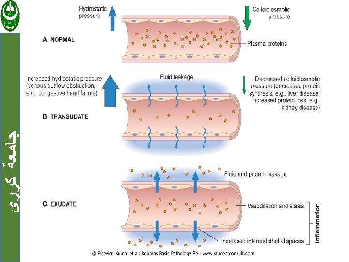
ﺟﺎﻣﻌﺔ ﻛﺮﺭﻱ Vascular Changes in acute inflammation: 1. Transient vasoconstriction (lasting only for seconds) 2. Vasodilation. 3. Increase vascular permeability leading to fluid leak and formation of exudates ( exudates = fluid rich in protein )
ﺟﺎﻣﻌﺔ ﻛﺮﺭﻱ Cellular Events in acute inflammation: Main cells involved are leukocytes (neutrophils) The cellular events are: (1) Margination of leukocytes. (2) Firm adhesion to the endothelium. (3) Transmigration between endothelial cells. (4) Migration in interstitial tissues toward infectious agent.
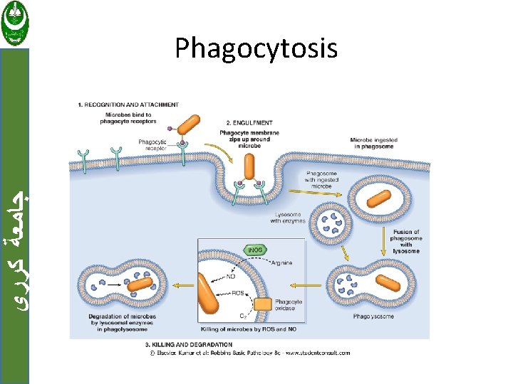
ﺟﺎﻣﻌﺔ ﻛﺮﺭﻱ Leukocyte Activation(Phagocytosis) Phagocytosis consists of three steps: (1) Recognition of the particle to the ingesting leukocyte. (2) Engulfment and formation of a phagocytic vacuole. (3) Killing and degradation of the ingested material.
ﺟﺎﻣﻌﺔ ﻛﺮﺭﻱ Mediators involved in acute inflammation : 1. Cell derived 2. Plasma protein derived.
Cell derived mediators: ﺟﺎﻣﻌﺔ ﻛﺮﺭﻱ 1. Vasoactive Amines: Histamine and serotonin are stored as preformed molecules in mast cells and other cells and are among the first mediators to be released in acute inflammatory reactions.
ﺟﺎﻣﻌﺔ ﻛﺮﺭﻱ Preformed histamine is released in response to a variety of stimuli: (1) physical injury such as trauma or heat. (2) immune reactions. (3) C 3 a and C 5 a fragments of complement, the so-called anaphylatoxins. (4) certain cytokines (e. g. , IL-1 and IL-8).
ﺟﺎﻣﻌﺔ ﻛﺮﺭﻱ Histamine causes arteriolar dilation and increased vascular permeability. Serotonin (5 -hydroxytryptamine) is a preformed vasoactive mediator, with effects similar to those of histamine.
ﺟﺎﻣﻌﺔ ﻛﺮﺭﻱ 2. Arachidonic Acid (AA) Metabolites: Prostaglandins, Leukotrienes, and Lipoxins. Their synthesis is increased at sites of inflammatory response. Leukocytes, mast cells, endothelial cells, and platelets are the major sources of AA metabolites in inflammation.
ﺟﺎﻣﻌﺔ ﻛﺮﺭﻱ AA metabolism proceeds along one of two major enzymatic pathways: 1. Cyclooxygenase stimulates the synthesis of prostaglandins and thromboxanes. 2. Lipoxygenase is responsible for production of leukotrienes and lipoxins.
ﺟﺎﻣﻌﺔ ﻛﺮﺭﻱ 3. Platelet-Activating Factor: Generated from the membrane phospholipids of neutrophils, monocytes, basophils, endothelial cells, and platelets by the action of phospholipase A 2.
ﺟﺎﻣﻌﺔ ﻛﺮﺭﻱ Platelet-Activating Factor cause: 1. vasoconstriction and bronchoconstriction. 2. enhanced leukocyte adhesion, chemotaxis, leukocyte degranulation. 3. stimulates the synthesis of other mediators.
ﺟﺎﻣﻌﺔ ﻛﺮﺭﻱ 4. Cytokines : The major cytokines in acute inflammation are: i. TNF and IL-1. ii. chemoattractant cytokines called chemokines (functions in inflammation is leukocyte recruitment). iii. Other cytokines that are more important in chronic inflammation (IFN-γ) and IL-12.
ﺟﺎﻣﻌﺔ ﻛﺮﺭﻱ TNF and IL-1 may enter the circulation and induce the systemic acute-phase reaction that is often associated with infection and inflammatory diseases: 1. Fever. 2. Lethargy. 3. Hepatic synthesis of various acute-phase proteins. 4. Metabolic wasting (cachexia). 5. Neutrophil release into the circulation
ﺟﺎﻣﻌﺔ ﻛﺮﺭﻱ 5. Reactive Oxygen Species : When secreted at low levels, ROS can increase chemokine, cytokine, and adhesion molecule expression, thus amplifying the cascade of inflammatory mediators.
ﺟﺎﻣﻌﺔ ﻛﺮﺭﻱ At higher levels, these mediators are responsible for tissue injury by : (1) endothelial damage, with thrombosis and increased permeability. (2) protease activation and antiprotease inactivation, with a net increase in breakdown of the ECM. (3) direct injury to the cell.
ﺟﺎﻣﻌﺔ ﻛﺮﺭﻱ 6. Nitric Oxide : Roles in inflammation, including (1) relaxation of vascular smooth muscle (vasodilation). (2) antagonism of platelet activation (adhesion, aggregation, and degranulation) (3) reduction of leukocyte recruitment at inflammatory sites. (4) action as a microbicidal agent in activated macrophages.
ﺟﺎﻣﻌﺔ ﻛﺮﺭﻱ 7. Lysosomal Enzymes of Leukocytes: The lysosomal granules of neutrophils and monocytes contain many molecules that can mediate acute inflammation. The most important of these lysosomal molecules are enzymes e. g acid proteases, neutral proteases.
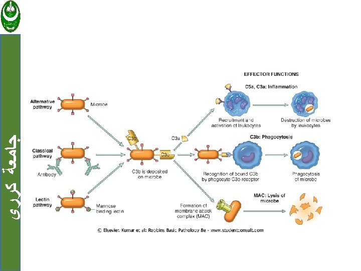
Plasma Protein-Derived Mediators: ﺟﺎﻣﻌﺔ ﻛﺮﺭﻱ 1. Complement: The complement system consists of plasma proteins that play role in host defense (immunity) and inflammation.
ﺟﺎﻣﻌﺔ ﻛﺮﺭﻱ • In inflammation complement proteins coat (opsonize) particles, such as microbes, for phagocytosis , and contribute to the inflammatory response by increasing vascular permeability and leukocyte chemotaxis
ﺟﺎﻣﻌﺔ ﻛﺮﺭﻱ • Also complement activation generates a porelike membrane attack complex (MAC) that punches holes in the membranes of invading microbes.
ﺟﺎﻣﻌﺔ ﻛﺮﺭﻱ 2. Coagulation and Kinin Systems : Activated Hageman factor (factor XIIa) initiates four systems involved in the inflammatory response: (1) the kinin system, producing vasoactive kinins (2) the clotting system, inducing the activation of thrombin, fibrinopeptides, and factor X, all with inflammatory properties (3) the fibrinolytic system, producing plasmin and inactivating thrombin (4) the complement system, producing the anaphylatoxins C 3 a and C 5 a.
ﺟﺎﻣﻌﺔ ﻛﺮﺭﻱ Role of Mediators in Different Reactions of Inflammation: Vasodilation Prostaglandins Nitric oxide Histamine Increased vascular permeability Histamine and serotonin C 3 a and C 5 a (by liberating vasoactive amines from mast cells, other cells) Bradykinin Leukotrienes C 4, D 4, E 4 PAF Substance P
TNF, IL-1 Chemokines C 3 a, C 5 a Leukotriene B 4 (Bacterial products, e. g. , N-formyl methyl peptides) Fever IL-1, TNF Prostaglandins Pain Prostaglandins Bradykinin Neuropeptides Tissue damage Lysosomal enzymes of leukocytes Reactive oxygen species Nitric oxide ﺟﺎﻣﻌﺔ ﻛﺮﺭﻱ Leukocyte recruitment and activation
MORPHOLOGIC PATTERNS OF ACUTE INFLAMMATION: ﺟﺎﻣﻌﺔ ﻛﺮﺭﻱ 1. Serous inflammation Characterized by watery, relatively proteinpoor fluid that , collecting in the site of injury , e. g; skin blister resulting from a burn or viral infection. Fluid in a serous cavity is called an effusion
ﺟﺎﻣﻌﺔ ﻛﺮﺭﻱ Serous inflammation of a skin blister showing the epidermis separated from the dermis by a focal collection of serous effusion
ﺟﺎﻣﻌﺔ ﻛﺮﺭﻱ 2. Fibrinous inflammation Occurs as a consequence of severe injuries, resulting in greater vascular permeability. Histologically, there is accumulated extravascular fibrin appears eosinophilic. A fibrinous exudate is characteristic of inflammation in the lining of body cavities, such as the meninges, pericardium, and pleura.
ﺟﺎﻣﻌﺔ ﻛﺮﺭﻱ Fibrinous pericarditis. A, Deposits of fibrin on the pericardium. B, A pink meshwork of fibrin exudate (F) overlies the pericardial surface (P).
ﺟﺎﻣﻌﺔ ﻛﺮﺭﻱ 3. Suppurative (purulent) inflammation : Charactrized by presence of large amounts of purulent exudate (pus) consisting of neutrophils, necrotic cells, and edema fluid. Certain organisms (e. g. , staphylococci) are more likely induce the reaction. Abscesses are focal collections of pus.
ﺟﺎﻣﻌﺔ ﻛﺮﺭﻱ Purulent inflammation. A, Multiple bacterial abscesses in the lung (arrows). B, The abscess contains neutrophils and cellular debris, and is surrounded by congested blood vessels
ﺟﺎﻣﻌﺔ ﻛﺮﺭﻱ 4. ulcer: Local defect, or excavation, of the surface of an organ or tissue that is produced by necrosis of cells and sloughing (shedding) of inflammatory necrotic tissue. It is most commonly occur in: (1) inflammatory necrosis of the mucosal surface. (2) tissue necrosis of extremities in persons who have circulatory disturbances.
ﺟﺎﻣﻌﺔ ﻛﺮﺭﻱ The morphology of an ulcer. A, A chronic duodenal ulcer. B, Low-power cross-section of a duodenal ulcer with an acute inflammatory exudate in the base.
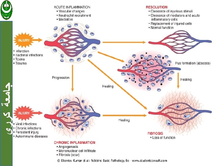
ﺟﺎﻣﻌﺔ ﻛﺮﺭﻱ Out come of acute inflammation: 1. Resolution 2. Healing by scarring and fibrosis 3. Progress to chronic inflammation