Common Pediatric Fractures Trauma Dr Kholoud AlZain Prof


 ratio •")


 Casting still the commonest")
 Casting still the commonest")
 K-wires • Most commonly used internal fixation (I. F) • Usually used in")
 Intramedullary wires (Elastic nails)")
 Screws")
 Plates specially in multiple trauma")
 I. M. N only in adolescents (>12 y)")
 Ex-fix usually in open #")


 ⅓ commonest middle")
: – No orthotics – Unite")


 • From the #: – Malunion – Nonunion –")


 may be: • Posteromedial (75%),")
 may be: • Posteromedial (75%),")


 – For 2")


: – Median and anterior interosseous")
 Myositis ossificans Angular deformity (cubitus")

")
 only one cortex is involved –")
 fracture: • Are stable • Immobilized for pain")
 fracture:")
: • Greater ability to remodel (why ? )")


- Slides: 100
Common Pediatric Fractures & Trauma Dr. Kholoud Al-Zain Prof. Zamzam Ass. Professor and Consultant Pediatric Orthopedic Surgeon Dec 2017
Objectives • • • Introduction Difference between Ped & adult Physis # Salter-Harris classification Indications of operative treatment Methods of treatment of Ped # & trauma Common Ped #: – U. L clavicle, humeral supracondylar, distal radius – L. L femur shaft • Example
Pediatric Fractures
Introduction • • Fractures account for ~15% of all injuries in children Boys > girls Rate increases with age Type of fractures vary in various age groups (infants, children, adolescents ) Mizulta, 1987
Difference Between A Child & Adult’s Fractures
Why are Children’s Fractures Different ? • Growth plate: – Perfect remodeling power – Injury of growth plate may cause: • Angular deformity • Or leg length inequality (L. L. I)
Why are Children’s Fractures Different ? • Bone: – Increased (collagen: bone) ratio • Less brittle • Deformation
Why are Children’s Fractures Different ? • Cartilage: – Difficult X-ray evaluation – Size of articular fragment often under-estimated
Why are Children’s Fractures Different ? • Periosteum: – Metabolically active • More callus, rapid union, increased remodeling – Thickness and strength • May aid reduction
Why are Children’s Fractures Different ? • Ligaments: – Functionally stronger than bone. – Higher proportion of injuries that produce sprains in adults result in fractures in children.
Why are Children’s Fractures Different ? • Age related fracture pattern: – Infants diaphyseal # – Children metaphyseal # – Adolescents epiphyseal
Why are Children’s Fractures Different ? • Physiology – Better blood supply rare delayed and non-union
Remodeling
Physis Fractures
Physis Injuries • • • Account for ~25% of all children’s # More in boys More in upper limb Most heal well rapidly with good remodeling Growth may be affected
Physis Injuries- Classifications Salter-Harris
Salter-Harris Classification
Salter-Harris Classification
Physis Injuries- Complications • Physeal bridging < 1% • Cause affecting growth (varus, valgus, or even L. L. I) • Keep in mind: – Small bridges (<10%) may lyse spontaneously – Central bridges more likely to lyse – Peripheral bridges more likely to cause deformity
Physis Injuries- Complications • Take care with: – Avoid injury to physis during fixation – Monitor growth over a long period (18 -24 m) – When suspecting physeal bar do MRI
Indications of Operative Treatment
General Management Indications for surgery • • • Open fractures Severe soft-tissue injury Fractures with vascular injury Compartment syndrome Multiple injuries Displaced intra articular fractures (Salter-Harris III-IV ) Failure of conservative means (irreducible or unstable #’s) Malunion and delayed union Adolescence Head injury Neurological disorder Uncooperative patient
Methods of Treatment of Pediatric Fractures & Trauma
1) Casting still the commonest
1) Casting still the commonest
2) K-wires • Most commonly used internal fixation (I. F) • Usually used in metaphyseal fractures
3) Intramedullary wires (Elastic nails)
4) Screws
5) Plates specially in multiple trauma
6) I. M. N only in adolescents (>12 y)
7) Ex-fix usually in open #
Methods of Fixation Co a n i mb n o i t
Common Pediatric Fractures
Common Pediatric Fractures • Upper limb: – Clavicle – Humeral supracondylar – Distal radius • Lower Limbs: – Femur shaft (diaphysis)
Clavicle Fractures
Clavicle # - Incidents • • • 8 -15% of all pediatric # 0. 5% of normal SVD 1. 6% of breech deliveries 90% of obstetric # The periosteal sleeve always remains in the anatomic position (remodeling is ensured)
Clavicle # - Mechanism Injury • Indirect fall onto an outstretched hand • Direct: – The most common mechanism – Has highest incidence of injury to the underlying: • N. V &, • Pulmonary structures • Birth injury
Clavicle # - Examination • Look Ecchymosis • Feel: – Tender # site – As a palpable mass along the clavicle (as in displaced #) – Crepitus (when lung is compromised) • Special tests Must assesse for any: – N. V injury – Pulmonary injury
Clavicle # - Reading XR • Location: – (medial, middle, lateral) ⅓ commonest middle ⅓ – Commonest # site middle/lateral ⅓ • Open or closed see air on XR • Displacement % • Fracture type
Clavicle # - Treatment • Newborn (< 28 days): – No orthotics – Unite in 1 w • 1 m – 2 y: – Figure-of-eight – For 2 w • 2 – 12 y: – Figure-of-eight or sling – For 2 -4 weeks
Clavicle # - Remodeling
Clavicle # - Treatment Indications of operative treatment: • Open #’s, or • Neurovascular compromise
Clavicle # - Complications (rare) • From the #: – Malunion – Nonunion – Secondary from healing: • Neurovascular compromise • Pulmonary injury • In the wound: – Bad healed scar – Dehiscence – Infection
Humeral Supracondylar Fractures
Supracondylar #- Incidences • • 55 -75% of all elbow # M: F 3: 2 Age 5 - 8 years Left (non-dominant) side most frequently #
Supracondylar #- Mechanism of Injury • Indirect: – Extension type – >95% • Direct: – Flexion type – < 3%
Supracondylar #- Clinical Evaluation • Look: – Swollen – S-shaped angulation – Pucker sign (dimpling of the skin anteriorly) – May have burses • Feel: – Tender elbow • Move: – Painful & can’t really move it • Neurovascular examination
Supracondylar #- Gartland Classification Type-III Complete displacement (extension type) may be: • Posteromedial (75%), or • Posterolateral (25%)
Supracondylar #- Gartland Classification Type-III Complete displacement (extension type) may be: • Posteromedial (75%), or • Posterolateral (25%)
Supracondylar #- Gartland Classification
Normal XR Lines • • Anterior Humeral Line Hour-glass appearance Fat-pad sign Radio-capitellar line
Type 1 • • Anterior Humeral Line Hour-glass appearance Fat-pad sign Radio-capitellar line
Type 2
Type 3
Supracondylar #- Treatment • Type-I: – Above elbow cast (or splint) – For 2 -3 weeks • Type-II: – Closed reduction & above elbow casting, or – Closed reduction with percutaneous pinning (if: unstable or sever swelling), & above elbow cast (splint) – For 4 -6 weeks • Type III: – Attempt closed reduction & percutaneous pinning – If fails open reduction & pinning (ORIF) – For 4 -6 weeks – Direct ORIF if open #
Supracondylar #- Treatment
Supracondylar #- Treatment
Supracondylar #- Complications • Neurologic injury (7% to 10%): – Median and anterior interosseous nerves (most common) – Most are neurapraxias – Requiring no treatment • Vascular injury (0. 5%): – Direct injury to the brachial artery, or – Secondary to swelling (compartment syndrome)
Supracondylar #- Complications • • Loss of motion (stiffness) Myositis ossificans Angular deformity (cubitus varus) Compartment syndrome
Supracondylar #- Flexion Type 3
Distal Radial Fractures (Metaphysis)
Classification • Depending on pattern: – Torus (buckle) only one cortex is involved – Incomplete (greenstick) – Complete
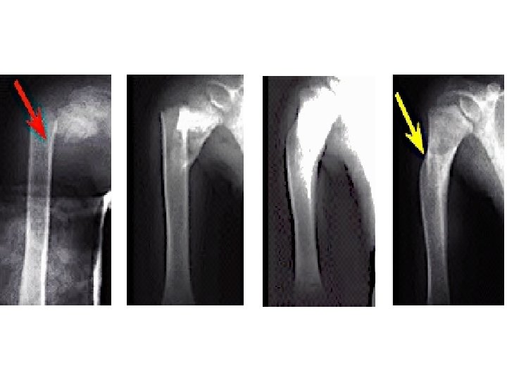
Distal Radius Metaphyseal Injuries Torus (buckle) fracture: • Are stable • Immobilized for pain relief in below elbow cast, 2 -3 weeks
Distal Radius Metaphyseal Injuries Torus (buckle) fracture:
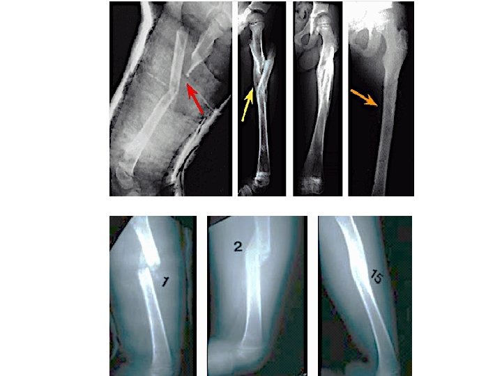
Distal Radius Metaphyseal Injuries Incomplete (greenstick): • Greater ability to remodel (why ? ) • Closed reduction and above elbow cast
Distal Radius Metaphyseal Injuries Complete fracture: • Closed reduction, then well molded above elbow cast for 6 -8 w • Or open reduction and fixation (internal or external)
Distal Radius Metaphyseal Injuries Complete fracture: • Closed reduction, then well molded above elbow cast for 6 -8 w
Distal Radius Metaphyseal Injuries Complete fracture: • Or open reduction and fixation (internal or external)
Distal Radius Metaphyseal Injuries Complete fracture: Indications for ORIF: • Irreducible fracture • Open fracture • Compartment syndrome
Distal Radius Meta. Injuries- Complications • Malunion Residual angulation may result in loss of forearm rotation • Nonunion Rare • Refracture With early return to activity (before 6 w) • Growth disturbance Overgrowth or undergrowth • Neurovascular injuries With extreme positions of immobilization
Examples of Distal Radial Fractures
Distal Radial Fractures Physeal Injuries
Distal Radial Physeal #- “S. H” Type I
Distal Radial Physeal #- “S. H” Type II
Distal Radial Physeal #- “S. H” Type III
Distal Radial Physeal #- Treatment Types I & II • Closed reduction followed by above elbow cast • We can accept deformity: – 50% translation – With no angulation or rotation • Growth arrest can occur in 25% with repeated closed reduction manipulations • Open reduction is indicated in: – Irreducible # – Open #
Distal Radial Physeal #- Treatment Types II AP Lat
Distal Radial Physeal #- Treatment Types II
Distal Radial Physeal #- Types III
Distal Radial Physeal #- Treatment Types III • Anatomic reduction necessary intra-articular • ORIF with smooth pins or screws
Distal Radial Physeal #- Treatment Types IV & V • Rare injuries • Need ORIF
Distal Radial Physeal #- Complications • Physeal arrest – Shortening – Angular deformity • Ulnar styloid nonunion • Carpal tunnel syndrome
Femoral Shaft Fractures
Femoral Shaft # • 1. 6% of all pediatric # • M>F • Age: – (2 – 4) years old – Mid-adolescence • Adolescence >90% due to RTA
Femoral Shaft #- Mechanism of Injury • Direct trauma: – RTA, – Fall, or • Indirect trauma: – Rotational injury • Pathologic #: – Osteogenesis imperfecta – Nonossifying fibroma – Bone cysts – Tumors
Femoral Shaft #- Clinical Evaluation • Look: – – – Pain, Swelling of the thigh, Inability to ambulate, and Variable gross deformity Careful O/E of the overlying soft tissues to rule out the possibility of an open fracture (puncture wound) • Feel: – Tender # site • Careful neurovascular examination is essential
Femoral Shaft #- Treatment < 6 m: • Pavlik Harness • Closed reduction & immediate hip spica casting • Or traction 1 -2 w, then hip spica casting
Femoral Shaft #- Treatment 6 m – 6 y: • Closed reduction & immediate hip spica casting (>95%) • Or traction 1 -2 w, then hip spica casting
Femoral Shaft #- Treatment 6 – 12 y: • Flexible I. M. N • Bridge Plating • External Fixation
Femoral Shaft #- Treatment 6 – 12 y: • Flexible IMN • Bridge Plating • External Fixation
Femoral Shaft #- Treatment 6 – 12 y: • Flexible IMN • Bridge Plating • External Fixation: – Multiple injuries – Open fracture – Comminuted # – Unstable patient
Femoral Shaft #- Treatment 12 y to skeletal maturity: • Intramedullary fixation with either: – Flexible nails, or – Locked I. M nail
Femoral Shaft #- Treatment Operative Indications: • • • Multiple trauma, including head injury Open fracture Vascular injury Pathologic fracture Uncooperative patient
Femoral Shaft #- Complications • Malunion – Remodeling will not correct rotational deformities • Leg length discrepancy – Secondary to shortening or overgrowth • Muscle weakness • Nonunion (rare)
Any Questions?
Remember …
Remember • Pediatric fractures have great remodeling potentials • The importance of growth plates & periosteum in remodeling • A good number of cases can be treated conservatively • Operative fixations aids in avoiding complications
Objectives • Difference between adult & pediatric # • Growth plate # Salter-Harris classification, treatments, & complications • Methods of treatment of pediatric # & there indications • Know the common pediatric #: mechanism of injury, evaluations (clinical & radiological), treatments, and complications