UROLOGICAL TRAUMA RENAL TRAUMA n Renal trauma occurs


")
 n Vascular injury")

,")


: the method allows to find damage of bones,")


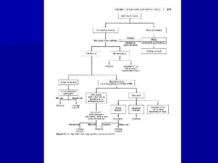
 n Staging begins with an abdominal CT scan, the most direct")

 internal bleedings in case of isolated")


- Slides: 54
UROLOGICAL TRAUMA
RENAL TRAUMA n Renal trauma occurs in approximately 1 -5% of all traumas. n Renal injuries are the most common injuries of the urinary system. n Blunt trauma directly to the abdomen, flank, or back is the most common mechanism, accounting for 80 -85% of all renal injuries.
Ethiology Trauma may result from motor vehicle accidents, fights, falls, and contact sports. n Fractured ribs and transverse vertebral processes may penetrate the renal parenchyma or vasculature. n n Gun-shot and knife wounds cause most penetrating injuries to the kidney.
Classification
Classification n Minor renal trauma n Major renal trauma (15% of cases)
Classification n Vascular injury (about 1% of all blunt trauma cases) n Vascular injury of the renal pedicle is rare but may occur, usually from blunt trauma.
Clinical Findings n The clinic of the closed damage of kidney depends on its degree. n Each kind of trauma is accompanied by characteristic manifestations and general signs, which are pain and intumescence in lumbar region, haematuria.
Signs • Shock symptoms • Flank ecchimosis, edema • Pain • Hematuria (mikro-, gross), bladder • Oligoanuria, Subileus tamponade
Clinical Findings n Symptoms: n Pain in lumbar region on the side of damage is observed in 80 - 95 % of cases of isolated traumas of kidney and in 10 -20 % of combined injuries. It is dull or acute with irradiation in inguinal region or external sexual organs.
Diagnostics n Laboratory Findings: n Microscopic or gross hematuria is usually present. n The hematocrit may be normal initially, but a drop may be found when serial studies are done. Persistent bleeding may necessitate operation.
Diagnostics: Chromocystoscopia n Chromocystoscopy, if possible, also helps to establish the correct diagnosis. n This method of research sometimes allows to find a location of bleeding (that it is very important in case of combined trauma), to analyse functions of damaged and opposite kidney, state of urinary bladder wall. n
X-Ray Findings n Observing radiography (KUB): the method allows to find damage of bones, to suspect presence of retroperitoneal hematoma (contours of kidney and lumbar muscles are absent). n Excretory urography gives an opportunity to define the side of damage, anatomical and function status of injured and opposite kidney.
X-Ray Findings n . X-ray signs of renal damage are weak and later spreading of X-ray contrast solution in calyces-bowling systems, subcapsular and retrorenal spreading of X-ray contrast, deformation of renal bowl and calyces.
• IVP
X-Ray Findings n On angiogramms one can see violation of arterial and venous circulation attached to marginal injuries, filling of pararenal tissue with X-ray contrast due to injuries of renal artery branches.
Ultrasonography n Ultrasound scans can detect renal lacerations but cannot definitely assess their depth and extent. In addition, they do not provide functional information.
Computed tomography (CT) n Staging begins with an abdominal CT scan, the most direct and effective means of staging renal injuries.
TREATMENT n Bed regimen is provided within 10 -20 day. n Measures to stop bleeding (administration of haemostatic agents, hemo- and plasmotransfusion), administration of analgetics, antibiotics of a wide spectrum of action, and also dynamical overseeing by arterial pressure. n Antibiotics are used for pyelonephritis prophylactics.
TREATMENT n Indications to operative treatment: n а) internal bleedings in case of isolated renal damage, which are accompanied by an anaemia, decrease of arterial pressure, fast pulse; b) hematuria within a day with worsening of general state of the patient; c) hematoma in lumbar region, which is slowly growing; d) combination of renal damage and organs of abdominal cavity or thorax. n n n
TREATMENT n The operation should be maximum savings and directed on the decision of two tasks - stopping of bleeding and normalization of urine outflow.
TREATMENT
INJURIES OF THE URINARY BLADDER Bladder injuries occur most often from external force and are often associated with pelvic fractures. (About 15% of all pelvic fractures are associated with concomitant bladder or urethral injuries. ) n Iatrogenic injury may result from gynecologic and other extensive pelvic procedures as well as from hernia repairs and transurethral operations. n
Classification closed and open n isolated and combined n intraperitoneal, retroperitoneal and mixed. n
Bladder injuries 1. Intraperitoneal n Blunt trauma in filled bladder Rising of intravesical pressure Hydraulic impact Intraperitoneal bladder rapture (dome, posterior wall) 2. Extraperitoneal
Clinical Findings n Symptoms: n There is usually a history of lower abdominal trauma. Blunt injury is the usual cause. Patients ordinarily are unable to urinate, but when spontaneous voiding occurs, gross hematuria is usually present. Most patients complain of pelvic or lower abdominal pain. n n n
Clinical Findings n Signs: Heavy bleeding associated with pelvic fracture may result in hemorrhagic shock, usually from venous disruption of pelvic vessels. n An acute abdomen indicates intraperitoneal bladder rupture. n A palpable mass in the lower abdomen usually represents a large pelvic hematoma. n On rectal examination, landmarks may be indistinct because of a large pelvic hematoma. n
Clinical Findings n Laboratory Findings: Catheterization usually is required in patients with pelvic trauma but not if bloody urethral discharge is noted. n When catheterization is done, gross or, less commonly, microscopic hematuria is usually present. n
X-Ray Findings n A plain abdominal film generally demonstrates pelvic fractures. There may be haziness over the lower abdomen from blood and urine extravasation. n An intravenous urogram should be obtained to establish whether kidney and ureteral injuries are present.
X-Ray Findings n Bladder disruption is shown on cystography. n The sign of retroperitoneal rupture is accumulation of X-ray contrast matter in perivesical fat tissue. n With intraperitoneal extravasation, free contrast medium is visualized in the abdomen, highlighting bowel loops.
X-Ray Findings
Treatment n n A. Emergency Measures: Shock and hemorrhage should be treated. n B. Surgical Measures: n The bladder should be opened in the midline and carefully inspected. After repair, a suprapubic cystostomy tube is usually left in place to ensure complete urinary drainage and control of bleeding.
Treatment n In a case of retroperitoneal complete rupture of the bladder it is exposed by suprapubic extraperitoneal access carefully inspected and is juncture by two-row catgut junctures.
Treatment n Intraperitoneal bladder ruptures should be repaired via a transperitoneal approach after careful transvesical inspection and closure of any other perforations.
INJURIES Of THE URETHRA n Urethral injuries are uncommon and occur most often in men, usually associated with pelvic fractures or straddle-type falls. n The urethra can be separated into 2 broad anatomic divisions: the posterior urethra, consisting of the prostatic and membranous portions, and the anterior urethra, consisting of the bulbous and pendulous portions
Clinical Findings n Signs: n Blood at the urethral meatus is the single most important sign of urethral injury (Urethroragia).
Clinical Findings n n Suprapubic tenderness and the presence of pelvic fracture are noted on physical examination. A large developing pelvic hematoma may be palpated. Perineal or suprapubic contusions are often noted. Rectal examination may reveal a large pelvic hematoma with the prostate displaced superiorly.
Clinical Findings n Instrumental Examination: n The only instrumentation involved should be for urethrography. n Catheterization or urethroscopy should not be done, because these procedures pose an increased risk of hematoma, infection, and further damage to partial urethral disruptions.
X-Ray Findings n Fractures of the bony pelvis are usually present.
X-Ray Findings
Treatment In case of ischuria instead of a high cystotomy it is possible to perform troacar epicystostomy. n Shock and hemorrhage should be n treated.
Treatment n Surgical Measures: n Urethral catheterization should be avoided. Initial management should consist of suprapubic cystostomy to provide urinary drainage. n n A midline lower abdominal incision should be made, care being taken to avoid the large pelvic hematoma. n The suprapubic cystostomy is maintained in place for about 3 months. This allows resolution of the pelvic hematoma, and the prostate and bladder will slowly return to their anatomic positions.
Treatment n Urethral reconstruction -Reconstruction of the urethra after prostatic disruption can be undertaken within 3 months.
Complications n Stricture, impotence, and incontinence as complications of prostatomembranous disruption.
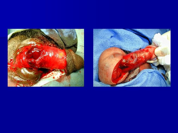
Penis fracture Tunica albuginea rapture
Penis trauma
Paraphimosis
Testicle torsion DD: Epididymitis acute begining Doppler sonography In doubt : always operation 4 -6 hours
Skrotal hematoma
Testicle trauma
Testicle trauma
Priapism 4 -6 h time limit Punction + aspiration from 50 to 200 ml of blood Injektion of 5 mg Alpha-Sympathomimetics (Effortil) Winter Shunting fistula
THANK YOU FOR ATTENTION