Tanya L Zakrison Ryder Trauma Center March 02


- Slides: 35
Tanya L. Zakrison Ryder Trauma Center March 02, 2010 Principles of Vascular Surgery - Popliteal injuries
Case 1 ID: 23 M RFA: GSW to right knee In trauma bay ATLS ▪ HD stable ▪ Base deficit -3 Right dorsalis pedis absent What is next? What if this was a SGW?
Case 1 Xray: Comminuted fracture distal femur Ortho wants to ex-fix Who goes first? what if there is associated nerve injury – should you bother with revascularization? What is the surgical approach? What type of incision will you make? How will you repair the popliteal artery?
Case 2 ID: 34 M RFA: MVC , belted passenger In trauma bay ATLS HD stable Left posterior knee dislocation ▪ Reduced by ortho ▪ Palpable DP, PT What is next?
Case 2 Left ABI = 0. 90 How do you calculate ABI? What is next? What is the risk of injury to the popliteal artery? What is the best investigative modality to rule out arterial injury? The injured leg is cold 4 hours later What happened?
Case 3 ID: 30 M RFA: multiple GSWs Torso, abdomen, extremities In trauma bay HD unstable Base deficit = -16 GSW to left distal thigh ▪ Expanding hematoma FAST is (+)
Case 3 What is next? Liver packed Abdomen is closed with a bogota bag Pt. stabilizing, base deficit still -10 What is next?
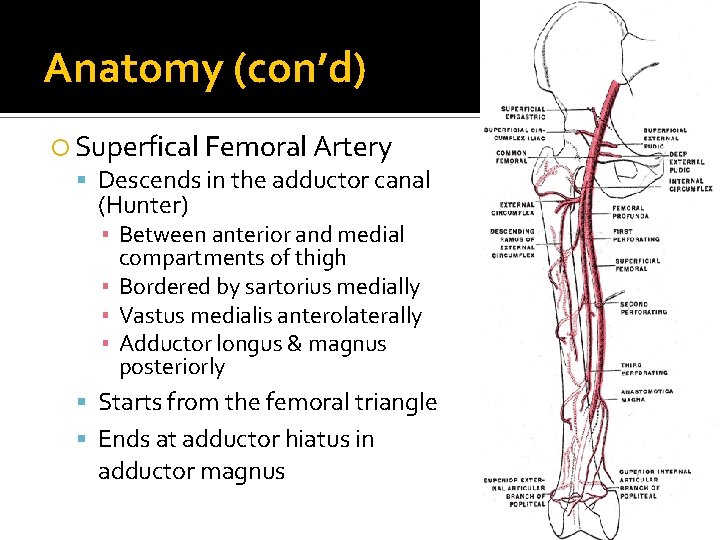
Anatomy Common femoral artery Superficial femoral artery Profunda femoris ▪ Major blood supply to thigh Femoral triangle bounded by: Inguinal ligament (superior) Medial border of sartorius (lateral) Medial border of adductor longus & pectineus (medial) Floor of triangle: Iliacus, psoas major, pectineus, adductor longus
Anatomy (con’d) Superfical Femoral Artery Descends in the adductor canal (Hunter) ▪ Between anterior and medial compartments of thigh ▪ Bordered by sartorius medially ▪ Vastus medialis anterolaterally ▪ Adductor longus & magnus posteriorly Starts from the femoral triangle Ends at adductor hiatus in adductor magnus
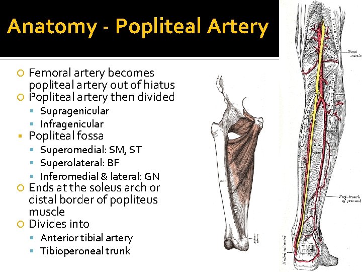
Anatomy - Popliteal Artery Femoral artery becomes popliteal artery out of hiatus Popliteal artery then divided Supragenicular Infragenicular Popliteal fossa Superomedial: SM, ST Superolateral: BF Inferomedial & lateral: GN Ends at the soleus arch or distal border of popliteus muscle Divides into Anterior tibial artery Tibioperoneal trunk
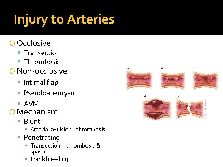
Injury to Arteries Occlusive Transection Thrombosis Non-occlusive Intimal flap Pseudoaneurysm AVM Mechanism Blunt ▪ Arterial avulsion - thrombosis Penetrating ▪ Transection – thrombosis & spasm ▪ Frank bleeding
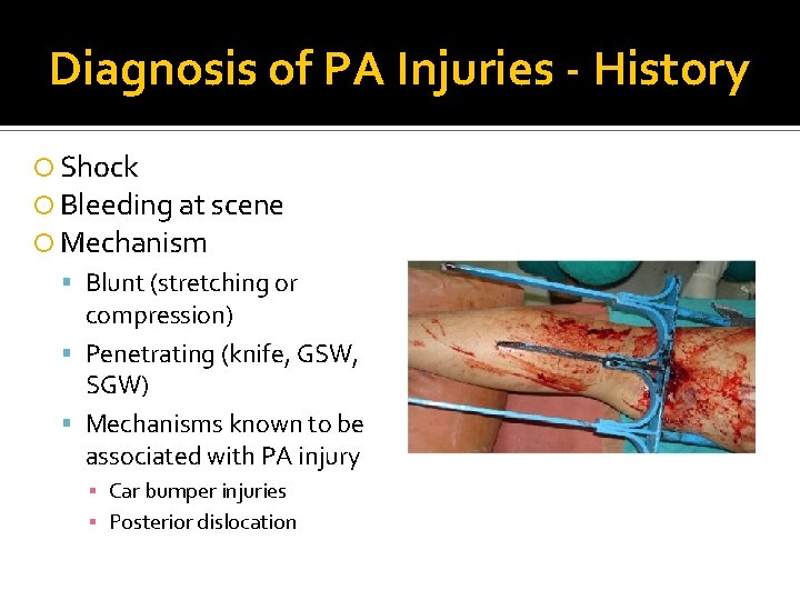
Diagnosis of PA Injuries - History Shock Bleeding at scene Mechanism Blunt (stretching or compression) Penetrating (knife, GSW, SGW) Mechanisms known to be associated with PA injury ▪ Car bumper injuries ▪ Posterior dislocation
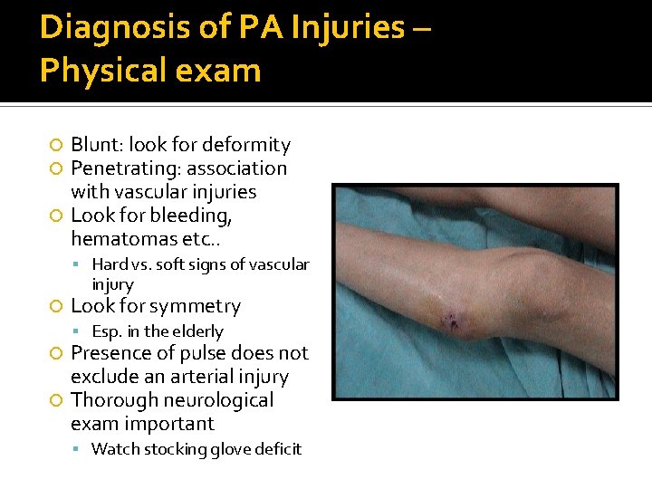
Diagnosis of PA Injuries – Physical exam Blunt: look for deformity Penetrating: association with vascular injuries Look for bleeding, hematomas etc. . Hard vs. soft signs of vascular injury Look for symmetry Esp. in the elderly Presence of pulse does not exclude an arterial injury Thorough neurological exam important Watch stocking glove deficit
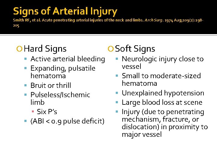
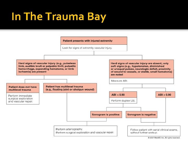
Signs of Arterial Injury Smith RF, et al. Acute penetrating arterial injuries of the neck and limbs. Arch Surg. 1974 Aug; 109(2): 198205 Hard Signs Soft Signs Active arterial bleeding Neurologic injury close to vessel Expanding, pulsatile hematoma Small to moderate-sized hematoma Bruit or thrill Unexplained hypotension Pulseless/Ischemic limb Large blood loss at scene ▪ Six P’s Injury (due to penetrating mechanism, fracture, or (ABI < 0. 9 pulse deficit) dislocation) in proximity to major vessel
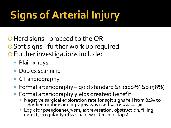
Signs of Arterial Injury Hard signs - proceed to the OR Soft signs - further work up required Further investigations include: Plain x-rays Duplex scanning CT angiography Formal arteriography – gold standard Sn (100%) Sp (98%) Formal arteriography yields greatest benefit ▪ Negative surgical exploration rate for soft signs fell from 84% to 2% when routine angiography was used Reid JDS, Arch Surg 1988 ▪ Look for pseudoaneurysm, extravasation, obstruction, filling defect, irregularity of vascular wall (intimal flaps)
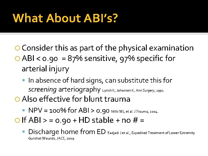
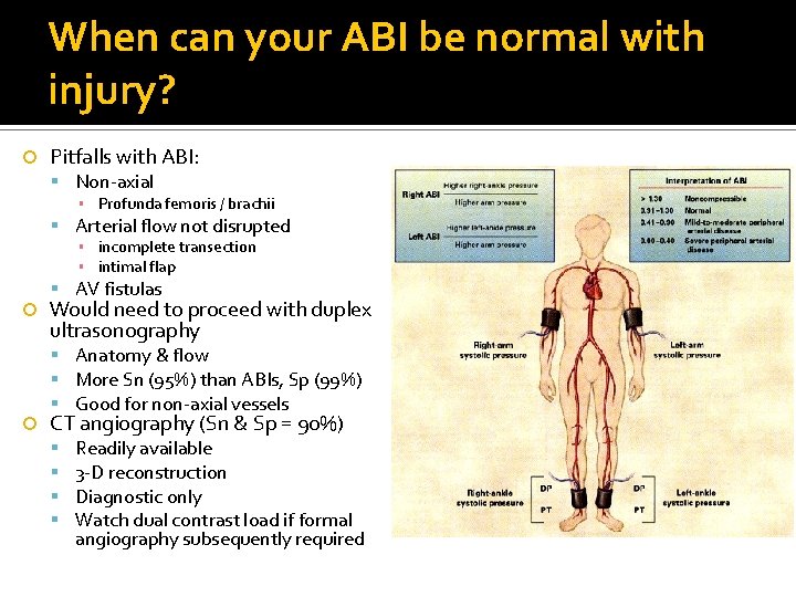
What About ABI’s? Consider this as part of the physical examination ABI < 0. 90 = 87% sensitive, 97% specific for arterial injury In absence of hard signs, can substitute this for screening arteriography Lynch K, Johansen K, Ann Surgery, 1991. Also effective for blunt trauma NPV = 100% for ABI > 0. 90 Mills WJ, et al. J Trauma, 2004. If ABI > = 0. 90 + HD stable + no # = Discharge home from ED Sadjadi J et al. , Expedited Treatment of Lower Extremity Gunshot Wounds, JACS, 2009.
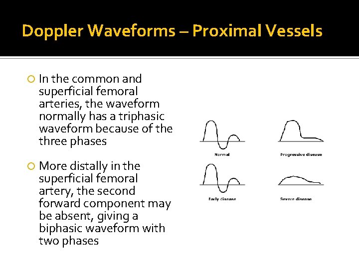
Doppler Waveforms – Proximal Vessels In the common and superficial femoral arteries, the waveform normally has a triphasic waveform because of the three phases More distally in the superficial femoral artery, the second forward component may be absent, giving a biphasic waveform with two phases
When can your ABI be normal with injury? Pitfalls with ABI: Non-axial ▪ Profunda femoris / brachii Arterial flow not disrupted ▪ incomplete transection ▪ intimal flap AV fistulas Would need to proceed with duplex ultrasonography Anatomy & flow More Sn (95%) than ABIs, Sp (99%) Good for non-axial vessels CT angiography (Sn & Sp = 90%) Readily available 3 -D reconstruction Diagnostic only Watch dual contrast load if formal angiography subsequently required
Indications for Arteriography Unclear location or extent of injury Extensive soft tissue injury Fracture or dislocation Trajectory parallel to an artery Multiple wounds Shotgun injuries Peripheral vascular disease Intraoperative angiography reduces warm ischemia times, reducing the likelihood of amputation in PA injuries Callcut R, et al. Impact of Intraoperative Arteriography on Limb Salvage for Traumatic Popliteal Artery Injury, J Trauma, 2009
Approach to Vascular Injury Ligate vs. repair vs. shunt? Repair Primary or secondary (graft)? ▪ Arteriorrhaphy ▪ End to end anastomosis Autogenous or prosthetic conduit? Autogenous ▪ Reversed greater saphenous vein ▪ Any other superficial vein Prosthetic ▪ PTFE ▪ Dacron ▪ other Shunt In line (Argyle, Javid etc. . . ) or looped (Pruitt-Inahara)
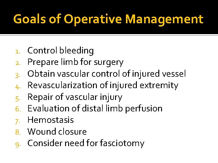
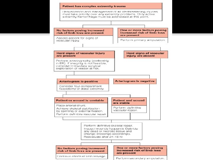
Goals of Operative Management 1. 2. 3. 4. 5. 6. 7. 8. 9. Control bleeding Prepare limb for surgery Obtain vascular control of injured vessel Revascularization of injured extremity Repair of vascular injury Evaluation of distal limb perfusion Hemostasis Wound closure Consider need for fasciotomy
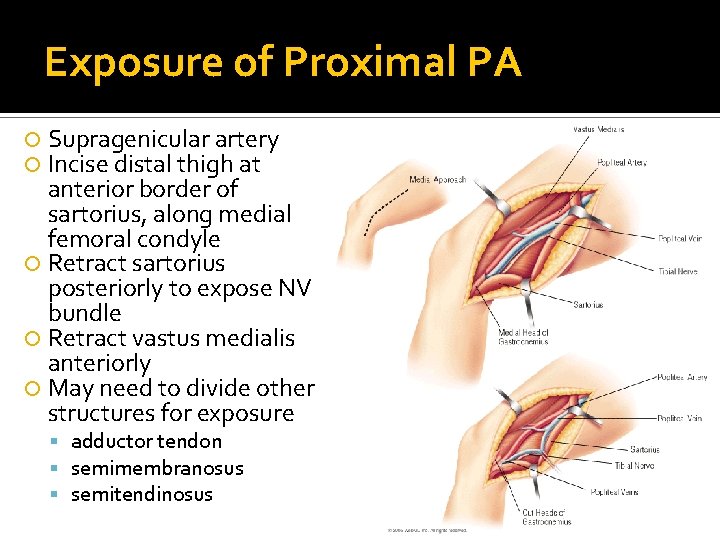
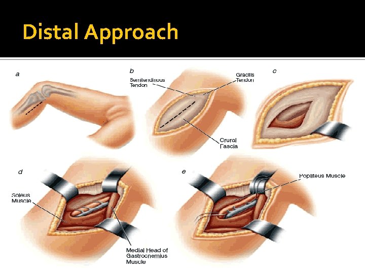
Exposure of Proximal PA Supragenicular artery Incise distal thigh at anterior border of sartorius, along medial femoral condyle Retract sartorius posteriorly to expose NV bundle Retract vastus medialis anteriorly May need to divide other structures for exposure adductor tendon semimembranosus semitendinosus
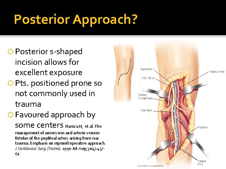
Posterior Approach? Posterior s-shaped incision allows for excellent exposure Pts. positioned prone so not commonly used in trauma Favoured approach by some centers Hamza N, et al. The management of aneurysms and arterio-venous fistulae of the popliteal artery arising from war trauma. Emphasis on sigmoid operative approach. J Cardiovasc Surg (Torino). 1990 Jul-Aug; 31(4): 45761.
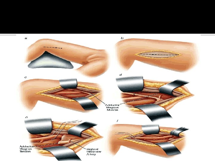
Distal Approach
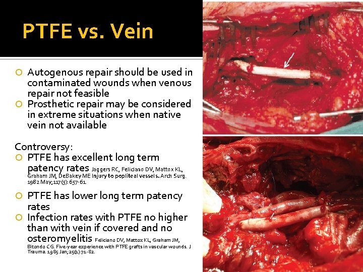
PTFE vs. Vein Autogenous repair should be used in contaminated wounds when venous repair not feasible Prosthetic repair may be considered in extreme situations when native vein not available Contr 0 versy: PTFE has excellent long term patency rates Jaggers RC, Feliciano DV, Mattox KL, Graham JM, De. Bakey ME Injury to popliteal vessels. Arch Surg. 1982 May; 117(5): 657 -61. PTFE has lower long term patency rates Infection rates with PTFE no higher than with vein if covered and no osteromyelitis Feliciano DV, Mattox KL, Graham JM, Bitondo CG. Five-year experience with PTFE grafts in vascular wounds. J Trauma. 1985 Jan; 25(1): 71 -82.
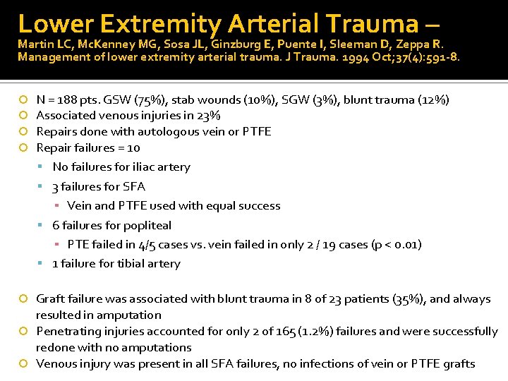
Lower Extremity Arterial Trauma – Martin LC, Mc. Kenney MG, Sosa JL, Ginzburg E, Puente I, Sleeman D, Zeppa R. Management of lower extremity arterial trauma. J Trauma. 1994 Oct; 37(4): 591 -8. N = 188 pts. GSW (75%), stab wounds (10%), SGW (3%), blunt trauma (12%) Associated venous injuries in 23% Repairs done with autologous vein or PTFE Repair failures = 10 No failures for iliac artery 3 failures for SFA ▪ Vein and PTFE used with equal success 6 failures for popliteal ▪ PTE failed in 4/5 cases vs. vein failed in only 2 / 19 cases (p < 0. 01) 1 failure for tibial artery Graft failure was associated with blunt trauma in 8 of 23 patients (35%), and always resulted in amputation Penetrating injuries accounted for only 2 of 165 (1. 2%) failures and were successfully redone with no amputations Venous injury was present in all SFA failures, no infections of vein or PTFE grafts
Lower Extremity Arterial Trauma – Martin LC, Mc. Kenney MG, Sosa JL, Ginzburg E, Puente I, Sleeman D, Zeppa R. Management of lower extremity arterial trauma. J Trauma. 1994 Oct; 37(4): 591 -8. Conclusions: PTFE and vein have equal graft patency for the repair of the iliac and femoral arteries The patency of PTFE was significantly worse in the popliteal location Graft failure and amputation were more common with popliteal and tibial injuries from blunt mechanisms
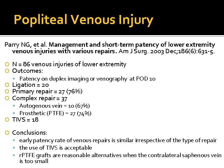
Popliteal Venous Injury Parry NG, et al. Management and short-term patency of lower extremity venous injuries with various repairs. Am J Surg. 2003 Dec; 186(6): 631 -5. N = 86 venous injuries of lower extremity Outcomes: Patency on duplex imaging or venography at POD 10 Ligation = 20 Primary repair = 27 (76%) Complex repair = 37 Autogenous vein = 10 (67%) Prosthetic (PTFE) = 27 (74%) TIVS = 18 Conclusions: early patency rate of venous repairs is similar irrespective of the type of repair the use of TIVS is acceptable r. PTFE grafts are reasonable alternatives when the contralateral saphenous vein is too small
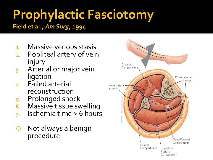
Prophylactic Fasciotomy Field et al. , Am Surg, 1994 1. 2. 3. 4. 5. 6. 7. Massive venous stasis Popliteal artery of vein injury Arterial or major vein ligation Failed arterial reconstruction Prolonged shock Massive tissue swelling Ischemia time > 6 hours Not always a benign procedure
Associated Nerve Injury Only 7% of patients with concomitant nerve and vascular injury will have a functioning limb, despite successful vascular repair, vs. 39% of patients with arterial injury alone Visser et al. Am J Surg, 1980 Spontaneous recovery may occur in 69% of patients with nerve injuries due to GSWs between 3 -9 months after injury Expected with axonotmesis and neuropraxia Omer GF. , Hand Clinic, 1986
When is Amputation a Concern? N = 24 civilian injuries 1975 -1998 Penetrating trauma – 10. 5% amputation Blunt trauma - 27. 5% - 71% amputation Frykberg E - Popliteal vascular injuries, Surgical clinics of North America, 2002. Factors associated with amputation Yahya et al. ANZ J Surg, 2005 ▪ ▪ Extensive soft-tissue injuries Associated skeletal trauma Knee dislocations Prolonged ischemic times MESS score > 6 predicts need for amputation Accuracy 100% Criteria include: age, shock, skeletal/soft tissue injury, limb ischemia Also determined by neurological injury, failure of arterial repair, associated fractures
In The Trauma Bay
Thank you!