
Head and Facial Injury Scott Marquis MD Overview
- Slides: 136
Head and Facial Injury Scott Marquis, MD
Overview n n n Head injury What to look for Appropriate management Facial injury Review
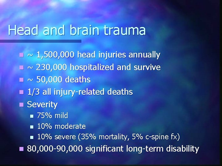
Head and brain trauma n n n ~ 1, 500, 000 head injuries annually ~ 230, 000 hospitalized and survive ~ 50, 000 deaths 1/3 all injury-related deaths Severity n n 75% mild 10% moderate 10% severe (35% mortality, 5% c-spine fx) 80, 000 -90, 000 significant long-term disability
Head & brain trauma n Risk Groups n n Highest: Males 15 -24 yrs of age Very young children: 6 mos to 2 yrs of age Young school age children Elderly >75 yrs
Head injury n Broad and Inclusive Term n n n Traumatic insult to the head that may result in injury to soft tissue, bony structures, and/or brain injury Blunt Trauma Penetrating Trauma
Brain injury “A traumatic insult to the brain capable of producing physical, intellectual, emotional, social and vocational changes” n Three broad categories n n Focal injury n Cerebral contusion n Intracranial hemorrhage n Epidural hemorrhage Subarachnoid hemorrhage Diffuse Axonal Injury n Concussion
Mechanisms of head injury n Motor vehicle crashes, MVC n n Most common cause of head trauma Most common cause of subdural hematoma Sports injuries n Falls n n Common in elderly and in presence of alcohol Associated with subdural hematomas Penetrating trauma n Missiles more common than sharp projectiles
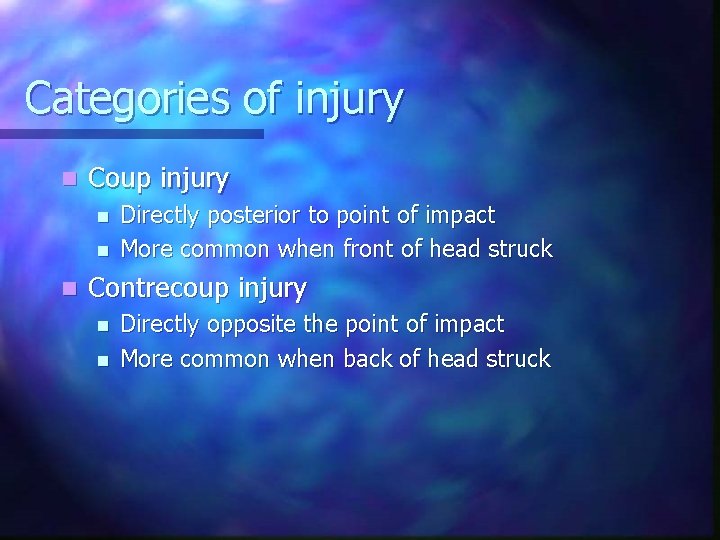
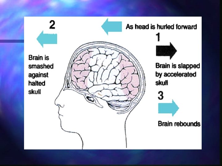
Categories of injury n Coup injury n n n Directly posterior to point of impact More common when front of head struck Contrecoup injury n n Directly opposite the point of impact More common when back of head struck
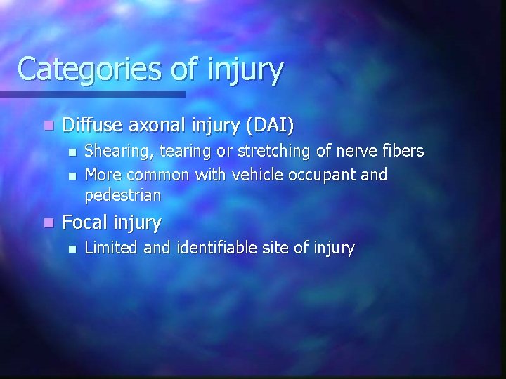
Categories of injury n Diffuse axonal injury (DAI) n n n Shearing, tearing or stretching of nerve fibers More common with vehicle occupant and pedestrian Focal injury n Limited and identifiable site of injury
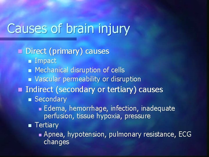
Causes of brain injury n Direct (primary) causes n n Impact Mechanical disruption of cells Vascular permeability or disruption Indirect (secondary or tertiary) causes n n Secondary n Edema, hemorrhage, infection, inadequate perfusion, tissue hypoxia, pressure Tertiary n Apnea, hypotension, pulmonary resistance, ECG changes
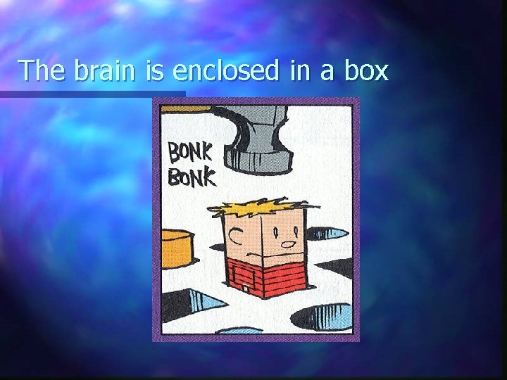
The brain is enclosed in a box
Brain anatomy Occupies 80% of intracranial space n Divisions n n Cerebrum Cerebellum Brain Stem
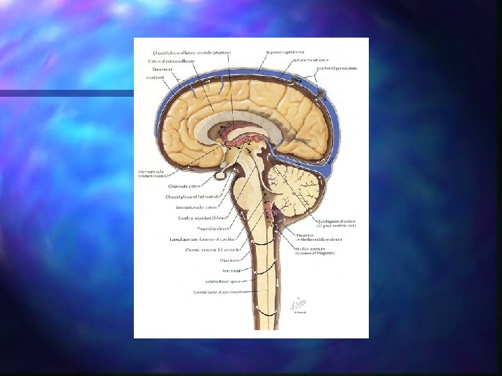
Brain anatomy n Cerebral spinal fluid, CSF n n Clear, colorless Circulates throughout brain and spinal cord Cushions and protects Ventricles n Center of brain n Secrete CSF by filtering blood n Forms blood-brain barrier
Brain anatomy n Blood Supply n n Vertebral arteries n Supply posterior brain (cerebellum and brain stem) Carotid arteries n Most of cerebrum
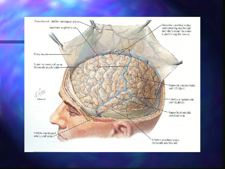
Brain anatomy n Meninges n n n Dura mater: tough outer layer, separates cerebellum from cerebral structures, landmark for lesions Arachnoid: web-like, venous vessels that reabsorb CSF Pia mater: directly attached to brain tissue
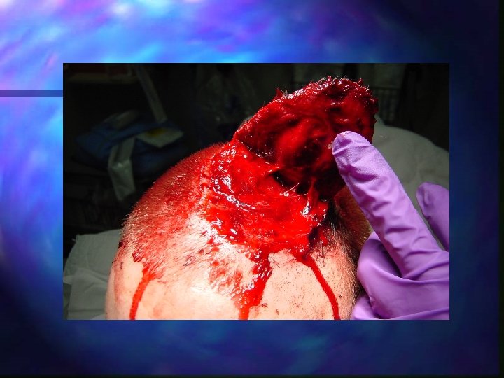
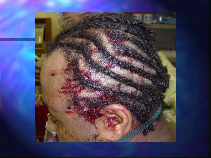
Scalp lacerations n Scalp laceration or avulsion n n Most common injury Vascularity = diffuse bleeding Generally does not cause hypovolemia in adults Can produce hypovolemia in children
Scalp anatomy n Scalp n n n S: skin C: connective tissue A: aponeurosis (galea) L: loose areolar tissue P: pericranium Scalp very vascular n n major blood loss watch kids and adults with prolonged extrication
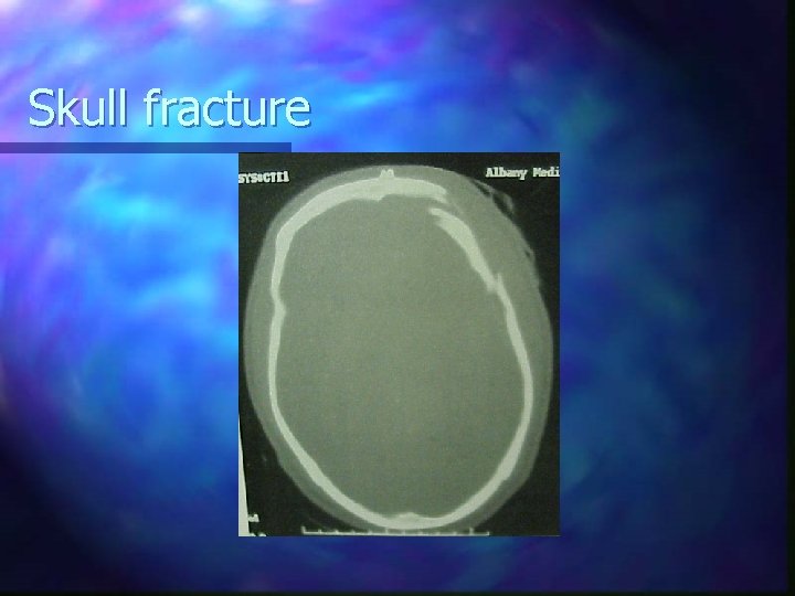
Skull fracture
Skull fracture Present in 60% of pts with severe head injury n Types: n n n Linear: usually incidental finding on CT Depressed: mechanism is usually intense blow to scalp with object of small surface area. Surgical repair needed if depressed more than 5 mm
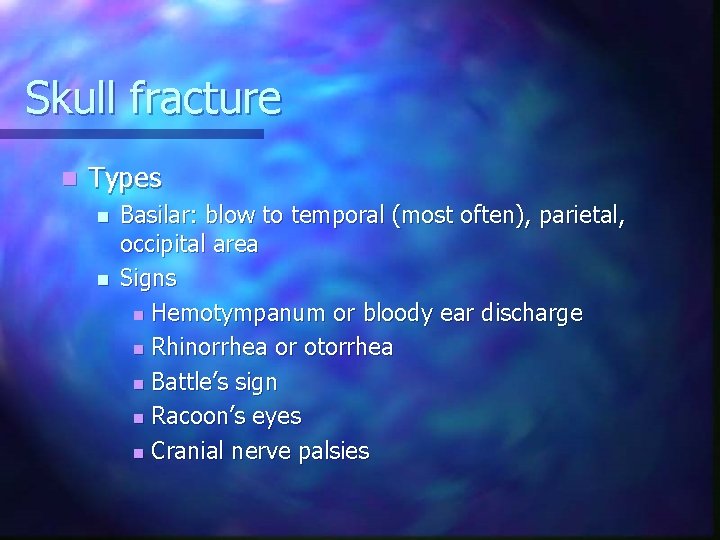
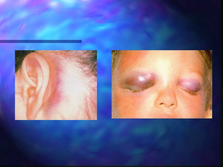
Skull fracture n Types n n Basilar: blow to temporal (most often), parietal, occipital area Signs n Hemotympanum or bloody ear discharge n Rhinorrhea or otorrhea n Battle’s sign n Racoon’s eyes n Cranial nerve palsies
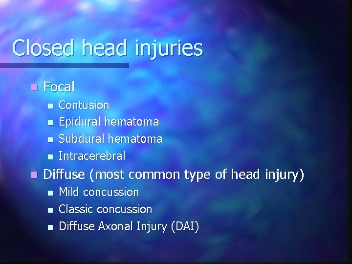
Closed head injuries n Focal n n n Contusion Epidural hematoma Subdural hematoma Intracerebral Diffuse (most common type of head injury) n n n Mild concussion Classic concussion Diffuse Axonal Injury (DAI)
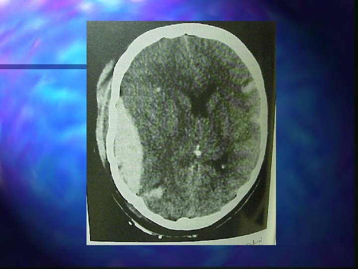
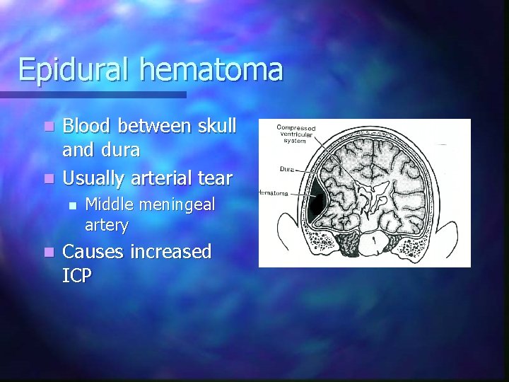
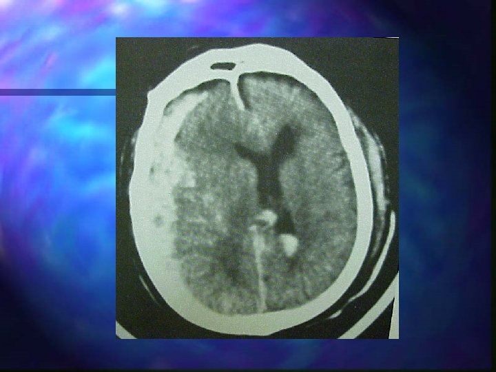
Epidural hematoma Blood between skull and dura n Usually arterial tear n n n Middle meningeal artery Causes increased ICP
Epidural hematoma n n n Unconsciousness followed by lucid interval Rapid deterioration Decreased LOC, headache, nausea, vomiting Hemiparesis, hemiplegia Unequal pupils (dilated on side of clot) Increase BP, decreased pulse (Cushing’s reflex)
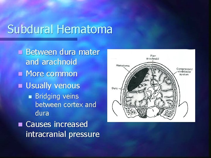
Subdural Hematoma Between dura mater and arachnoid n More common n Usually venous n n n Bridging veins between cortex and dura Causes increased intracranial pressure
Subdural hematoma n n n Slower onset Increased ICP Headache, decreased LOC, unequal pupils Increased BP, decreased pulse Hemiparesis, hemiplegia
Intracerebral hematoma Usually due to laceration of brain n Bleeding into cerebral substance n Associated with other injuries n n n Neuro deficits depend on region involved and size n n DAI Repetitive with frontal lobe Increased ICP
Concussion n n Transient loss of consciousness Retrograde amnesia, confusion Resolves spontaneously without deficit Usually due to blunt head trauma
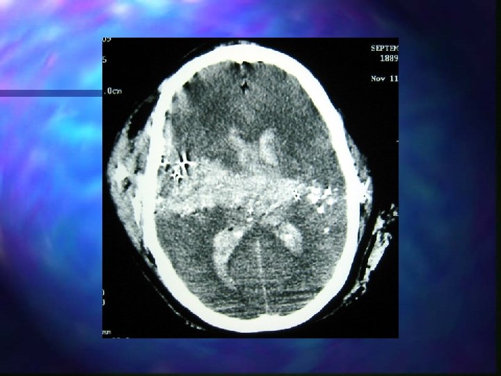
Diffuse axonal injury n n n Tearing or shearing of nerve fibers at time of impact Rapid acceleration-deceleration injury (MVA) Functional or physiologic dysfunction Not gross anatomic abnormality Most common CT finding after severe head trauma
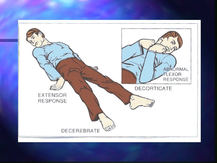
Diffuse axonal injury Prolonged post-traumatic coma not due to mass lesion or ischemic insults n Coma begins at time of trauma n Usually evidence of decorticate or decerebrate posturing, autonomic dysfunction (HTN, fever) n
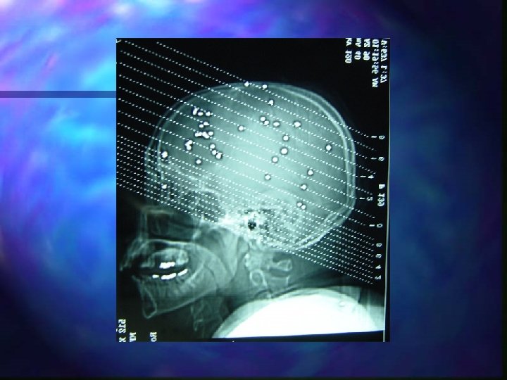
Penetrating head injury n Severity depends on n n Energy of missile Path Amount of scatter of bone and metal fragments Presence of mass lesion Accompanied by n n Severe face and neck injuries Significant blood loss Difficult airway Spinal instability
What the brain needs n High metabolic rate n n Consumes 20% of body’s oxygen Largest user of glucose Requires thiamine Can not store nutrients
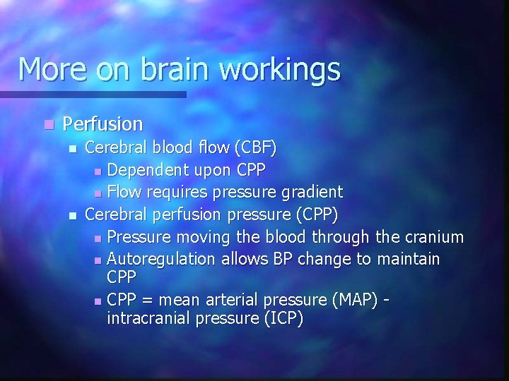
More on brain workings n Perfusion n n Cerebral blood flow (CBF) n Dependent upon CPP n Flow requires pressure gradient Cerebral perfusion pressure (CPP) n Pressure moving the blood through the cranium n Autoregulation allows BP change to maintain CPP = mean arterial pressure (MAP) intracranial pressure (ICP)
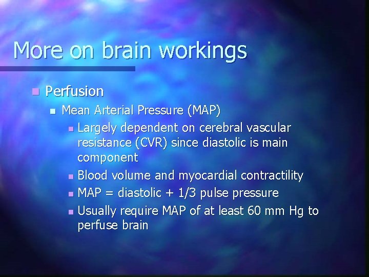
More on brain workings n Perfusion n Mean Arterial Pressure (MAP) n Largely dependent on cerebral vascular resistance (CVR) since diastolic is main component n Blood volume and myocardial contractility n MAP = diastolic + 1/3 pulse pressure n Usually require MAP of at least 60 mm Hg to perfuse brain
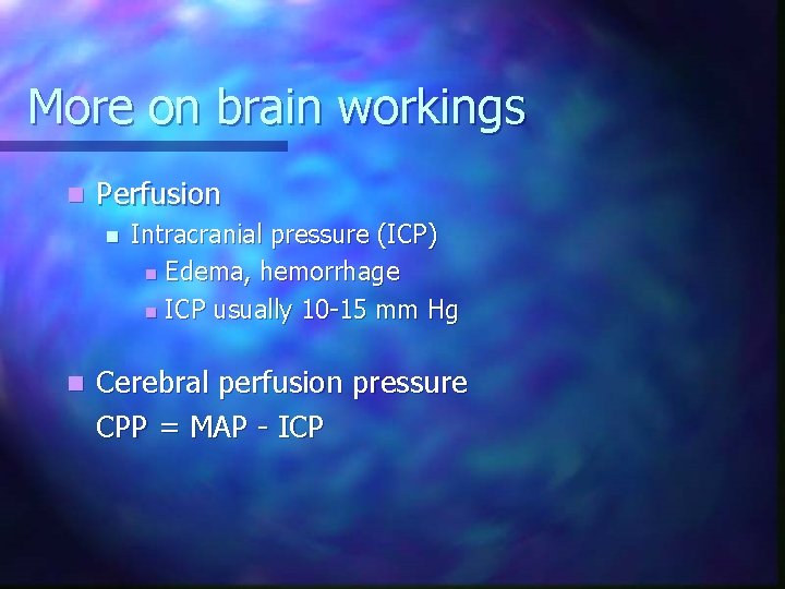
More on brain workings n Perfusion n n Intracranial pressure (ICP) n Edema, hemorrhage n ICP usually 10 -15 mm Hg Cerebral perfusion pressure CPP = MAP - ICP
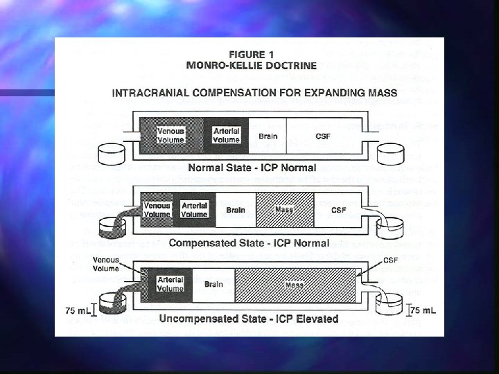
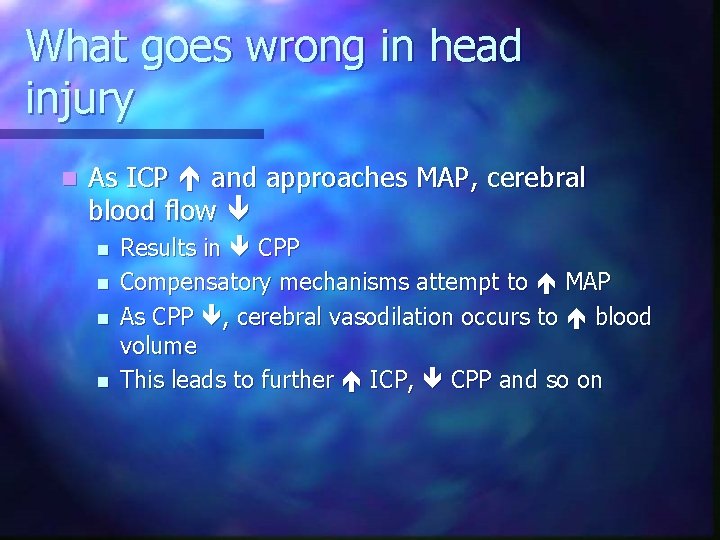
What goes wrong in head injury n As ICP and approaches MAP, cerebral blood flow n n Results in CPP Compensatory mechanisms attempt to MAP As CPP , cerebral vasodilation occurs to blood volume This leads to further ICP, CPP and so on
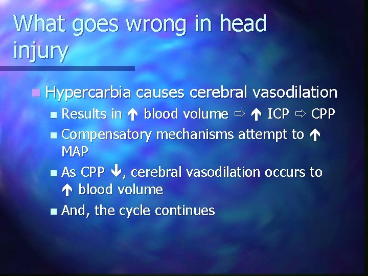
What goes wrong in head injury n Hypercarbia causes cerebral vasodilation Results in blood volume ICP CPP n Compensatory mechanisms attempt to MAP n As CPP , cerebral vasodilation occurs to blood volume n And, the cycle continues n
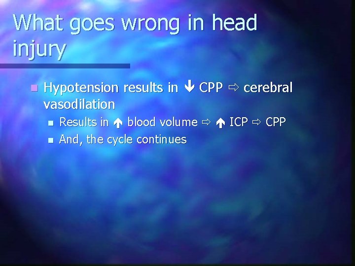
What goes wrong in head injury n Hypotension results in CPP cerebral vasodilation n n Results in blood volume ICP CPP And, the cycle continues
What goes wrong in head injury n Pressure exerted downward on brain n n Cerebral cortex or RAS n Altered level of consciousness Hypothalamus n Vomiting
What goes wrong in head injury n Pressure exerted downward on brain n n Brain stem n BP and bradycardia 2° vagal stimulation n Irregular respirations or tachypnea n Unequal/unreactive pupils 2° oculomotor nerve paralysis n Posturing Seizures dependent on location of injury
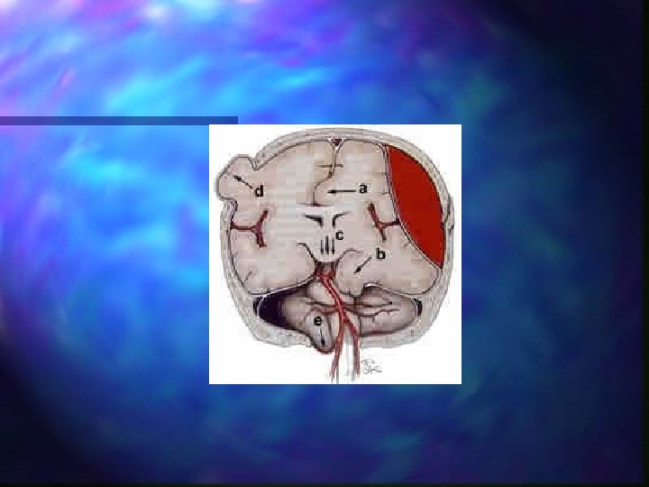
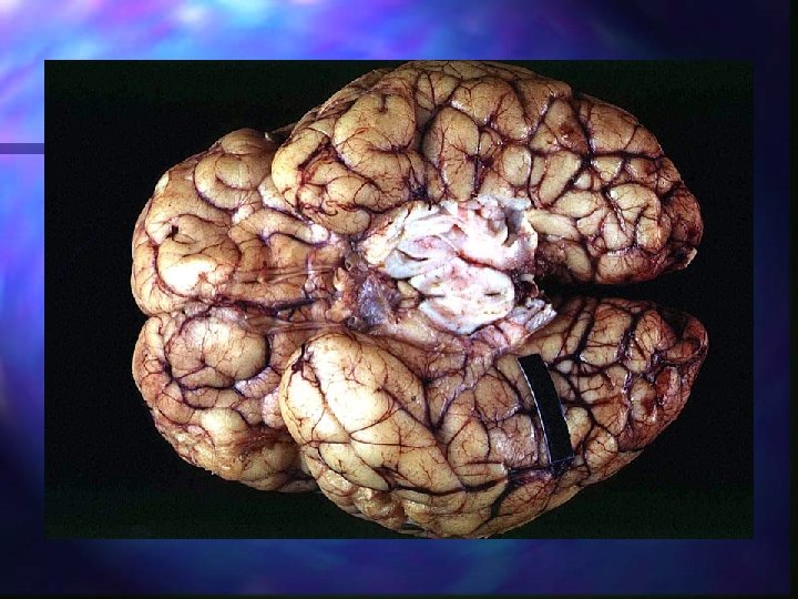
Herniation Transtentorial n Uncal n
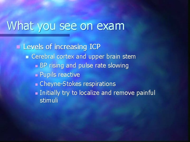
What you see on exam n Levels of increasing ICP n Cerebral cortex and upper brain stem n BP rising and pulse rate slowing n Pupils reactive n Cheyne-Stokes respirations n Initially try to localize and remove painful stimuli
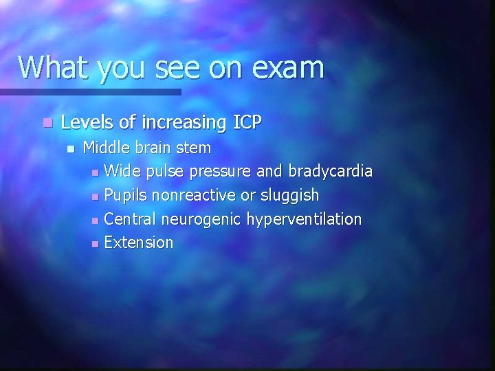
What you see on exam n Levels of increasing ICP n Middle brain stem n Wide pulse pressure and bradycardia n Pupils nonreactive or sluggish n Central neurogenic hyperventilation n Extension
What you see on exam n Levels of increasing ICP n Lower brain stem / medulla n Pupil blown (side of injury) n Ataxic or absent respirations n Flaccid n Irregular or changing pulse rate n Decreased BP n Usually not survivable
Global function: assessment n LOC = best indicator n n Altered LOC = Intracranial trauma UPO Trauma patient unable to follow commands = chance of intracranial injury needing surgery
Global function n AVPU scale n n A = Alert V = Responds to Verbal stimuli P = Responds to Painful stimuli U = Unresponsive
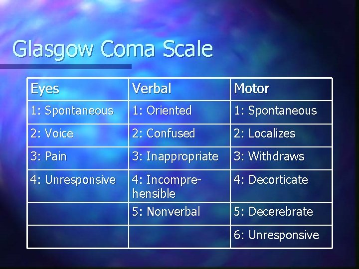
General brain function n Glasgow Coma Scale, GCS n n Eye opening Verbal response Motor response Reliable measure, repeatable
Glasgow Coma Scale Eyes Verbal Motor 1: Spontaneous 1: Oriented 1: Spontaneous 2: Voice 2: Confused 2: Localizes 3: Pain 3: Inappropriate 3: Withdraws 4: Unresponsive 4: Incomprehensible 5: Nonverbal 4: Decorticate 5: Decerebrate 6: Unresponsive
Eyes Window to soul and CNS n Pupil size, equality, and response to light n
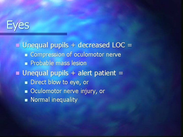
Eyes n Unequal pupils + decreased LOC = n n n Compression of oculomotor nerve Probable mass lesion Unequal pupils + alert patient = n n n Direct blow to eye, or Oculomotor nerve injury, or Normal inequality
Movement Is patient able to move all extremities? n How do they move? n n n Decorticate Decerebrate Hemiparesis or hemiplegia Paraplegia or quadraplegia
Movement Lateralized or focal signs = lateralized or focal deficits n Altered motor function may be due to fracture or dislocation n
Vital Signs Cushing’s triad n Suggests increased intracranial pressure n n Increased BP Decreased pulse Irregular respiratory pattern
Vital Signs Isolated head injury will not cause hypotension in adults n Look for another life threatening injury n n n Chest Abdomen Pelvis Multiple long bone fractures Large scalp lacerations
Summary for assessment n n Most important sign = LOC Direction of changes more important than single observations Importance lies in continued reassessment compared with initial exam UPO, altered LOC in trauma = intracranial injury
Goals for treatment Maintain adequate oxygenation n Maintain sufficient BP for good brain perfusion n Avoid secondary brain damage n
Blood pressure n A single episode of hypotension = doubles patient mortality
Oxygenation n Hypoxemia is a strong predictor of poor outcome
Airway management n Open n Clear n n Assume C-spine trauma Jaw thrust with C-spine control Suction as needed Maintain or secure n n n Intubation if no gag reflex RSI, lidocaine and vecuronium Avoid nasal intubation
Breathing Oxygenate - 100% O 2 n Ventilate n No routine hyperventilation n n Adults 10 -12 BPM Children 12 -16 BPM Infants 16 -20 BPM
Breathing n Respiratory Patterns n n n Cheyne Stokes n Diffuse injury to cerebral hemispheres Central neurological hyperventilation n Injury to mid-brain Apneustic n Injury to pons
Ventilation § Hyperventilation recommended only for signs of cerebral herniation! § § Posturing Pupillary abnormalities Neurologic deterioration after correction of hypotension or hypoxemia Decrease in GCS of more than two points in patients with initial GCS less than 9 § Adults 16 -20 BPM § Children 20 -24 BPM § Infants 24 -28 BPM
Hyperventilation n Benefits n n Decreased Pa. CO 2 Vasoconstriction Decreased ICP Risks n n n Decreased cerebral blood flow Decreased oxygen delivery to tissues Increased edema
Circulation Maintain adequate BP and perfusion n IV of LR/NS TKO if BP normal or elevated n If BP decreased n n LR/NS bolus titrated to SBP ~ 90 mm Hg Consider PASG/MAST if SBP below 80 Monitor EKG -- Do not treat bradycardia
Immobilization Spinal motion restriction n If BP normal or elevated, spine board head elevated 300 n
Intravenous therapy n Drug therapy considerations n Only after: n n Management of ABC’s Controlled hyperventilation
Useful drugs n Diazepam n n n Anticonvulsant Give if patient experiences seizures 5 mg IV May mask changes in LOC May depress respirations May worsen hypotension
Useful drugs n Vecuronium n n n RSI Defasciculating dose Decrease brain oxygen demand
Useful drugs n Lidocaine n n n RSI, few minutes prior 1. 5 mg/kg IV Prevents increases in ICP
Useful drugs n Mannitol n n n n Decreases cerebral edema Improves cerebral blood flow and oxygen delivery Plasma expander Osmotic diuretic 1 g/kg IV May cause hypotension May worsen intracranial hemorrhage Don’t have it!
Glucose n n Assess blood glucose Administer only if hypoglycemic Hyperglycemia can harm injured brain secondary to osmotic shifts Consider thiamine in malnourished
Transport of head injuries Choose trauma center n Any moderate and severe (GCS 3 -13) need to go to trauma center where neurosurgery is available n Air medical transport if needed n Severe injuries need to be recognized quickly and transported rapidly as early surgical intervention is often only truly lifesaving treatment n
Helmet removal n Immediate removal if interferes with priorities n n n Access to airway or airway management Ventilation Cervical spine motion restriction May only need to remove face piece to access airway n Technique n n Requires adequate assistance Training in the procedure Padding if shoulder pads left on
Summary n n n n Spinal precautions Avoid hypoxia Consider intubation early Avoid hypotension Frequent reassessment Hyperventilate for herniation Triage wisely
Any questions?
Resources n www. braintrauma. org
Facial injuries n Mortality n n n Primarily associated with brain and spine injury Severe facial fractures may interfere with airway and breathing Morbidity n n Disability concerns Cosmetic concerns
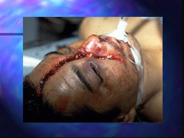
Facial trauma n n n Seldom life-threat unless injury involves the airway Spinal motion restriction Airway is the most difficult and most critical priority Consider early intubation Suction and control bleeding Critical trauma patient - transport accordingly
Facial trauma n Causes n n MVC, home accidents, athletic injuries, animal bites, violence, industrial accidents… Soft tissue n n Lacerations, abrasions, avulsions Vascular area supplied by internal and external carotids
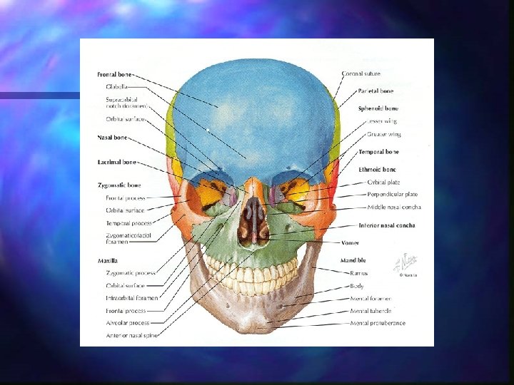
Facial bone anatomy n n n Frontal Nasal Zygoma / zygomatic arch Maxilla Mandible
Facial fractures Mandible, maxilla, nasal bones, zygoma & rarely the frontal bone n Signs and symptoms n n Pain, swelling, deep lacerations, limited ocular movement, facial asymmetry, crepitus, deviated nasal septum, bleeding, depression on palpation, malocclusion, blurred vision, diplopia, broken or missing teeth
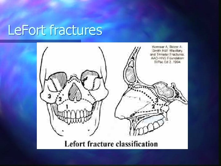
Midface fractures n n n May be significant hemorrhage C-spine precautions Avoid nasotracheal intubation, if possible Le. Fort fracture Tripod fracture
Midface fractures n Appearance n n “Donkey face” (lengthening) “Pumpkin face” (edema) Nasal flattening Often associated with orbital fractures
Le. Fort fractures
Mouth injuries MVC n Blunt injury to the mouth or chin n Penetrating injury due to GSW, laceration, or puncture n
Mouth injuries n Primary concerns n n Airway compromise secondary to bleeding FB aspiration secondary to broken or avulsed teeth Impaled object Management n n n ABCs n Suction prn Stabilize impaled object Collect tissue: tongue or tooth
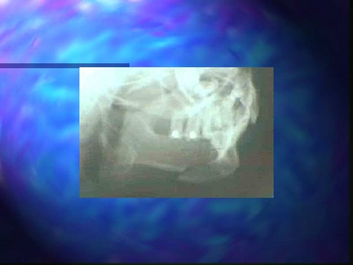
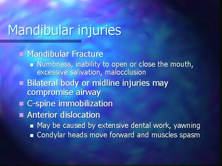
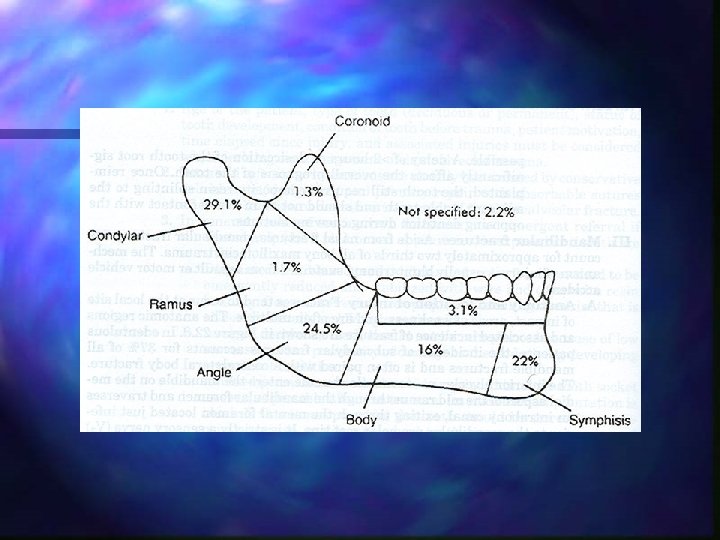
Mandibular injuries n Mandibular Fracture n Numbness, inability to open or close the mouth, excessive salivation, malocclusion Bilateral body or midline injuries may compromise airway n C-spine immobilization n Anterior dislocation n May be caused by extensive dental work, yawning Condylar heads move forward and muscles spasm
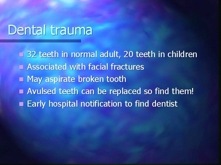
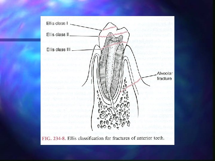
Dental trauma n n n 32 teeth in normal adult, 20 teeth in children Associated with facial fractures May aspirate broken tooth Avulsed teeth can be replaced so find them! Early hospital notification to find dentist
Dental trauma n n <15 minutes, may be asked to replace the tooth in socket Do not rinse or scrub (removes periodontal membrane and ligament) Preserve in fresh whole milk Saline OK for less than 1 hour
Nasal injuries Variety of mechanisms including blunt or penetrating trauma n Swelling, deformity, crepitance n Most common injury n n n Adults - Epistaxis Children - Foreign bodies
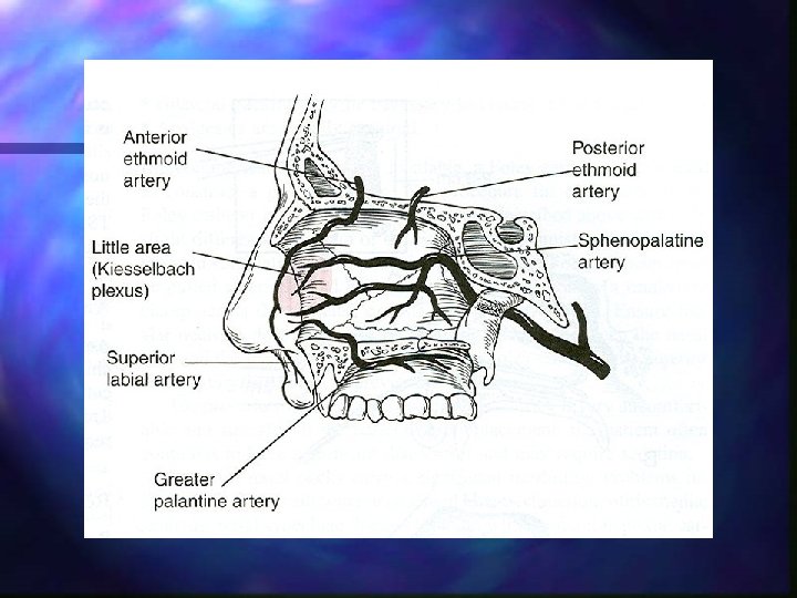
Nasal injuries n Epistaxis n n n Anterior bleeding from septum n Usually venous Posterior bleeding n Often drains to airway May be associated with n Sphenoid and/or ethmoid fractures n Basilar skull fracture
Nasal injury: management n Epistaxis n n n Direct pressure over septum Upright position, leaning forward or in lateral recumbent position If CSF present, do not apply direct pressure n n Allow to drain Needs neurosurgical consult
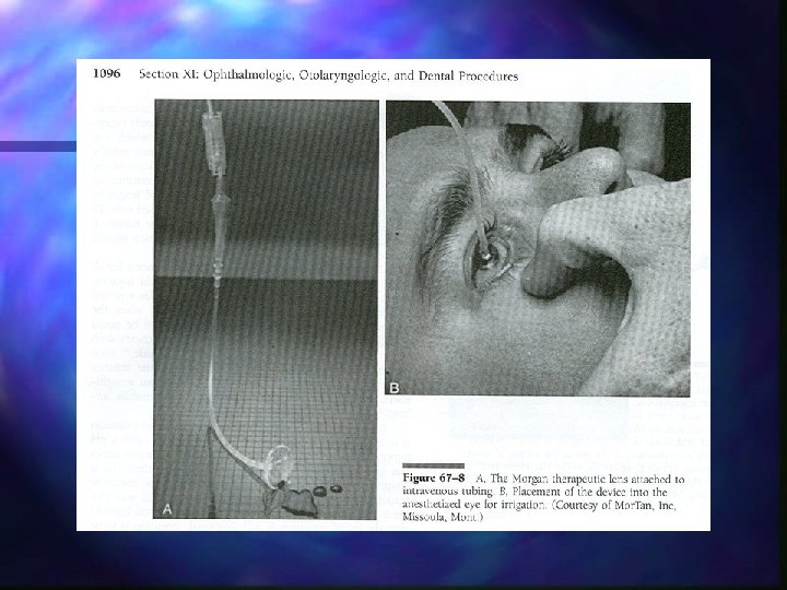
Eye injury types n Penetrating n n Abrasions Foreign bodies (deep, superficial, impaled) Lacerations (deep or superficial, eyelid) Burns n n Flash Acid/alkali
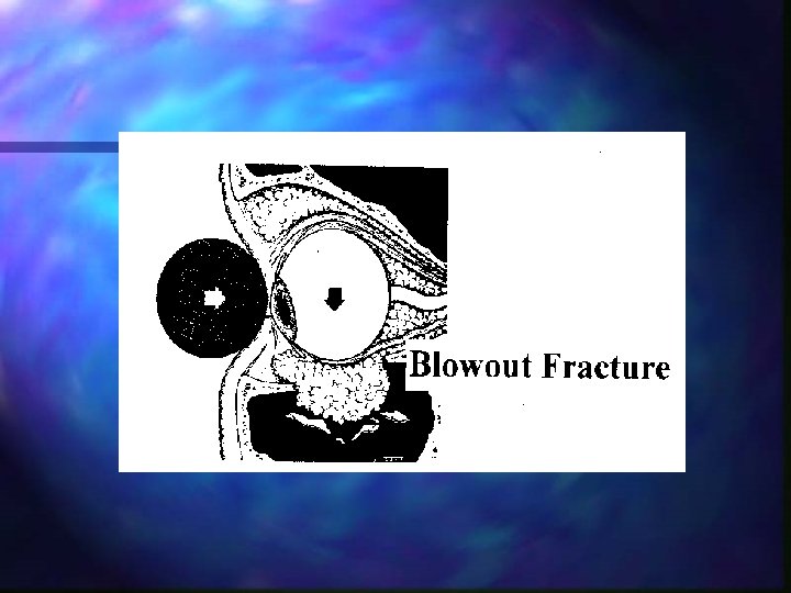
Eye injury types n Blunt n n n Swelling Conjunctival hemorrhage Hyphema Ruptured globe Blow-out fracture of orbit Retinal detachment
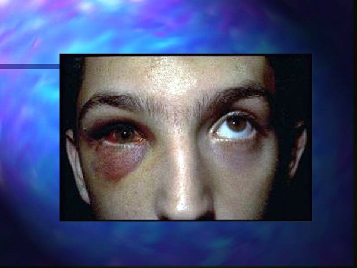
Blow-out orbital fracture n n n n Usually result of a direct blow to the eye Flattened face, numbness Epistaxis, altered vision Periorbital swelling Diplopia Inophthalmos Impaired ocular movement
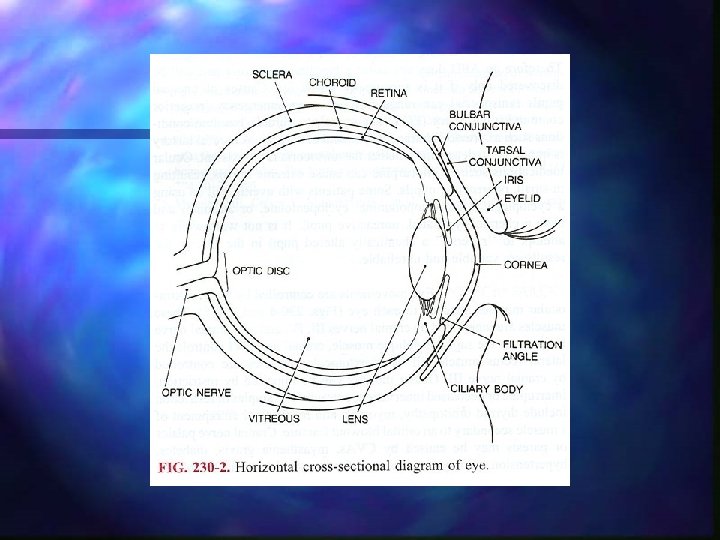
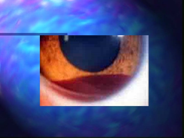
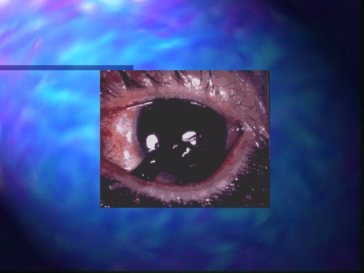
Globe injuries Contusion, laceration, hyphema, globe or scleral rupture n Signs and symptoms - loss of visual acuity, blood in anterior chamber, dilation or constriction of pupil, pain, soft eye, pupil irregularity n
Globe injuries Consider C-spine precautions due to forces required for injury n No pressure to globe for dressing, cover both eyes for protection n Avoid activities that increase intra-ocular pressure n
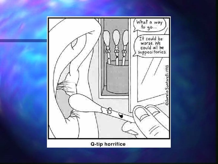
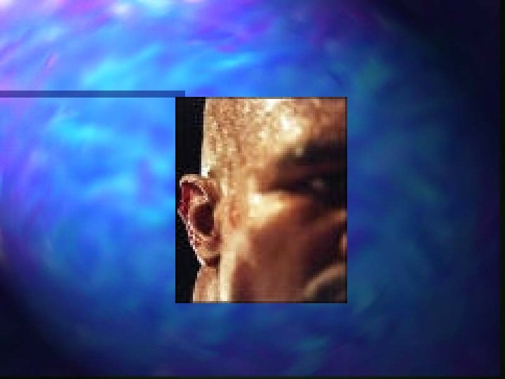
Ear injury n External injuries n n n Lacerations, avulsions, amputations, frostbite Control bleeding with direct pressure Internal injuries n n Spontaneous rupture of eardrum will usually heal spontaneously Penetrating objects should be stabilized, not removed! n Removal may cause deafness or facial paralysis n Hearing loss may be result of otic nerve damage in basilar skull fracture
Ear injury n Separation of ear cartilage n n Treat as an avulsion Dress and bandage Consider disability and cosmetic concerns Bleeding from ear canal n Cover with loose dressing only
Summary n n n n Control bleeding Manage airway accordingly Avoid nasal tracheal intubation when possible Assume c-spine injury is present Gather parts and stabilize objects Trauma survey for other life-threats Transport accordingly
Questions?