
Common pediatric Fractures and Trauma DR Khalid A


- Slides: 76
Common pediatric Fractures and Trauma DR. Khalid A Bakarman Assistant Prof, Pediatric Orthopedic Consultant
objectives § At the end of this lecture the students should be able to: Ø know most of the mechanism of injury Ø make the diagnosis of common pediatric fractures Ø request and interpret the appropriate x-rays Ø initiate the proper management of fractures Ø know which fractures can be treated by conservative or operative methods and the ways of fixation Ø know the possible complications of different fractures and how to avoid them.
The different between adult and pediatric bones • Pediatric bone has a higher water content and lower mineral content per unit volume than adult bone so less brittle than adult bone. • The physis (growth plate) is a unique cartilaginous structure is frequently weaker than bone in torsion, shear, and bending, predisposing the child to injury through it. • The physis is traditionally divided into four zones that the injury through it can cause shortening, angular deformities.
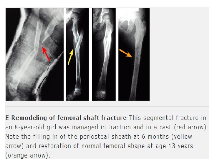
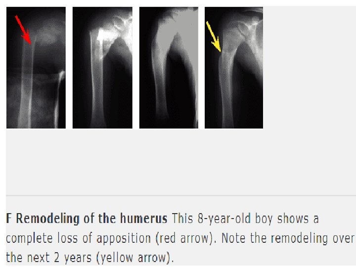
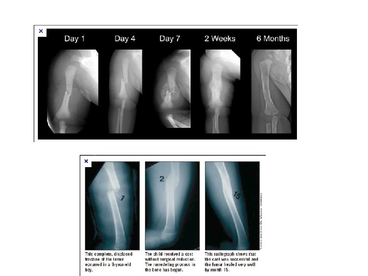
• The periosteum in a child is a thick fibrous structure than adult bone so there is high remodeling rate • ligaments in children are functionally stronger than bone. Therefore, a higher proportion of injuries that produce sprains in adults result in fractures in children.
Common Pediatric Fractures • Upper limb a. Clavicle. b. Supracondylar Fracture. c. Distal Radius. • Lower Limbs a. Femur fractures
CLAVICLE FRACTURES • 8% to 15% of all pediatric fractures • 0. 5% of normal deliveries and in 1. 6% of breech deliveries • 90% of obstetric fractures • 80% of clavicle fractures occur in the shaft • The periosteal sleeve always remains in the anatomic position. Therefore, remodeling is ensured.
Mechanism of Injury • Indirect: Fall onto an outstretched hand • Direct: This is the most common mechanism, it carries the highest incidence of injury to the underlying neurovascular and pulmonary structures • Birth injury
Clinical Evaluation • Birth fractures an asymmetric, palpable mass overlying the fractured clavicle • typically present with a painful, palpable mass along the clavicle, Tenderness, there may be tenting of the skin, crepitus, and ecchymosis. • Neurovascular , the brachial plexus and upper extremity vasculature may injured. • Pulmonary status must be assessed.
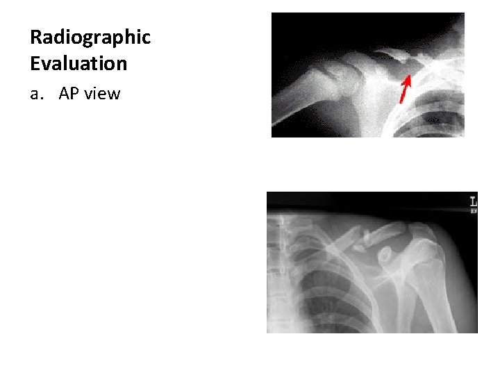
Radiographic Evaluation a. AP view
Classification Descriptive • • • Location Open versus closed Displacement Angulation Fracture type: segmental, comminuted, greenstick
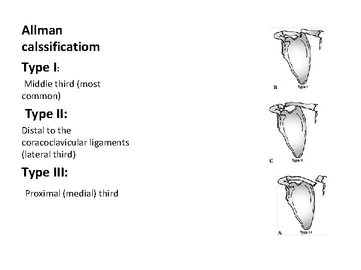
Allman calssificatiom Type I: Middle third (most common) Type II: Distal to the coracoclavicular ligaments (lateral third) Type III: Proximal (medial) third
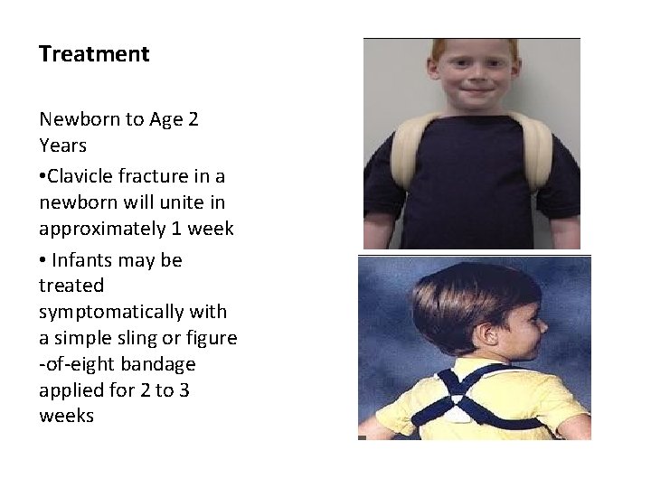
Treatment Newborn to Age 2 Years • Clavicle fracture in a newborn will unite in approximately 1 week • Infants may be treated symptomatically with a simple sling or figure -of-eight bandage applied for 2 to 3 weeks
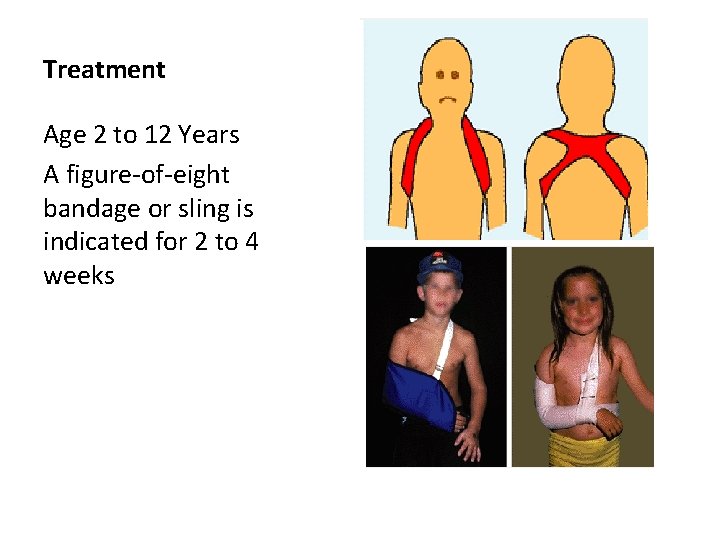
Treatment Age 2 to 12 Years A figure-of-eight bandage or sling is indicated for 2 to 4 weeks
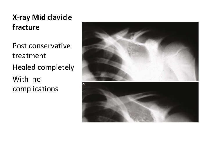
X-ray Mid clavicle fracture Post conservative treatment Healed completely With no complications
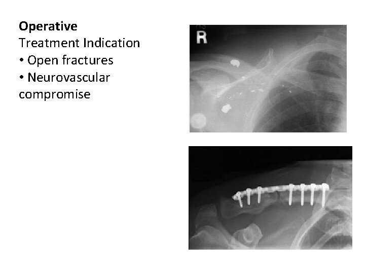
Operative Treatment Indication • Open fractures • Neurovascular compromise
Complications Rare • • Neurovascular compromise Malunion Nonunion Pulmonary injury
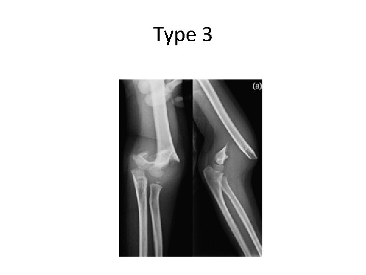
Supracondylar Fracture • • 55% to 75% of all elbow fractures. The male-to-female ratio is 3: 2. 5 to 8 years, The left, or nondominant side, is most frequently injured
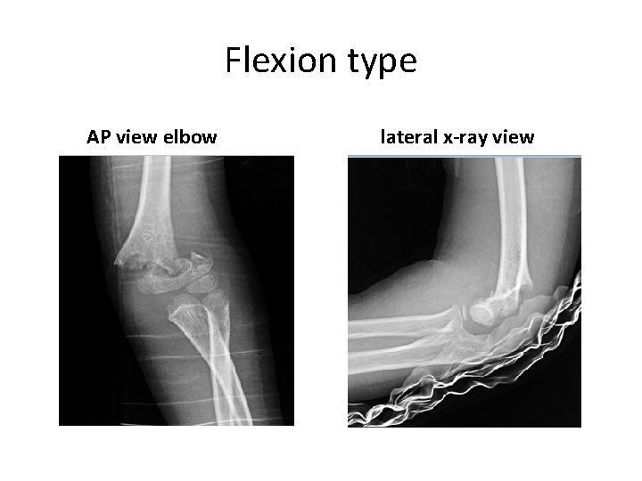
MECHANISM OF INJURY • Indirect most commonly a result of a fall onto an outstretched upper extremity. (Extension type >95%) • Direct a fall onto a flexed elbow or from an object striking the elbow (e. g. , baseball bat, automobile)---(Flexion type < 3%)
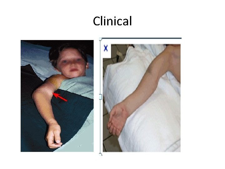
Clinical Evaluation • a swollen, tender elbow with painful range of motion. • S-shaped angulation at the elbow • Pucker sign (dimpling of the skin anteriorly ) • Neurovascular examination the median, radial, and ulnar nerves as well as their terminal branches. Capillary refill and distal pulses should be documented.
Clinical
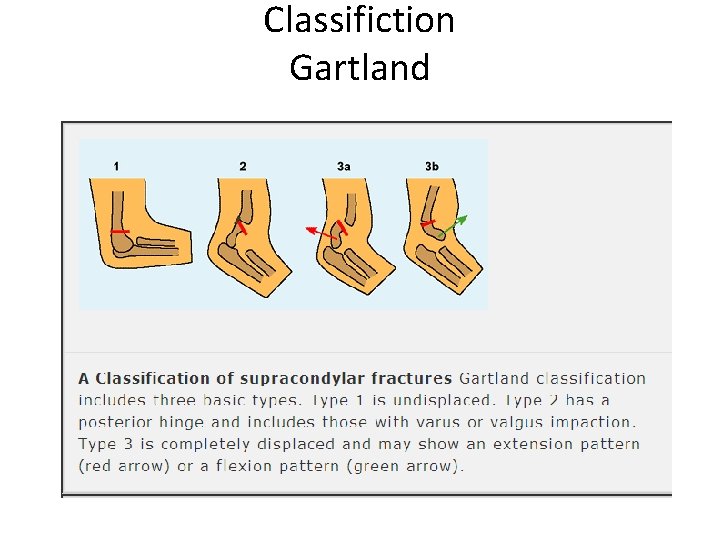
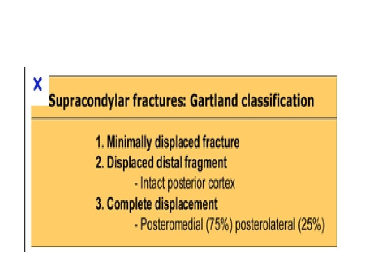
Classifiction Gartland
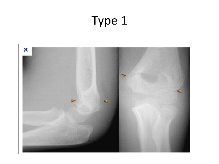
Type 1
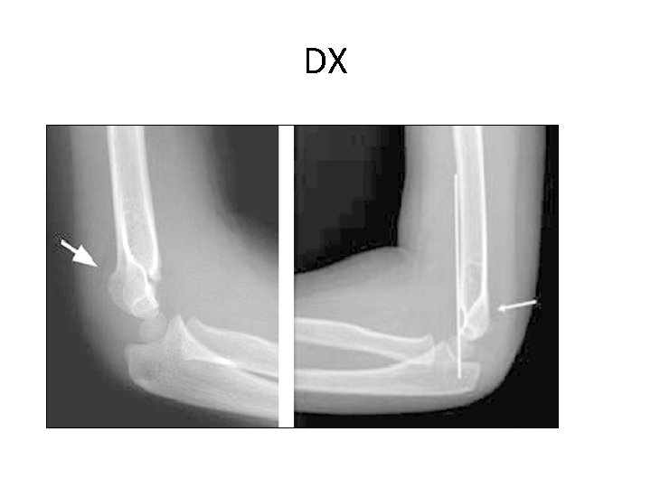
DX
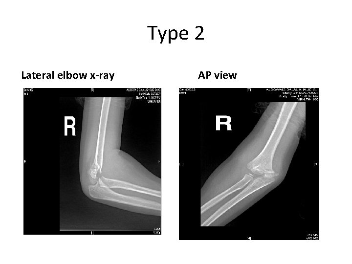
Type 2 Lateral elbow x-ray AP view
Type 3
Flexion type AP view elbow lateral x-ray view
Treatment of extension type • Type I: Immobilization in a long arm cast or splint at 60 to 90 degrees of flexion is indicated for 2 to 3 weeks • Type II: reduce by closed methods followed by casting; it may require pinning if unstable , sever swelling , tilting,
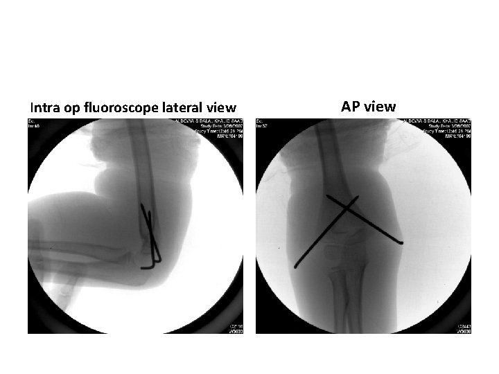
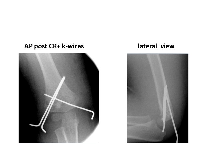
• Type III: § Attempt closed reduction and pinning § Open reduction and internal fixation may be necessary for rotationally unstable fractures, open fractures, and those with neurovascular injury
Intra op fluoroscope lateral view AP view
AP post CR+ k-wires lateral view
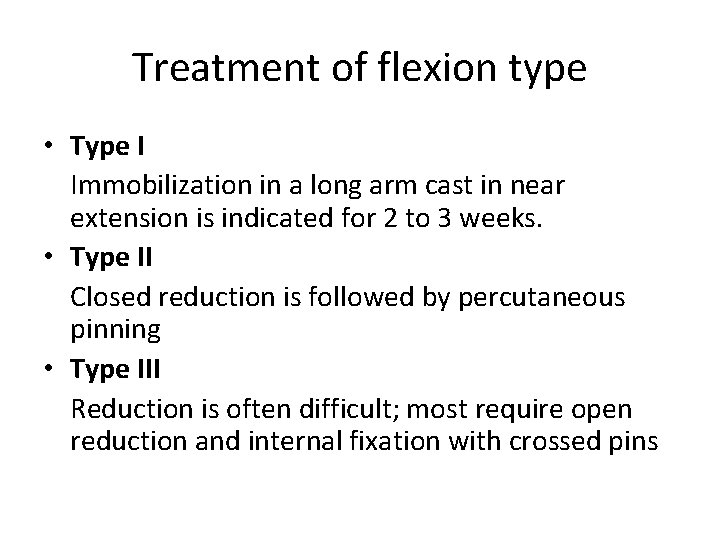
Treatment of flexion type • Type I Immobilization in a long arm cast in near extension is indicated for 2 to 3 weeks. • Type II Closed reduction is followed by percutaneous pinning • Type III Reduction is often difficult; most require open reduction and internal fixation with crossed pins
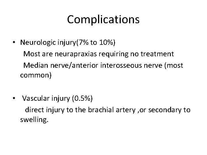
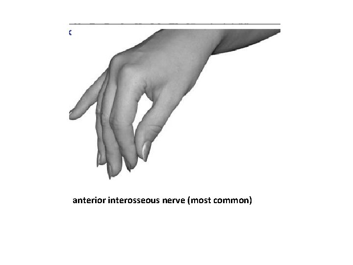
Complications • Neurologic injury(7% to 10%) Most are neurapraxias requiring no treatment Median nerve/anterior interosseous nerve (most common) • Vascular injury (0. 5%) direct injury to the brachial artery , or secondary to swelling.
anterior interosseous nerve (most common)
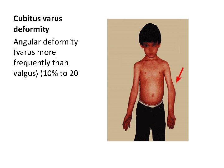
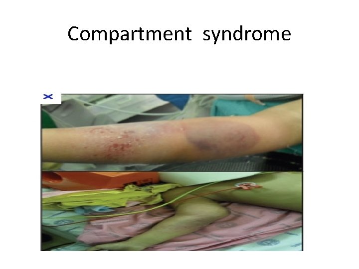
• Loss of motion • Myositis ossificans • Angular deformity (varus more frequently than valgus) (10% to 20) • Compartment syndrome (<1%)
Cubitus varus deformity Angular deformity (varus more frequently than valgus) (10% to 20
Compartment syndrome
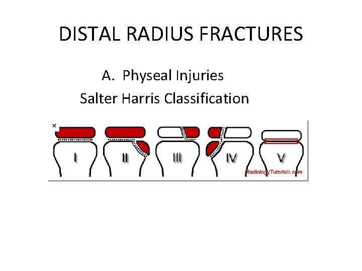
DISTAL RADIUS FRACTURES A. Physeal Injuries Salter Harris Classification
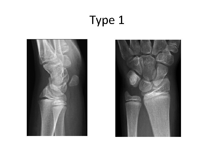
Type 1
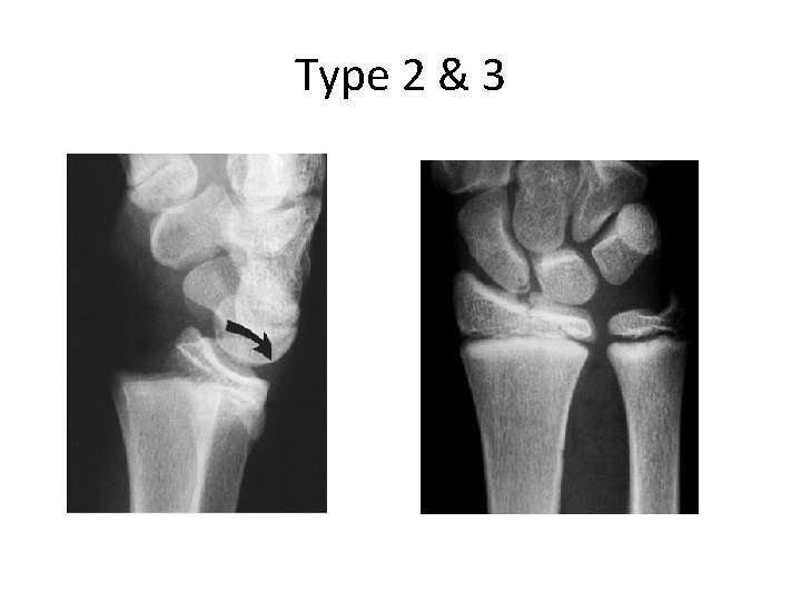
Type 2 & 3
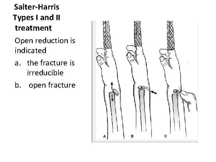
Treatment § Salter-Harris Types I and II o closed reduction is followed by application of a long arm cast or sugar tong splint with the forearm pronated o 50% apposition with no angular or rotational deformity is acceptable. Growth arrest can occur in 25% of patients if two or more manipulations are attempted
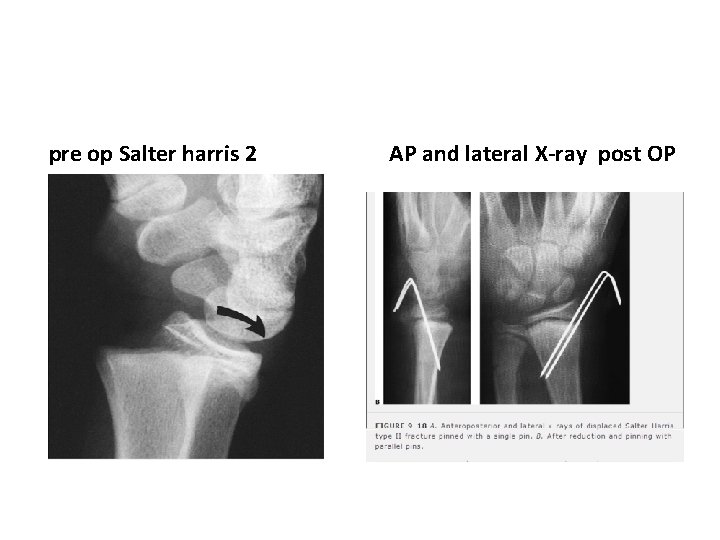
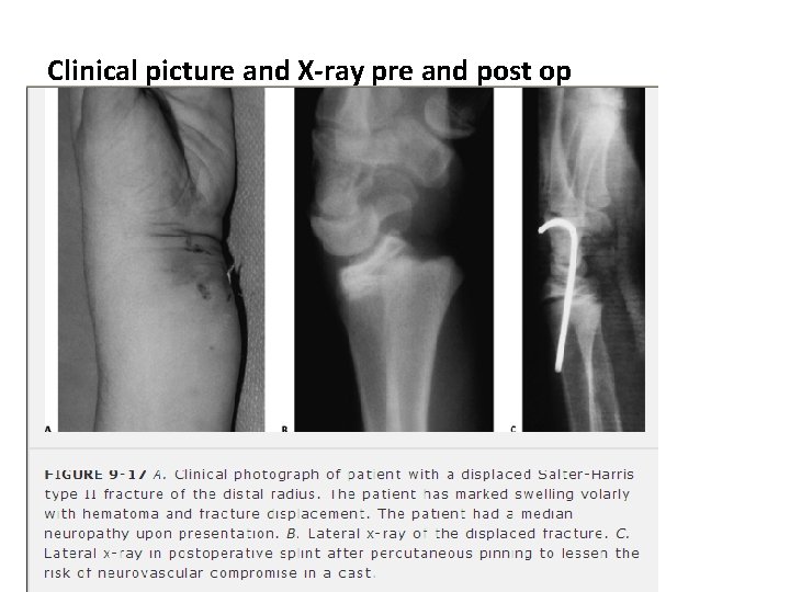
Salter-Harris Types I and II treatment Open reduction is indicated a. the fracture is irreducible b. open fracture
pre op Salter harris 2 AP and lateral X-ray post OP
Clinical picture and X-ray pre and post op
• Salter-Harris Type III Anatomic reduction is necessary Open reduction and internal fixation with smooth pins or screws parallel to the physis is recommended if the fracture is inadequately reduced. • Salter-Harris Types IV and V Rare injuries, need ORIF
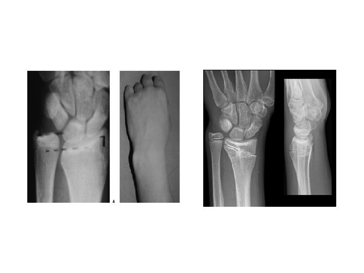
Complications • Physeal arrest lead to shortening , an angular deformity. • Ulnar styloid nonunion • Carpal tunnel syndrome
B. Metaphyseal Injuries Classification o the direction of displacement o involvement of the ulna o biomechanical pattern a. torus (only one cortex is involved) b. incomplete (greenstick) c. complete
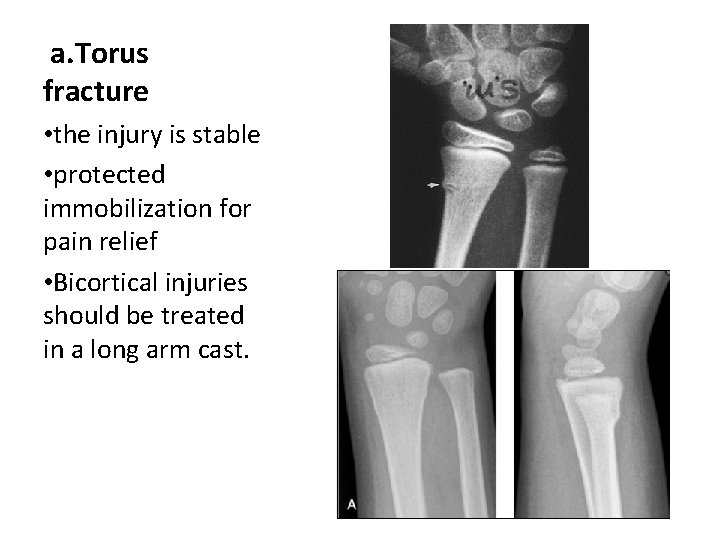
a. Torus fracture • the injury is stable • protected immobilization for pain relief • Bicortical injuries should be treated in a long arm cast.
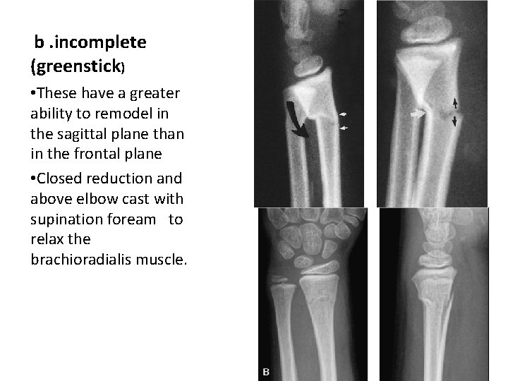
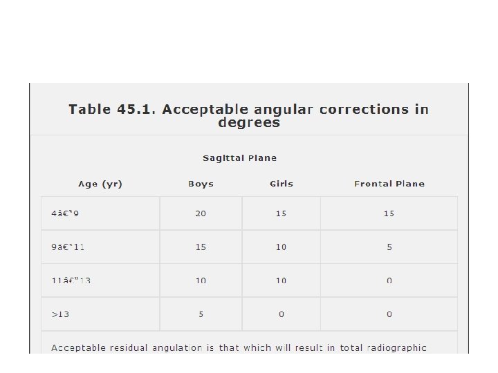
b. incomplete (greenstick) • These have a greater ability to remodel in the sagittal plane than in the frontal plane • Closed reduction and above elbow cast with supination foream to relax the brachioradialis muscle.
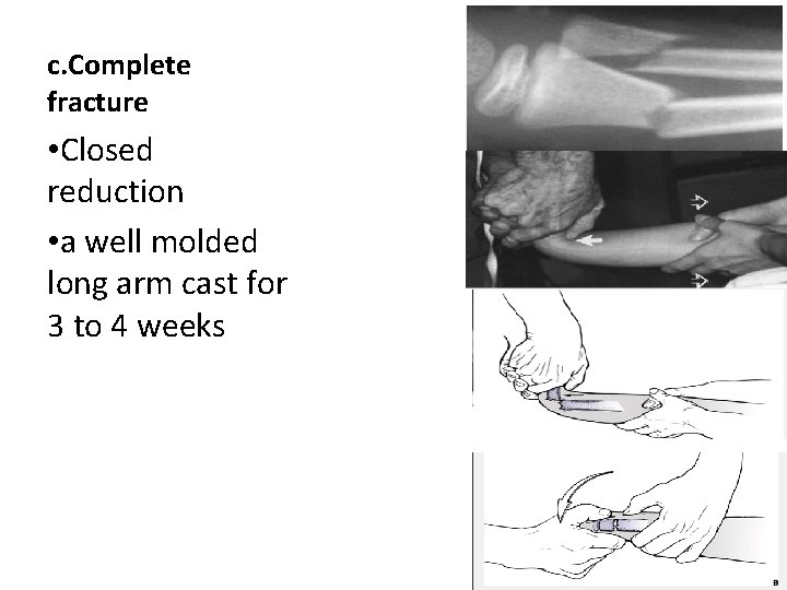
c. Complete fracture • Closed reduction • a well molded long arm cast for 3 to 4 weeks
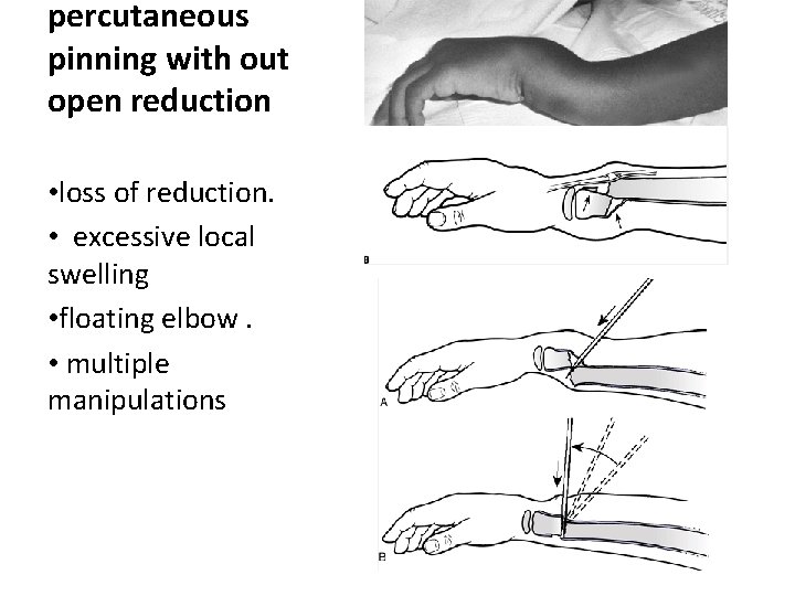
percutaneous pinning with out open reduction • loss of reduction. • excessive local swelling • floating elbow. • multiple manipulations
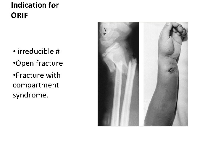
Indication for ORIF • irreducible # • Open fracture • Fracture with compartment syndrome.
Complications • Malunion Residual malangulation of more than 20% may result in loss of forearm rotation. • Nonunion – rare • Refracture an early return to activity (before 6 weeks) • Growth disturbance (overgrowth or undergrowth) 3 mm/9 -12 Y • Neurovascular injuries avoid extreme positions of immobilization.
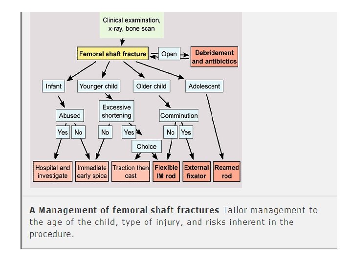
Pediatric Femoral Shaft • 1. 6% of all pediatric fractures. • Boys > girls • Bimodal distribution of incidence 2 to 4 years of age, mid-adolescence. • In children younger than walking age, 80% of these injuries are caused by child abuse; this decreases to 30% in toddlers. • In adolescence, >90% due to RTA
MECHANISM OF INJURY • Direct trauma Motor vehicle accident, pedestrian injury, fall, and child abuse • Indirect trauma Rotational injury • Pathologic fractures osteogenesis imperfecta, nonossifying fibroma, bone cysts, and tumors
CLINICAL EVALUATION • an inability to ambulate, with extreme pain, variable swelling, and variable gross deformity • A careful neurovascular examination is essential • a careful examination of the overlying soft tissues to rule out the possibility of an open fracture
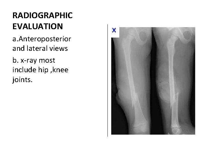
RADIOGRAPHIC EVALUATION a. Anteroposterior and lateral views b. x-ray most include hip , knee joints.
CLASSIFICATION • Descriptive o Open versus closed o Level of fracture: proximal, middle, distal third o Fracture pattern: transverse, spiral, oblique, butterfly fragment o Comminution o Displacement o Angulation • Anatomic Subtrochanteric Shaft Supracondylar
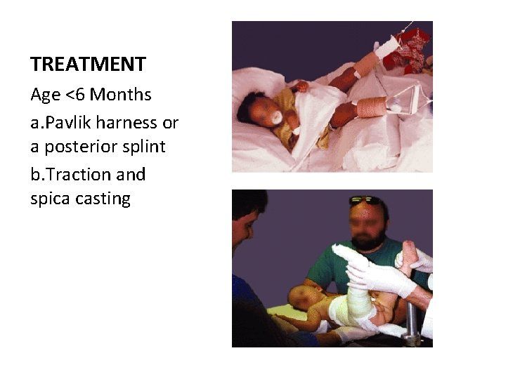
TREATMENT Age <6 Months a. Pavlik harness or a posterior splint b. Traction and spica casting
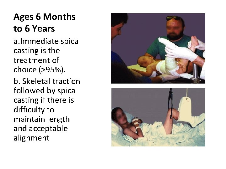
Ages 6 Months to 6 Years a. Immediate spica casting is the treatment of choice (>95%). b. Skeletal traction followed by spica casting if there is difficulty to maintain length and acceptable alignment
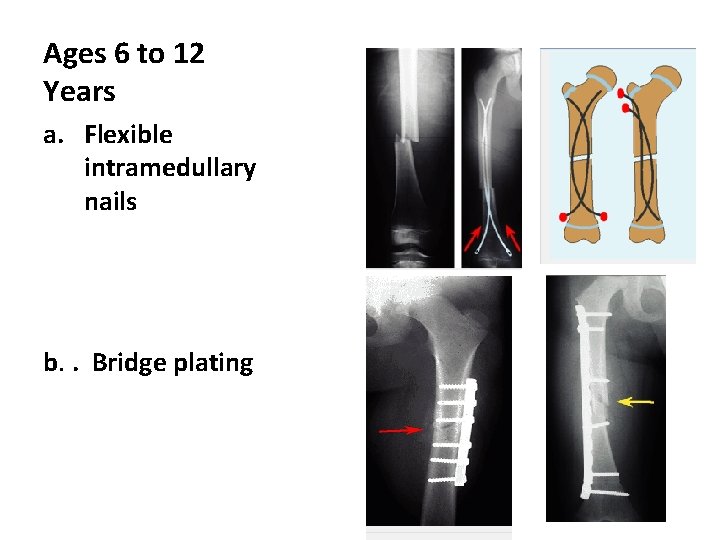
Ages 6 to 12 Years a. Flexible intramedullary nails b. . Bridge plating
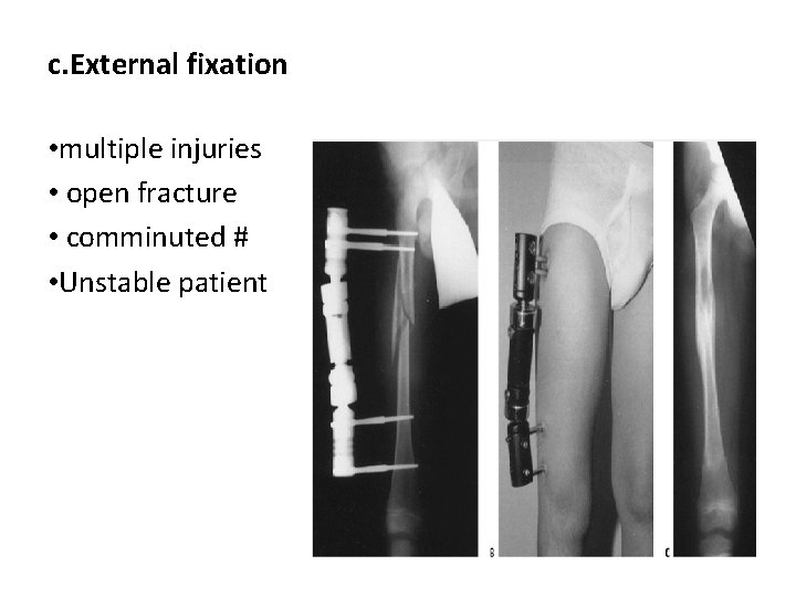
c. External fixation • multiple injuries • open fracture • comminuted # • Unstable patient
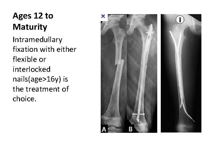
Ages 12 to Maturity Intramedullary fixation with either flexible or interlocked nails(age>16 y) is the treatment of choice.
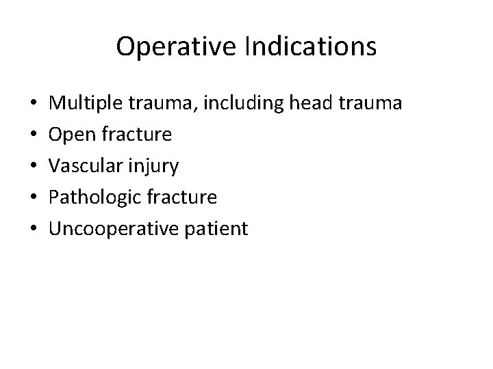
Operative Indications • • • Multiple trauma, including head trauma Open fracture Vascular injury Pathologic fracture Uncooperative patient
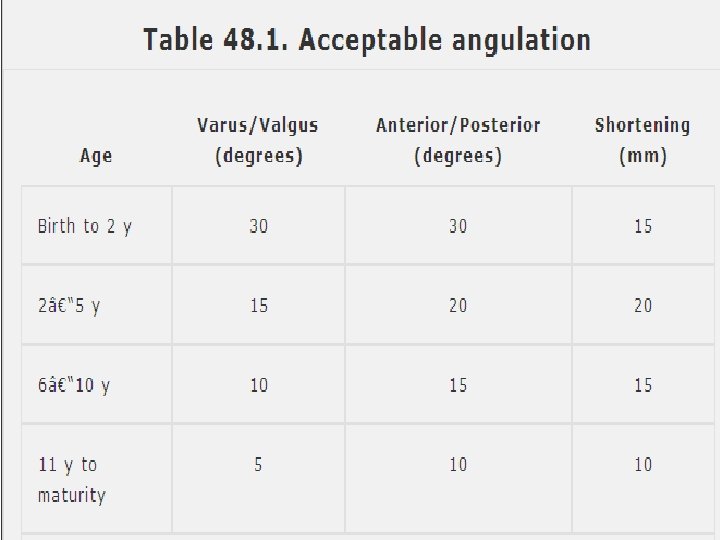
COMPLICATIONS • Malunion Remodeling will not correct rotational deformities • Nonunion –rare • Muscle weakness • Leg length discrepancy Secondary to shortening or overgrowth Overgrowth of 1. 5 to 2. 0 cm is common in the 2 - to 10 -year age • Osteonecrosis with antegrade IM nail<16 year.