Lower Extremity Trauma Lower Extremity Trauma Hip Fractures


 occur with axial load on femur, typically with hip")

 Type II")


 Early mobilization")


 Total Hip Replacement")
 Elderly Patients ORIF possible (higher")


 due to hyperextension injury")


 is most common complication (38%) more common with delayed mobilization")


 Tibial Shaft Fractures Distal Tibia Fractures (Tibial")

")


 Soft Tissue Complications Infection Potential Amputation")


 Based on cadaveric study First word refers to position")
 A: Fibula Fracture distal to mortise B: Fibula Fracture")
 Splint Delayed fixation")


 Talar neck fractures = 50%")


")


 Axillary")


")


- Slides: 126
Lower Extremity Trauma
Lower Extremity Trauma Hip Fractures / Dislocations Femur Fractures Patella Fractures Knee Dislocations Tibia Fractures Ankle Fractures Foot Fracture
Hip Fractures n Hip Dislocations n Femoral Head Fractures n Femoral Neck Fractures n Intertrochanteric Fractures n Subtrochanteric Fractures
Hip Dislocations Significant trauma, usually MVA Posterior: Hip flexion, IR, Add Anterior: Extreme ER, Abd/Flex
Anatomic classification posterior dislocation (90%) occur with axial load on femur, typically with hip flexed and adducted axial load through flexed knee (dashboard injury) § associated with osteonecrosis posterior wall acetabular fracture femoral head fractures sciatic nerve injuries ipsilateral knee injuries (up to 25%) anterior dislocation § associated with femoral head impaction or chondral injury § occurs with the hip in abduction and external rotation §
Anterior Dislocation 7 -10% of hip dislocations Mechanism: Forced abduction with external rotation of hip. Anterior hip capsule is torn or avulsed. Femoral head is levered out anteriorly.
Epstein Classification of Anterior Hip Dislocations Type I Superior (pubic and subspinous) Type II Inferior (obturator and perineal) A No associated fracture B Associated fracture of the femoral head/neck C Associated fracture of the acetabulum
Thompson and Epstein Classification of Hip Dislocations Type II Pure dislocation with at most a small posterior wall Dislocation with large posterior wall Type III Dislocation with comminuted posterior wall Type IV Dislocation with “acetabular floor” fracture Type V Dislocation with femoral head fracture
Hip Dislocations Emergent Treatment: Closed Reduction Dislocated hip is an emergency Goal is to reduce risk of AVN and DJD Allows restoration of flow through occluded or compressed vessels Literature supports decreased AVN with earlier reduction Requires proper anesthesia Requires “team” (i. e. more than one person)
Hip Dislocations Emergent Treatment: Closed Reduction General anesthesia with muscle relaxation facilitates reduction, but is not necessary Conscious sedation is acceptable Attempts at reduction with inadequate analgesia/ sedation will cause unnecessary pain, cause muscle spasm, and make subsequent attempts at reduction more difficult
Hip Dislocations Emergent Treatment: Closed Reduction Allis Maneuver Assistant stabilizes pelvis with pressure on ASIS Surgeon stands on stretcher and gently flexes hip to 90 deg, applies progressively increasing traction to the extremity with gentle adduction and internal rotation Reduction can often be seen and felt Insert hip Reduction Picture
Hip Dislocations Following Closed Reduction Check stability of hip to 90 deg flexion Repeat AP pelvis Judet views of pelvis (if acetabulum fx) CT scan with thin cuts through acetabulum R/O bony fragments within hip joint (indication for emergent OR trip to remove incarcerated fragment of bone)
Hip Dislocations Following Closed Reduction No flexion > 60 deg (Hip Precautions) Early mobilization with P. W. B. for 4 -6 weeks MRI at 3 months (follow risk of AVN)
Complications n Post-traumatic arthritis up to 20% for simple dislocation, markedly increased for complex dislocation n Femoral head osteonecrosis 5 -40% incidence Increased risk with increased time to reduction n Sciatic nerve injury 8 -20% incidence associated with longer time to reduction n Recurrent dislocations less than 2%
Femoral Head Fractures Concurrent with hip dislocation due to shear injury
Femoral Head Fractures Pipkin Classification I: Fracture inferior to fovea II: Fracture superior to fovea III: Femoral head + acetabulum fracture IV: Femoral head + femoral neck fracture
Femoral Head Fractures Treatment Options Type I Nonoperative: non-displaced ORIF if displaced Type II: ORIF Type III: ORIF of both fractures Type IV: ORIF vs. hemiarthroplasty
Epidemiology 250, 000 Hip fractures annually Expected to double by 2050 At risk populations Elderly: poor balance & vision, osteoporosis, inactivity, medications, malnutrition Young: high energy trauma
Femoral Neck Fractures Garden Classification I Valgus impacted II Non-displaced III Complete: Partially Displaced IV Complete: Fully Displaced I II IV Functional Classification Stable (I/II) Unstable (III/IV)
Femoral Neck Fractures Treatment Options Operative ORIF Hemiarthroplasty (Endoprosthesis) Total Hip Replacement
Femoral Neck Fractures Young Patients Urgent ORIF (<6 hrs) Elderly Patients ORIF possible (higher risk AVN, non-union, and failure of fixation) Hemiarthroplasty Total Hip Replacement
Hemi ORIF THR
Intertrochanteric Hip Fx Intertrochanteric Femur Fracture Extra-capsular femoral neck To inferior border of the lesser trochanter
Intertrochanteric Hip Fx Intertrochanteric Femur Fracture Physical Findings: Shortened / ER Posture Obtain Xrays: AP Pelvis, Cross table lateral
Intertrochanteric Hip Fx Classification # of parts: Head/Neck, GT, LT, Shaft Stable Resists medial & compressive Loads after fixation Unstable Collapses into varus or shaft medializes despite anatomic reduction with fixation Reverse Obliquity
Intertrochanteric Hip Fx Stable Unstable Reverse Obliquity
Intertrochanteric Hip Fx Treatment Options Stable: Dynamic Hip Screw Unstable/Reverse: IM Recon Nail
Subtrochanteric Femur Fx Classification Located from LT to 5 cm distal into shaft Intact Piriformis Fossa? Treatment IM Nail Cephalomedullary IM Nail ORIF
Femoral Shaft Fx Winquist n n n Type 0 - No comminution Type 1 - Insignificant butterfly fragment with transverse or short oblique fracture Type 2 - Large butterfly of less than 50% of the bony width, > 50% of cortex intact Type 3 - Larger butterfly leaving less than 50% of the cortex in contact Type 4 - Segmental comminution
Femoral Shaft Fx Treatment Options IM Nail with locking screws ORIF with plate/screw construct External fixation Consider traction pin if prolonged delay to surgery
Distal Femur Fractures Distal Metaphyseal Fractures Look for intra-articular involvement Plain films CT
Distal Femur Fractures Treatment: Retrograde IM Nail ORIF open vs. MIPO Above depends on fracture type, bone quality, and fracture location
Knee Dislocations High association of injuries Ligamentous Injury ACL, Posterolateral Corner LCL, MCL Vascular Injury Intimal tear vs. Disruption Obtain ABI’s (+) Arteriogram Vascular surgery consult with repair within 8 hrs Peroneal >> Tibial N. injury
KNEE DISLOCATION n Devastating injury resulting from high or low energyhighenergy usually from MVC or fall from height commonly a dashboard injury resulting in axial load to flexed knee n low-energy often from athletic injury generally has a rotational component morbid obesity is a risk-facto
Associated Injuries n vascular injury 5 -15% in all dislocations 40 -50% in anterior/posterior dislocations due to tethering at the politeal fossa § proximal - fibrous tunnel at the adductor hiatus § distal - fibrous tunnel at soleus muscle n nerve injury usually common peroneal nerve injury (25%) tibial nerve injury is less common n fractures present in 60% tibia and femur most common
Classification n Anterior most common type of dislocation (30 -50%) due to hyperextension injury arterial injury is generally an intimal tear due to traction n posterior 2 nd most common type (25%) highest rate of complete tear of popliteal artery n lateral 13% of knee dislocations due to varus or valgus force highest rate of peroneal nerve injury Medial n rotational
Schenck Classification KD I Multiligamentous injury with involvement of ACL or PCL KD II Injury to ACL and PCL only (2 ligaments) KD III Injury to ACL, PCL, and PMC or PLC (3 ligaments) KD IV Injury to ACL, PMC, and PLC (4 ligaments) KD V Multiligamentous injury with periarticular fracture
vascular exam n Rule out vascular injury on exam both before and after reduction palpate the dorsalis pedis and posterior tibial pulses n if pulses are present and normal measure Ankle-Brachial Index (ABI) § if ABI >0. 9 then monitor with serial examination (100% Negative Predictive Value) § if ABI <0. 9 perform arterial duplex ultrasound or CT angiography if arterial injury confirmed then consult vascular surgery n If pulses are absent or diminished confirm that the knee joint is reduced or perform immediate reduction and reassessment immediate surgical exploration if pulses are still absent following reduction § ischemia time >8 hours has amputation rates as high as 86% if pulses present after reduction angiography
Initial Treatment reduce knee and re-examine vascular status § considered an orthopedic emergency § splint knee in 20 -30 degrees of flexion § confirm reduction is held with repeat radiographs in brace/splint § vascular consult indicated if
Complications n Stiffness (arthrofibrosis) is most common complication (38%) more common with delayed mobilization n Laxity and instability (37%) n Peroneal nerve injury (25%) most common in posterolateral dislocations n Vascular compromise in addition to vessel damage, claudication, skin changes, and muscle atrophy can occur
Patella Fractures History MVA, fall onto knee, eccentric loading Physical Exam Ability to perform straight leg raise against gravity (ie, extensor mechanism still intact? ) Pain, swelling, contusions, lacerations and/or abrasions at the site of injury Palpable defect
Patella Fractures Radiographs AP/Lateral/Sunrise views Treatment ORIF if ext mechanism is incompetent Non-operative treatment with brace if ext mechanism remains intact
Tibia Fractures Proximal Tibia Fractures (Tibial Plateau) Tibial Shaft Fractures Distal Tibia Fractures (Tibial Pilon/Plafond)
Tibial Plateau Fractures MVA, fall from height, sporting injuries Mechanism and energy of injury plays a major role in determining orthopedic care Examine soft tissues, neurologic exam (peroneal N. ), vascular exam (esp with medial plateau injuries) Be aware for compartment syndrome Check for knee ligamentous instability
Tibial Plateau Fractures Xrays: AP/Lateral +/- traction films CT scan (after ex-fix if appropriate)
Schatzker Classification of Plateau Fxs Lower Energy Higher Energy
Tibial Plateau Fractures Treatment Definitive ORIF for patients with varus/valgus instability, >5 mm articular stepoff Non-operative in nondisplaced stable fractures or patients with poor surgical risks
Tibial Plateau Fractures Treatment Spanning External Fixator may be appropriate for temporary stabilization and to allow for resolution of soft tissue injuries Insert blister Pics of ex-fix here
Tibial Shaft Fractures Mechanism of Injury Can occur in lower energy, torsion type injury (e. g. , skiing) More common with higher energy direct force (e. g. , car bumper) Open fractures of the tibia are more common than in any other long bone
Tibial Shaft Fractures n Open Tibia Fx n Priorities – ABC’S – Associated Injuries – Tetanus – Antibiotics – Fixation
Tibial Shaft Fractures Gustilo and Anderson Classification of Open Fx Grade 1 <1 cm, minimal muscle contusion, usually inside out mechanism Grade 2 1 -10 cm, extensive soft tissue damage Grade 3 3 a: >10 cm, adequate bone coverage 3 b: >10 cm, periosteal stripping requiring flap advancement or free flap 3 c: vascular injury requiring repair
Tibial Shaft Fractures Management of Open Fx Soft Tissues ER: initial evaluation wound covered with sterile dressing and leg splinted, tetanus prophylaxis and appropriate antibiotics OR: Thorough I&D undertaken within 6 hours with serial debridements as warranted followed by definitive soft tissue cover
Tibial Shaft Fractures n Definitive Soft Tissue Coverage – Proximal third tibia fractures can be covered with gastrocnemius rotation flap – Middle third tibia fractures can be covered with soleus rotation flap – Distal third fractures usually require free flap for coverage
Tibial Shaft Fractures Treatment Options IM Nail ORIF with Plates External Fixation Cast or Cast-Brace
Tibial Shaft Fractures Advantages of IM nailing Lower non-union rate Smaller incisions Earlier weightbearing and function Single surgery
Tibial Shaft Fractures IM nailing of distal and proximal fx Can be done but requires additional planning, special nails, and advanced techniques
Tibial Pilon Fractures involving distal tibia metaphysis and into the ankle joint Soft tissue management is key! Often occurs from fall from height or high energy injuries in MVA “Excellent” results are rare, “Fair to Good” is the norm outcome Multiple potential complications
Tibial Pilon Fractures Initial Evaluation Plain films, CT scan Spanning External Fixator Delayed Definitive Care to protect soft tissues and allow for soft tissue swelling to resolve
Tibial Pilon Fractures Treatment Goals Restore Articular Surface Minimize Soft Tissue Injury Establish Length Avoid Varus Collapse Treatment Options IM nail with limited ORIF External Fixator
Tibial Pilon Fractures Complications Mal or Non-union (Varus) Soft Tissue Complications Infection Potential Amputation
Ankle Fractures Most common weight-bearing skeletal injury Incidence of ankle fractures has doubled since the 1960’s Highest incidence in elderly women Unimalleolar Bimalleolar Trimalleolar Open 68% 25% 7% 2%
n Osseous Anatomy
n Lateral Ligamentous Anatomy
n Medial Ligamentous Anatomy
n Syndesmosis Anatomy
Ankle Fractures History Mechanism of injury Time elapsed since the injury Soft-tissue injury Has the patient ambulated on the ankle? Patient’s age / bone quality Associated injuries Comorbidities (DM, smoking)
Ankle Fractures Physical Exam Neurovascular exam Note obvious deformities Pain over the medial or lateral malleoli Palpation of ligaments about the ankle Palpation of proximal fibula, lateral process of talus, base of 5 th MT Examine the hindfoot and forefoot
Ankle Fractures AP Ankle Tibiofibular overlap <10 mm is abnormal and implies syndesmotic injury Tibiofibular clear space >5 mm is abnormal - implies syndesmotic injury Talar tilt >2 mm is considered abnormal
Ankle Fractures Ankle Mortise View Foot is internally rotated and AP projection is performed Abnormal findings: Medial joint space widening Talocural angle <8 or >15 degrees (compare to normal side) Tibia/fibula overlap <1 mm
Ankle Fractures Lateral View Posterior malleolar fractures Anterior/posterior subluxation of the talus under the tibia Displacement/Shortening of distal fibula Associated injuries
Ankle Fractures Classification Systems (Lauge-Hansen) Based on cadaveric study First word refers to position of foot at time of injury Second word refers to force applied to foot relative to tibia at time of injury
Ankle Fractures Classification Systems (Weber-Danis) A: Fibula Fracture distal to mortise B: Fibula Fracture at the level of the mortise C: Fibula Fracture proximal to mortise
Ankle Fractures Initial Management Closed reduction (conscious sedation may be necessary) Splint Delayed fixation until soft tissues stable Pain control Monitor for possible compartment syndrome in high energy injuries
Ankle Fractures n Indications for non-operative care: – Nondisplaced fracture with intact syndesmosis and stable mortise – Less than 3 mm displacement of the isolated fibula fracture with no medial injury – Patient whose overall condition is unstable and would not tolerate an operative procedure n Management: – WBAT in short leg cast or CAM boot for 4 -6 weeks – Repeat x-ray at 7– 10 days to r/o interval displacement
Ankle Fractures n Indications for operative care: – Bimalleolar fractures – Trimalleolar fractures – Talar subluxation – Articular impaction injury – Syndesmotic injury
Ankle Fractures n ORIF: – Fibula § Lag Screw if possible + Plate § Confirm length/rotation – Medial Malleolus § Open reduce § 4 -0 cancellous screws vs. tension band – Posterior Malleolus § Fix if >30% of articular surface – Syndesmosis § Stress after fixation § Fix with 3 or 4 cortex screws
Hindfoot Fractures: Talus Calcaneus
Talar fractures: Rare Poor blood supply high incidence of AVN Can be major or minor
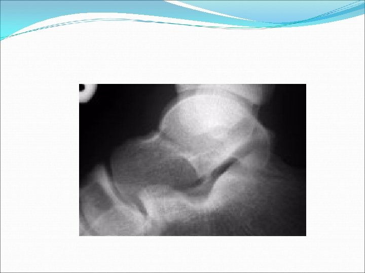
Major Talar fractures: Neck, head, body (& lat process) Talar neck fractures = 50% Hawkins type 1= non displaced + no joint inv. Type II = displaced with subluxation or dislocation of the subtalar joint BUT ankle joint is OK Type III = Type II +dislocation of ankle joint Type IV = Type III + talar head dislocation
Talar Neck #
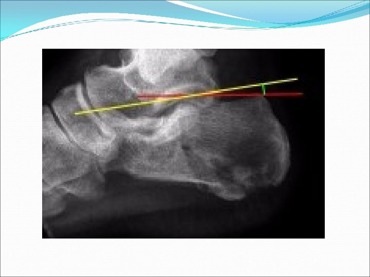
R calcaneus x-ray:
Bohler’s angle (30 -40 deg)
Treatment: Extraarticular= 25 -35% Anterior process, tuberosity, medial process, sustenaculum tali, and body If not displaced nor involving subtalar jt may treat with compressive dressings/casting * Intraarticular= post facet involved - well padded post splint or surgery
Calcaneal fractures: More than 50% are associated with other extremity or spinal fractures
Midfoot Fractures: Navicular Cuboid Lisfranc
Navicular fractures: -Most common midfoot fracture but still rare -treatment= non-displaced=short-leg walking cast x 6 wks displaced : surgery
Cuboid Fractures: Treat as per navicular fractures r/o Lisfranc injury
Lisfranc Joint: Formed by the articulations of metatarsals 1 -3 with the cuneiforms and metatarsals 4 & 5 with the cuboid The metatarsal bases of digits 2 -5 are joined by strong ligaments
Normally, medial aspect of metatarsals 1 -3 should align with medial borders of cuneiforms Metatarsals should be aligned dorsally with tarsals on lateral view Medial 4 th metatarsal should align with medial cuboid Any fracture or dislocation of the navicular or cuneiforms or widening between metatarsals 1 -3 Proximal 2 nd metatarsal # is pathogpneumonic
Normal Lisfranc joint
Treatment: May try closed reduction with traction but post reduction displacement of >2 mm or tarso-metatarsal angle> 15 degrees requires surgery
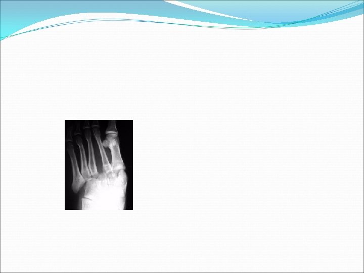
Forefoot fractures: Metatarsal Phalangeal
Treatment: Nondisplaced or min displaced fractures of metatarsal 2 -4 stiff shoe, casting, or fracture brace. Non displaced 1 st metatarsal NWB BK walking cast Displaced 1 st or 5 th metatarsal Attempt closed reduction if >3 mm displacement or 10 degrees angulation
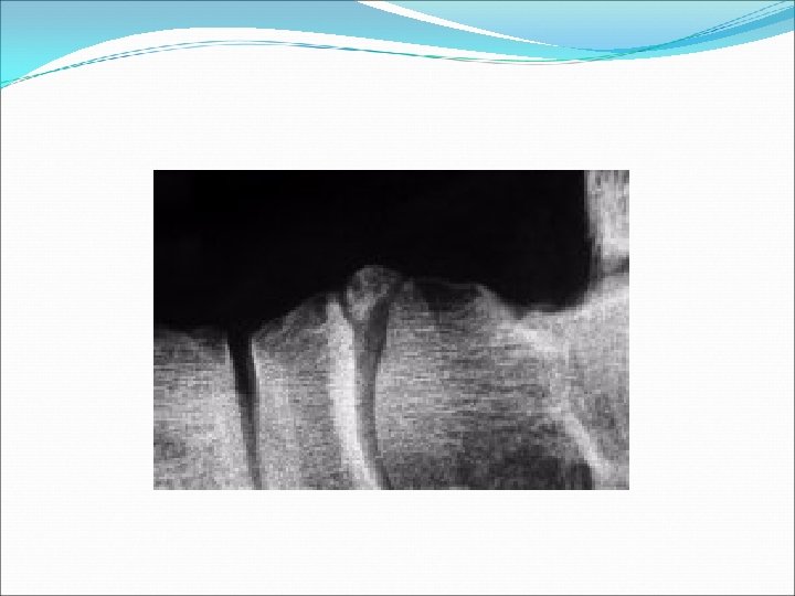
Treatment cont. Metatarsal base # r/o LF injury Jones Fracture=5 th metatarsal base fracture. Tx=non displaced NWB BK cast x 6 -8 wks = displaced surgery
Jones #
Dislocations of the Upper Limb
Dislocation of the Shoulder Mostly Anterior > 95 % of dislocations Posterior Dislocation occurs < 5 % True Inferior dislocation (luxatio erecta) occurs < 1% Habitual Non traumatic dislocation may present as Multi directional dislocation due to generalized ligamentous laxity and is Painless
Mechanism of anterior shoulder dislocation Usually Indirect fall on Abducted and extended shoulder May be direct when there is a blow on the shoulder from behind
Anterior Shoulder dislocation Usually also inferior Bankart’s Lesion
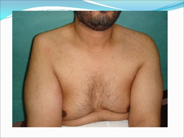
Clinical Picture Patient is in pain Holds the injured limb with other hand close to the trunk The shoulder is abducted and the elbow is kept flexed There is loss of the normal contour of the shoulder
Clinical Picture Loss of the contour of the shoulder may appear as a step Anterior bulge of head of humerus may be visible or palpable A gap can be palpated above the dislocated head of the humerus
X Ray anterior Dislocation of Shoulder
Associated injuries of anterior Shoulder Dislocation Injury to the neuro vascular bundle in axilla ( rare ) Injury of the Axillary or Circumflex Nerve ( Usually stretching leading to temporary neuropraxia ) Associated fracture
Axillary Nerve Injury Also called circumflex nerve It is a branch from posterior cord of Brachial plexus It hooks close round neck of humerus from posterior to anterior It pierces the deep surface of deltoid and supply it and the part of skin over it
Axillary nerve injury
Management of Anterior Shoulder Dislocation Is an Emergency It should be reduced in less than 24 hours or there may be Avascular Necrosis of head of humerus Following reduction the shoulder should be immobilised strapped to the trunk for 3 -4 weeks and rested in a collar and cuff
Methods of Reduction of anterior shoulder Dislocation Hippocrates Method ( A form of anesthesia or pain abolishing is required ) Stimpson’s technique ( some sedation and analgesia are used but No anesthesia is required ) Kocher’s technique is the method used in hospitals under general anesthesia and muscle relaxation
Hippocrates Method
Stimpson’s technique
Kocher’s Technique
SHOULDER REDUCTION Sedation Apply traction and counter traction Lift humeral head into the glenoid
Complications of anterior Shoulder Dislocation : Early Neuro vascular injury ( rare ) Axillary nerve injury Associated Fracture of neck of humerus or greater or lesser tuberosities
Elbow Dislocation n incidence elbow dislocations are the most common major joint dislocation posterolateral is the most common type of dislocation (80%) n demographics predominantly affects patients between age 10 -20 years old
n Simple no associated fracture account for 50 -60% of elbow dislocations n complex associated fracture present may take form of terrible triad injury involves a disruption of the LUCL, a radial head fracture, a coronoid tip fracture and a dislocation of the elbow
Fracture dislocation
complications n Varus Posteromedial instability n Loss of motion n Neurovascular injuries (ulnar/median nerves) n Compartment syndrome n Damage to articular surface n Chronic instability n Heterotopic ossification n Contracture/stiffness correlated with immobilization beyond 3 weeks
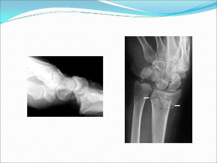
MONTEGGIA FRACTUREDISLOCATION
MONTEGGIA FRACTUREDISLOCATION
GALEAZZI FRACTUREDISLOCATION