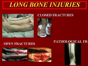
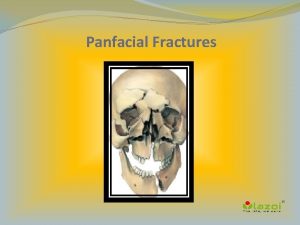
Radiographic Pathology Fractures and Related Injuries Fractures Fractures


- Slides: 52

Radiographic Pathology Fractures and Related Injuries

Fractures • Fractures may be recognized or suspected by the following signs • Fracture line • A step in the cortex • Bulging in cortex • Soft tissue swelling • Joint effusion

dislocation • Dislocation of the joint surfaces no longer maintain their normal relation • Usually associated with fractures

Dislocation shoulder

Posterior elbow dislocation

Simple Fractures Alignment & displacement of fragments Medial dislocation Lateral dislocation Medial angulations Lateral angulations Internal rotation External rotation

Fractures of the forearm

Colles • Colles fracture Fractures of the forearm • Fractures to the distal radius, usually with lateral or dorsal displacement of the distal fragment

Alignment & displacement of fragments

Uncommon fractures Stress and pathologic etiologies

CT Pathological fracture

CT

Other terms: fracture line not seen Impaction, depression, and compression

Directions of the fracture lines Transverse Oblique Spiral longitudinal

Types of Fractures ! Incomplete vs Complete

Green stick incomplete

• Figure 11. Avulsion of the medial epicondyle. The anteroposterior radiograph demonstrates displacement of the medial epicondyle into the joint (arrow). There is also fracture of the radial head.

Fractures involving the growth plate ! Salter-Harris Classification

Injuries of non-articulating joints

Acromioclavicular & coracoclavicular separation Sprain or tears in the AC and (or) CC ligaments, resulting in AC separation with inferior displacement of the scapula and extremity

Distal Humeral Fractures

Smith fractures • Smith fractures (reverse Colle’s fractures) • Fractures of the distal radiuswithdisplacement and angulation of the distal fragment

Fractures of the wrist-scaphoid

Bennett fracture-dislocation

Fractures of the Proximal Femur ! Intra- and Extracapsular fractures

Knee AP& lat • (a) The lateral shows a lipohaemarthrosis and the depressed tibial plateau. • (b) The anteroposterior radiograph confirms the depressed lateral tibial plateau.

KNEE • A 22 -year-old man who sustained a twisting injury to his left knee. • (A) Anteroposterior radiograph of the • left knee shows no obvious fracture. • (B) T 1 -weighted coronal MR image of the left knee shows fracture lines • (arrows).

Rad vs mri

RADIOGRAPHY & CT

Acetabular fracture Posterior dislocation of the femoral head

The diagnosis of a hip fracture most frequently HIP is made on physical examination and plain radiographs A large proportion of these fractures may be missed on plain radiography, however particularly in osteoporotic hips or in the presence of degenerative osteophytes. The incidence of occult hip fracture is estimated to be 2% to 10% in the patients presenting with painful hip after trauma Occult fractures may be secondary to trauma or secondary to stress

Normal Impaction

C-spine fractures

Cervical Spine Trauma Dr. Martin Leahy PGY-1 Dr. Norah Duggan - Faculty Dr. Martin Leahy PGY-1 Dr. Norah Duggan

Plain Film Radiology • The standard 3 view plain film series is the lateral, antero-posterior, and open-mouth view • The lateral cervical spine film must include the base of the occiput and the top of the first thoracic vertebra • The lateral view alone is inadequate and will miss up to 15% of cervical spine injuries.

• If lower cervical spine difficult to see, caudal traction on the arms may be used to improve visualisation • Repeated attempts at plain radiography are usually unsuccessful • If the lower cervical spine is not visible, a CT scan of the region is then indicated

• The anterior vertebral line , posterior vertebral line, and spinolaminar line should have a smooth curve with no steps or discontinuities • Malalignment of the posterior vertebral bodies is more significant than that anteriorly, which may be due to rotation • A step of >3. 5 mm is significant anywhere

• Anterior subluxation of one vertebra on another indicates facet dislocation • Less than 50% of the width of a vertebral body implies unifacet dislocation • Greater than 50% implies bilateral facet dislocation • This is usually accompanied by widening of the interspinous and interlaminar spaces

CT Scanning • Thin cut CT scanning should be used to evaluate abnormal, suspicious or poorly visualised areas on plain radiology • The combination of plain radiology and directed CT scanning provides a false negative rate of less than 0. 1%

Imaging technique • A p- lateral views • Oblique's • Stress films ( force inversion or eversion of the ankle) • Flexion and extension ( cervical spine) • X-ray of the other side for comparison • Delayed films

Chest trauma • Flail chest. Multiple comminuted displaced rightsided rib fractures are observed. There is diffuse increase in the density of the right lung due to contusion. Soft-tissue air is superimposed over the right chest

Chest trauma • Extrapleural hematoma with active intercostal bleed. A. Supine chest radiograph shows a large fluid collection in nondependent position with straight margin toward the lung indicating an extrapleural collection. Note displaced posterior left 5 th rib fracture. The patient has already had a left thoracotomy for intrathoracic bleeding on the left. B. CT image shows a source of active bleeding (arrow

Chest trauma • Supine chest radiograph of blunt trauma patient shows a left tension pneumothorax displacing the heart and mediastinum to the right. There are multiple left-sided rib fractures

Chest trauma • Large hemothorax. A 45 -yearold woman sustained a stab wound to the left hemithorax. A large hemothorax produces a meniscus and causes shift of the heart and mediastinum to the right

Abdominal trauma • Abdominal trauma is an injury to the abdomen. It may be blunt or penetrating and may involve damage to the abdominal organs. Signs and symptoms include abdominal pain, tenderness rigidity, and bruising of the external abdomen. Abdominal trauma presents a risk of severe blood loss and infection. Diagnosis may involve ultrasonography, computed tomography, and peritoneal lavage, and treatment may involve surgery. [1] Injury to the lower chest may cause splenic or liver injuries

Blunt trauma

Abdomen trauma • Abdominal trauma resulting in a right kidney contusion (open area) and blood surround the kidney (closed arrow) as seen on CT

• Female patient with rightsided colon perforation. Axial CT through the abdomen shows focal gas bubbles (red arrow) and anextraluminal fluid collection (blue arrow) adjacent to the contrast-filled colon.

• Image 3 a and 3 b (Computed Tomography): Coronal and axial views showing small bowel perforation with adjacent collection (arrow).

Other modalities • Radionuclide bone scanning in bone trauma fracture side shows increase activity within 2 - 3 days

CT SCANNING THE major advantages over plain films • Better assessment of fractures in bone of complex shape • Better assessment of the extend of tissue damage • Less manipulation of the patient is required

MRI • Even that the cortical bone does not produce an MR signal but a dark line across the bright signal of the fat in the bone marrow on can be seen • MRI is useful in demonstration soft tissue injury in muscles, tendons and ligaments • MRI is particularly useful in knee joint imaging
 What are the two types of physical components
 Health-related skill
 Orbitomeatal line
 Barium enema positioning
 Grid ratio formula
 Loading bench in darkroom
 Labial mounting
 Interpupillary line
 What is recorded detail in radiography
 Vertical bone loss
 Radiographic film
 Guide shoe marks
 Reverse townes view positioning
 Radiographic films
 Geometric unsharpness of margins in radiographic image:
 Dacrocystogram
 Unit 15:7 providing first aid for heat exposure
 Chapter 28 head and spine injuries
 Chapter 21 caring for head and spine injuries
 Emr chapter 15 injuries to muscles and bones
 Chapter 14:1 using body mechanics
 Jones and bartlett learning
 Chapter 13:2 preventing accidents and injuries
 Chapter 11 assessment and evaluation of sports injuries
 Chapter 12 lesson 3 planning a personal activity program
 Common track injuries
 How are sports injuries classified and managed
 Concentric fracture in glass
 Glass analysis in forensic science
 Beck weathers injuries
 Chapter 22 hunger games questions
 Sentinel injuries
 Westfield sports injuries
 Stobhill miu
 Human crutch method
 Chapter 17:10 providing first aid for specific injuries
 Chapter 11 injuries to the shoulder region
 Tnt swimming
 Pallet jack injuries
 The cause-specific mortality rate from roller-skating was:
 Climatic injuries
 Jsp 539
 Chapter 4 preventing injuries through fitness
 Chapter 13 worksheet recognizing different sports injuries
 Chapter 4 basics of tissue injuries
 Bo taoshi injuries
 Discoid miniscus
 Preventing hand injuries
 Intentional injury
 Glycerin and gelatin fake skin
 What is intentional fallacy
 Injury prevention safety and first aid
 Injuries first aid
