Radiographic Quality Chapter 5 Radiographic quality o Refers


- Slides: 33
Radiographic Quality Chapter 5
Radiographic quality o Refers to how easily details can be perceived on a radiograph. n Need to obtain as much diagnostic information as possible about the internal structures of the patient.
Quality depends on o Radiographic Density o Contrast o Geometric Factors that affect detail
Radiographic Density o Defined as the degree of blackness or darkness on a radiograph. o Black areas on a developed radiograph are produced by deposits of metallic silver in the film emulsion that result from exposure to x-rays and their subsequent processing.
Density continued …. o X-rays make radiographic film black n Degree of blackness on a radiograph depends on the amount of x-rays reaching the film. o Density is influenced by the quantity and quality of the x-ray beam, as well as the type and thickness of the tissue under examination.
Factors Affecting Radiographic Density. o Greater radiographic density may be produced by increasing: n n Total # of x-rays that reach the film The penetrating power of the x-rays The developing time The temperature of the developer
Let’s Review o m. As- number of x-rays leaving the x-ray tube in a set period of time. o When k. Vp increases, the penetrating power increases as well. This means more x-rays will reach the film causing a darker radiograph.
Other Density Influences o Thickness and type of tissue being radiographed. n Increase in thickness, means that the patient can absorb more x-rays which will results in a lighter image. p Large animals absorb more x-rays, so less xrays reach the film, so the lighter the image. To compensate, we increase the k. Vp.
Type of Tissue o Type of tissue affects density as well. Higher density tissues will cause less xrays to reach film, therefore have lighter areas on the film at that place.
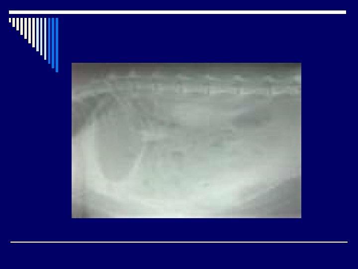
Gray film, lacks good density
Contrast o Defined as visible difference between two adjacent radiographic densities o Is divided into n n Radiographic Contrast Subject Contrast
Radiographic Contrast o Density between two adjacent areas on a radiograph. n n n Many black and white areas means you have contrast. If a radiograph has many grays and a small density difference between 2 adjacent areas, then it has low contrast. Need right amount of contrast. Not desirable to have to high or too low of contrast. Need it to be just right. Want grays, blacks and whites so eye can easily see detail.
Contrast is influenced by: o k. Vp level o Subject contrast o Scatter radiation o Film type o Film fog
Subject Contrast o Defined as the difference in density and mass between two adjacent anatomic structures o Depends on: n n Thickness of the anatomic part. Density of the anatomic part.
Contrast Continued o Bone will have more white on the film than soft tissue. Bone -> high contrast -> low k. Vp Soft tissue -> low contrast -> high k. Vp
Exposure factors o Poor contrast is due to inappropriate exposure factors. o m. As- affects contrast when too little or too much is used. n n Primarily affects density so should not affect contrast if proper k. Vp is used. If m. As is insufficient, contrast is reduced because overall density of the radiograph is reduced. If quantity of x-rays reaching the film is too low, film will be pale. If too much m. As is applied, overall film will be blacker but less effect on contrast.
How should m. As be adjusted?
Kilovoltage o Affects both contrast and density o Increase in k. Vp = Increase in penetrating power. o Increase in k. Vp = shorter wavelengths are produced. o As penetrating power increases, so does scatter radiation. This can alter radiographic contrast.
k. Vp o If too low, will have gray and white appearance and image will be imperceptible. o Will have low density because unable to reach film. o Causes difficulty in distinguishing anatomic organs
Scatter Radiation o Radiation that does not form an image and is scattered in all directions. o Contrast is decreased because inappropriate areas of the film are being exposed.
Sources of Scatter Radiation o From the Patient o Table o Film Tray
Backscatter o Backscatter- radiation arising from sources behind the image plane that are scattered back to the image. o Avoid backscatter by limiting the size of the x-ray beam so that the field does not exceed the image receptor. o Cassettes contain lead- foil backing to prevent backscatter from reaching the film.
Grids o When thick body parts are being radiographed, you want to minimize scatter radiation, you do this by using a grid. o Grid- device placed between the patient and the radiographic film designed to absorb non-image forming x-rays. o Composed of alternating strips of lead and spacer material.
Grids Continued o Spacer material usually consists of fiber, alumininum, or plastic because they have low x-ray absorption o Grids may be n n n Placed on top of the cassette. Built into the cassette. Placed under the table between the patient and the cassette.
Grid Pattern o Linear Grid- most table top machines are equipped with a linear grid- Lead strips parallel in their longitudinal axis. o Crossed Grid- 2 superimposed linear grids absorbs maximum amount of scatter radiation
Grids continued o Generally grids now are a part of the cassette or the x-ray table. o Potter Bucky Diaphragm- usually found in large animal situations. o Grids must be adequately cared for, can not be dropped because will result in permanent damage.
Radiographic Detail and Definition o Terms used to describe image sharpness, clarity, distinctness, and perceptibility. o Lack of detail factors n 1. Geometric unsharpness- loss of detail due to geometric distortion p Large focal spot size p Decreased SID p Motion p Screens and films
o 2. Geometric Distortion and Magnification - variation in size and shape of anatomic structures due to their position in relation to the x-ray source and film. n Important to keep areas being radiographed parallel to image receptor to avoid distortion. p Distortions include: Magnification, elongation, foreshortening.