LONG BONE INJURIES CLOSED FRACTURES OPEN FRACTURES PATHOLOGICAL

LONG BONE INJURIES CLOSED FRACTURES OPEN FRACTURES PATHOLOGICAL FR

FOREARM FRACTURES

FOREARM FRACTURES *SINGLE BONE FRs *BOTH BONES FRs *FRACTURE -DISLOCATION

SINGLE BONE FOREARM FRs SHAFT COLLES HEAD RADIUS Green Stick Fr

SINGLE BONE FOREARM FRs ULNA SHAFT LOWER 1/3 OLECRANON

BOTH BONES FOREARM FRs

FOREARM FRACTURE-DISLOCATION MONTEGGIA FRACTURE-DISLOCATION GALEAZZI FRACTURE-DISLOCATION
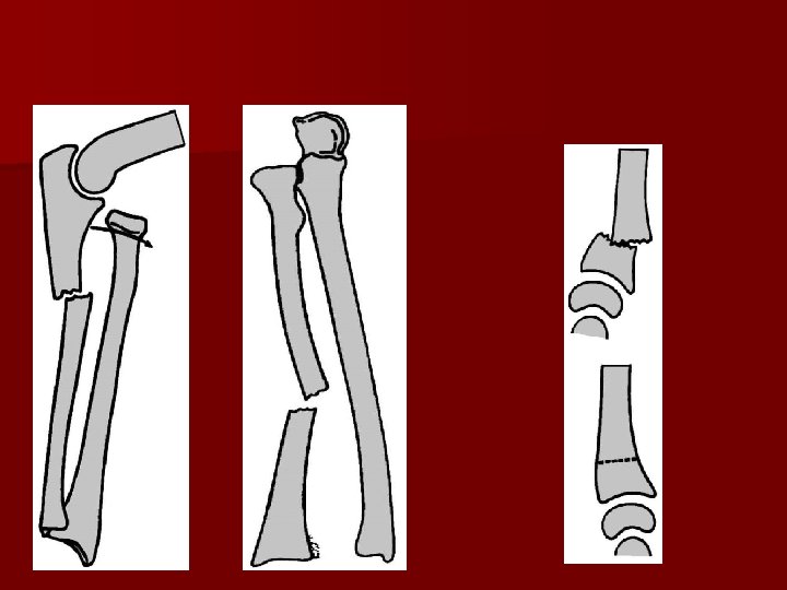

MONTEGGIA FRACTURE-DISLOCATION Definition: It is an angulated, displaced or comminuted fracture of the upper 1/3 ulna accompanied by a dislocated head radius at the superior radio-ulnar joint. Diagnosis: -Deformity and swelling of forearm and elbow. -Limited movements of elbow and pronation/ supination. -X-ray showing forearm and elbow. Treatment: Open reduction and internal fixation of the ulna after closed reduction of the radial head.

GALEAZZI FRACTURE-DISLOCATION Definition It is an angulated, displaced or comminuted fracture of distal 1/3 the radius accompanied by a dislocated lower end ulna at the inferior radio-ulnar joint. Diagnosis: Same as the Monttaeggia fracture except that the swelling is at the wrist. Treatment: Open reduction and internal fixation of the radial fracture after closed reduction of head of ulna.

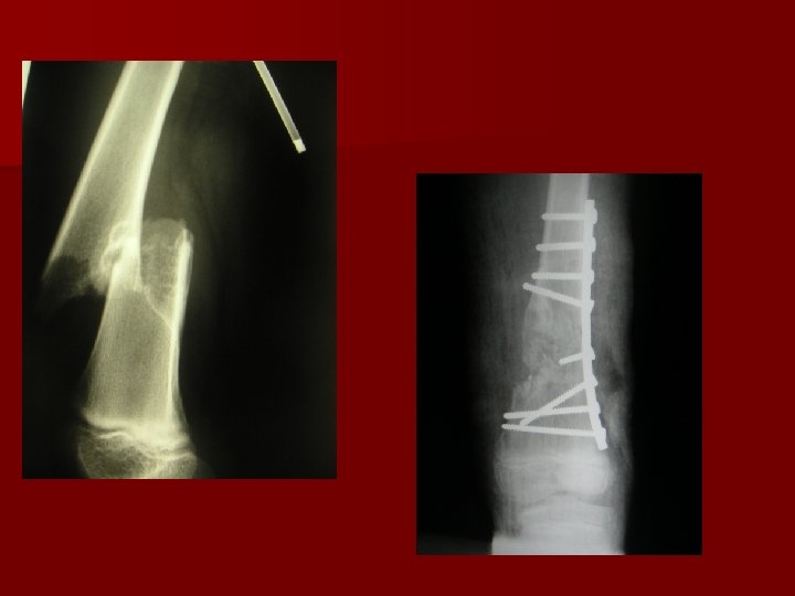
FRACTURE OF RADIUS & ULNA Mechanism of injury : Fall on outstretched hand Direct trauma Radiographic Evaluation: Plain X-ray two views at least required (A-P & lateral) with visualization of joint above and joint below fracture site. TREATMENT Non-operative treatment The rare undisplaced fracture of both radius and the ulna may be treated with well-molded long arm (above-elbow) cast in neutral rotation with elbow flexed to 90º. Operative treatment Open reduction + internal fixation is the treatment of choice for displaced forearm fractures involving radius and ulna in adults.

FRACTURE OF RADIUS & ULNA

COMPLICATIONS OF FOREARM FRACTURES Non union and malunion. Neurovascular injury. Compartmental syndromes. Volkman ischaemic contracture. Post traumatic radioulnar synostosis "uncommon". Post operative infection.
 RADIUS Epidemiology: Most common fracture of the upper")
FRACTURES OF THE DISTAL (LOWER END) RADIUS Epidemiology: Most common fracture of the upper extremity. Its incidence increases with age due to osteopenia & osteoporosis. Mechanism of injury: Fall on the outstretched hand is the most common. Clinical evaluation: - Patient present with deformity of the wrist and variable displacement of the hand in relation to the wrist: Dorsal: Colles fracture (dinner-fork deformity) Volar: in Smith fracture -There is tenderness, ecchymosis, lost active movement & limited painful passive movement. - Careful neurovascular assessment should be done (examine radial artery & median nerve). Radiographic evaluation. - Anteroposterior and lateral views to the wrist

Colles’ fracture: It is the most common of all fractures in older people. The patient is usually a post menopausal woman. Mechanism of injury: Fall on the out stretched hand. It occurs within 2 cm of the articular surface and may extend into the distal radio-carpal joint or the distal radio-ulnar joint. The distal fragment shows: Dorsal angulation (Dinner fork deformity), dorsal displacement, radial angulation, radial displacement, impaction (shortening) and There is often an accompanying fracture of the ulnar styloid, which may signify avulsion of the TFC insertion.

Clinical features: There is a dinner fork deformity, with prominence on the back of the wrist and a depression in front with local tenderness and pain on wrist movements. X-ray: There is a transverse fracture of the radius at the corticocancellous junction, and often the ulnar styloid process is broken off. The distal fragment is impacted into radial and backwards tilt. Sometimes it is comminuted or severely crushed. .
, cast is applied below elbow (if")
Treatment: Undisplaced fractures (or only very slightly displaced), cast is applied below elbow (if ulnar styloid is intact) or above elbow if there is fracture ulnar styloid. If the patient’s wrist is markedly swollen a splint is first applied till the sweling has resolved, then cast is applied. Displaced fractures must be reduced under anaesthesia then cast is applied either below or above elbow according to the presence of ulnar styloid fracture. X-rays are taken after one week, redisplacement is not uncommon to which re-reduction is done. The fracture usually unites in about 6 weeks.

Comminuted Colles’ fractures can not be treated sufficiently with cast immobilization, this is supplemented by K-wire fixation. Cast and wires are removed after 6 weeks. Severely comminuted fractures external fixator is needed.

Complications: Early Circulation in the fingers should be checked; cast may need to be split. Nerve injury is rare, but compression of the median nerve in the carpal tunnel is fairly common. Mild symptoms may resolve by elevation and release of cast. Persistent and severe symptoms require carpal tunnel release. Reflex sympathetic dystrophy is common but usually it does not progress to full picture of Sudek’s atrophy. This is avoided by finger exercises. Triangular fibrocartilage complex (TFCC) injury: As the distal radius displaces dorsally, the TFCC is damaged.

Late Malunion is common either because reduction was not complete or because displacement within the plaster was overlooked. Osteotomy to correct the deformity is needed if there is painful movement. Delayed union and non union of the distal radius are rare, but the ulnar styloid process often joins by fibrous tissue only and remains painful for several months. Stiffness of the shoulder, elbow and fingers from patient neglect is a common complication that can be avoided by exercises. Tendon rupture of extensor pollicis longus occasionally occurs a few weeks after an apparently trivial undisplaced fracture of the lower radius.

SMITH FR

BARTON FRACTURE This is an intra-articular fracture. A fracture in which the dorsal or volar rim of the distal radius is displaced with the hand.

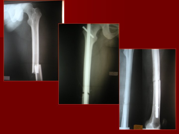
FRACTURE SHAFT FEMUR

E UR L A C I N I L C LOCAL: T C PI SIGNS Swelling, Ecchymosis, Tenderness, Limited movements Deformity, Length discrepancy, Abnormal movements, Crepitus SYMPTOMS: History of trauma, Pain, Swelling, Limited movements. Inability to put wt. SYSTEMIC: SHOCK HYPOVOLAEMIC NEUROGENIC SPINAL DISTAL: NEURO-VASCULAR

FRACTURE SHAFT FEMUR - IN CHILDREN - IN ADULTS

FRACTURE SHAFT FEMUR SITE

TREATMENT OF CLOCED FRACTURES IN CHILDREN UNDISPLACED REDUCIBLE CONSERVATIVE TREATMENT 1 -TRACTION BALANCED SKIN TRACTION +/- THOMAS SPLINT 2 - HIP SPICA GALLOW, s TRACTION

TREATMENT OF CLOCED FRACTURES UNDISPLACED REDUCIBLE CONSERVATIVE TREATMENT 1 -TRACTION GALLOW, s TRACTION SKIN TRACTION

Inter-locking Nail

EXTERNAL FIXATOR
 -MALUNION (angulation, rotation, shortening) -NONUNION -IMPLANT FAILURE -INFECTION")
COMPLICATIONS - MISSED INJURY (hip, knee) -MALUNION (angulation, rotation, shortening) -NONUNION -IMPLANT FAILURE -INFECTION -JOINT PROBLEMS
- Slides: 33