Anatomy LEOCT review 1 ANATOMY the study of
 of body")

 cells. The chromosome number stays")


 between cells. Types Adipose")


 Taste Buds • organs of taste • located on papillae")
 • Vibrate in response to tympanic membrane")
 Coats of the eye ball: Outer tunic: Cornea • anterior portion •")

. 2/3 of")


- Slides: 31
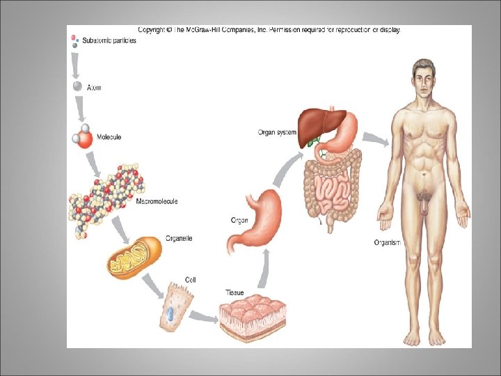
Anatomy LEOCT review 1. ANATOMY -the study of the structure (morphology, form) of body parts PHYSIOLOGY - the study of the function of body parts. 2. Atom – C, H, O Molecule - H 2 O, etc Macromolecule – Proteins, Nucleic acids Organelle – Ribosomes, Nucleus, etc Cell – Nerve cells, Skin cells Tissue – Epithelial, Connective, Muscle, etc Organ – Stomach, Brain, etc Organ system – Nervous, Digestive, etc Organism
3. Synthesis – The making of a compound. Also called anabolism The joining of amino acids to form a protein. The joining of glycerol and fatty acids to make a fat molecule. (Dehydration synthesis reaction) Decomposition – The breaking down of molecules or Compounds to form smaller molecules. Also called catabolism The breaking down of a complex carbohydrate (starch) into monosaccharides (glucose molecules) (Hydrolysis reaction)
4. Mitosis – Cell division in all somatic (body) cells. The chromosome number stays the same. (46 46) Meiosis – Cell division that takes place in the reproductive organs to form sperm or egg cells. The chromosome number is reduced to half. (46 23) 5. They catalyze chemical reactions, they are substrate specific. They bind to the substrate and is released when the chemical reaction is completed.
6. • Types of transport Weeee!! ! Passive Transport cell doesn’t use energy 1. Diffusion 2. Facilitated Diffusion 3. Osmosis • • Animations of Active Transport & Passive Transport high low Active Transport cell does use energy 1. Protein Pumps 2. Endocytosis 3. Exocytosis This is gonna be hard work!! high low
7. Skeletal, Cardiac and Smooth muscle Heavily striated Voluntary control, found in skeletal muscles, Help with movement, maintaining of posture, Produces heat. Finely striated Involuntary control, found in wall or heart, causes heart to contract and pump blood. Nonstriated Involuntary control, found in walls of hollow organs and blood vessels, causes movement of internal organs, blood vessels
8. Soma – body of neuron Axon – carries impulses away from neuron Dendrite – carries impulses to neuron
9. Cells far apart Have matrix (intercellular material-fluids, fibers, etc…) between cells. Types Adipose Cartilage Bone Blood 10. Temp rises - dermal blood vessels dilate, Sweat glands secrete sweat (heat from the blood escape to the outer environment, evaporation of sweat cools body off) – leads to a drop in body temp to normal. Temp drops – dermal blood vessels constrict, sweat glands remain inactive, arrector pili muscles contract causing goose bumps and allow the hair follicles to stand on end – leads to a rise in body temp to normal.
. Epidermis • Dermis - “true skin” • Subcutaneous layer • 11 12. By the type of tissue that binds the bones at each junction. Also according to the degree of movement possible at the bony junction.
Axial: 80 bones Skull - face and cranium Earbones - 3 Hyoid bone - in neck, not attached to any other bone helps in tongue movement Spinal column Sternum and ribs Appendicular skeleton - 126 bones Upper extremities and Lower extremities that connect to the: Pectoral girdle - scapula and clavicle (shoulder) and Pelvic girdle - hips
14. Diaphysis Main shaft Strong support Hollow = decrease in weight Epiphysis Ends of long bone Bulbous shape allows for muscle attachment and gives stability to joints Contains spongy tissue 15. Epiphysis of the long bone
16. Skull – Male is larger and heavier, forehead is shorter, Facial are less round, jaw larger. Pelvis – Female hips are broader, Pelvic cavity is wider, Pelvis bones of female are lighter and more delicate. Coccyx – Male coccyx is less movable than female. • 17. A nerve impulse reaches the end of an axon at the neuromuscular junction – forms a motor end plate. • The synaptic cleft separates the membrane of the neuron and the membrane of the muscle fiber. • When nerve impulse reaches the end of the axon, Ach (Acetylcholine) is released into the synaptic cleft. This stimulates the muscle fiber and causes a muscle impulse. • Sarcolemma is stimulated. • Calcium channels open and Ca ions are released • Specific sites on actin fibers are exposed. • Actin and myosin form linkages • Actin filaments are pulled toward the center of the sarcomere by myosin cross bridges. • Muscle fiber shortens and contracts
For animation of muscle contraction – See this website http: //www. dnatube. com/video/5034/Contraction-of-musclefunction-of-neuromuscular-junction 18. Trapezius
Anterior Muscles
Posterior Muscles
• Central Nervous System 19. • brain • spinal cord • Peripheral Nervous System • nerves • cranial nerves • spinal nerves • Neurons 20 • Neuroglial cells Different types of neurons: (By function) • Afferent (sensory) - to cord or brain • Efferent (motor) - away from cord or brain • Interneurons (synapse between 1 and 2) - from afferent to efferent (from sensory to motor) AND (By Type) Bipolar Unipolar Multipolar Neuroglial cells – May stimulate neurons in the embryo to specialize. Produce growth factors that nourish neurons, remove ions and Neurotransmitters that accumulate between neurons enabling them to continue transmitting information.
21. Afferent – carry nerve impulses from the peripheral body parts to the brain or spinal cord Efferent – carry nerve impulses from the brain or spinal cord to the effectors (muscles or glands). 22. Resting potential – difference in electrical charge between the inside and outside of an undisturbed nerve cell membrane. Action potential – sequence of electrical changes that occurs in a portion of a nerve cell membrane that is exposed to a stimulus that exceeds the membrane’s threshold.
23. Taste (Gustatory senses) Taste Buds • organs of taste • located on papillae of tongue, roof of mouth, linings of cheeks, and walls of pharynx Taste Receptors • chemoreceptors • taste cells – modified epithelial cells that function as receptors • taste hairs – microvilli that protrude from taste cells; sensitive parts of taste cells • Hearing • Auricle • Collects sounds waves • Flap on the side of the head • External auditory meatus • Ear canal • Carries sound to tympanic membrane • Terminates with tympanic membrane • Tympanic membrane • Ear drum • Vibrates in response to sound waves • Separates external from middle ear
Middle ear: • Three auditory ossicles (bones) • Vibrate in response to tympanic membrane • Malleus, incus, and stapes Inner ear: • Cochlea • Functions in hearing • Semicircular canals • Functions in equilibrium • Vestibule • Functions in equilibrium Sight: Visual Accessory Organs • Eyelids • Lacrimal apparatus • Extrinsic eye muscles • Lacrimal gland (tear gland) • lateral to eye • secretes tears
Sight (continue) Coats of the eye ball: Outer tunic: Cornea • anterior portion • transparent No blood vessels Sclera • posterior portion • opaque • protection Middle tunic: Iris • anterior portion • pigmented • controls light intensity Ciliary body • anterior portion • pigmented • holds lens • moves lens for focusing Choroid coat • provides blood supply • pigments absorb extra light
Inner tunic: • Retina • contains visual receptors - Rods and Cones • continuous with optic nerve • ends just behind margin of the ciliary body • composed of several layers Smell: Olfactory Receptors • chemoreceptors • respond to chemicals dissolved in liquids Olfactory Organs • contain olfactory receptors and supporting epithelial cells • cover parts of nasal cavity, superior nasal conchae, and a portion of the nasal septum
24. Location: Lies within the mediastinum - behind sternum (rests on diaphragm). 2/3 of the heart is on the left side of the body. Structure: • Coverings Pericardium - sac around the heart - loose-fitting Fibrous - tough white outer covering, Serous parietal - smooth lining of the fibrous sac. Epicardium - covers surface of the heart - smooth Also called serous visceral pericardium Pericardial space - area between pericardium and epicardium Contains fluid - pericardial fluid Lubricates beating heart • The wall of the heart: Epicardium – reduces friction. Myocardium – thick contractile middle layer, high in mitochondria. Endocardium – delicate inner layer. • Cavities Upper – atria Lower – ventricles
• Valves and Openings Blood flows in one direction. Cuspid valves – between cavities. Bicuspid (mitral) valve - left cavities. Tricuspid valve - right cavities. The semi-lunar valve moves blood out of the heart to the aorta and pulmonary artery. Function: Pump blood through the body. Blood flow (remember ‘Pump your blood’ song!) • CO 2 rich blood enters right atrium • Tricuspid valve to right ventricle • Pulmonary artery • CO 2 is given off in lungs, exchanged with O 2 • O 2 rich blood enters left atrium through the pulmonary veins • Bicuspid valve • Left ventricle • Aorta • Arteries • Aterioles • Capillaries – gives off O 2, picks up CO 2 – venules – veins – Vena cavae – right atrium
25. Arteries – strong, elastic vessels, adapted for carrying blood away from the heart under high pressure. Walls have 3 distinct layers Subdivide into thinner tubes and form aterioles Capillaries – smallest blood vessels. Connect smallest arterioles and the smallest venules. Venules – microsopic vessels that continue from capillaries and merge to form veins. Veins - carry blood back to the heart. Walls of veins are similar to those of arteries in that they compose of 3 distinct layers, but the middle layer is poorly developed and they consequently have thinner walls that contain less smooth muscle.
26. Plasma = fluid portion of blood. 55% of the blood’s volume 90% water, 8% proteins, and 2% acids and salts. Blood Cells: Erythrocytes – red blood cells (rbc) (99%) Leukocytes – white blood cells (. 2%) Thrombocytes – platelets (. 6 – 1%) 27. There are four different types of blood A, B, AB, O They are determined by the protein (antigen) found on the red blood cell membrane.
28. Respiratory system: v v v Air Distributor Gas exchanger Filters, warms, and humidifies air Influences speech Allows for sense of smell Urinary system: Waste removal Maintains water and electrolyte balance Regulates p. H Digestive system: Preparation of food for absorption and use by the millions of body cells Endocrine system releases hormones that: Regulate metabolic processes Control rates of certain chemical reactions Transport across cell membrane Regulate water and electrolyte balance Roles in reproduction, development, and growth
Lymphatic system: Transports excess fluid away from tissues and return it to the bloodstream. Also help defend the body against infections. Reproductive system: Makes gametes – sperm and ova 32. Brain is covered with the hard bony cranium The brain has 3 membranes (meninges)covering it: Dura Mater - outer, white fibrous tissue Arachnoid Membrane - cobwebby, middle Pia Mater - adheres to brain, transparent Divisions of the brain: Brainstem Midbrain Pons Medulla oblongata Cerebellum Diencephalon Cerebrum
oblongata Brain stem 28
33. Anatomical position - standing erect, face forward, palms forward It is helpful in as much as they allow medical staff to speak to each other and view images (X-ray or MRI) without having to continuously clarify meanings. 34. What is a reflex arc? Conduction of an impulse to and from the brain and spinal cord. Types: Types Two neuron arc - simplest form Consists of afferent and efferent neurons Three neuron arc - must common Consists of afferent, interneurons, and efferent Path: Begins with a sensory receptor at dendrite of sensory neuron, impulses enter the central nervous system. May have sinapses with interneurons, but will eventually connect with motor neurons which take the impulse to the effectors (muscles or glands) where the reaction takes place.
Two Neuron Arc 30
Three Neuron Arc 31