Anatomy Physiology II Anatomy II Review Lecture Anatomy


- Slides: 47

Anatomy & Physiology II • Anatomy II Review Lecture • Anatomy II Review Packet • EOC Review • Homework: Anatomy II EOC Review Thursday 5/24 • Homework due: • Anatomy Review Packet (Friday) • EOC Exam (Finals Week) • Tutoring Available Anatomy & Physiology II EOC Exam Review Daily Learning Target: By the end of class, students will be able to demonstrate an understanding of the anatomical structures & physiological processes of major body systems as evidenced by review question responses.

Anatomy & Physiology II EOC Exam Review Daily Learning Target: By the end of class, students will be able to demonstrate an understanding of the anatomical structures & physiological processes of major body systems as evidenced by review question responses. Anatomy/Physiology II • Sensory Organs – Ear, eye, tongue • Endocrine System – Organs & hormones • Lymphatic System – Lines of defense • Cardiovascular System – Heart, blood vessels, & blood • Respiratory, Digestive & Urinary Systems • Reproductive Systems

Anatomy & Physiology II EOC Exam Review Daily Learning Target: By the end of class, students will be able to demonstrate an understanding of the anatomical structures & physiological processes of major body systems as evidenced by review question responses. STD. 8 SENSORY ORGANS REVIEW Sensory Organs • Sensory Receptors • Ear Anatomy & Hearing – Structure & function • Eye Anatomy & Vision – Structure & function • Tongue & Taste – Structure & Function

GENERAL VS. SPECIAL SENSES General Senses – Receptors are widely distributed – Skin, muscles, tendons, joints, and viscera • Include: Special Senses – Receptors limited to the head – Innervated by cranial nerves • Include: – Touch, pressure, stretch, – Vision, hearing, heat, cold, and pain, taste, and smell blood pressure

SENSORY RECEPTORS • Mechanoreceptors • Physical force → touch and blood pressure • Photoreceptors: • Light → vision • Chemoreceptors: • Dissolved chemicals (taste/smell) & internal body chemistry

EAR ANATOMY

EAR ANATOMY • Outer Ear & Auditory Canal – Funnel sound to eardrum • Tympanic Membrane – Eardrum transfers vibrations to middle ear • Auditory Ossicles – Malleus, incus, stapes – Transmit vibration to oval window of inner ear

EAR ANATOMY Inner Ear • Semicircular Canals – Bony labyrinth & Membranous labyrinth – Maintain balance/ equilibrium • Cochlea – Houses spiral organ of Corti • Spiral Organ – Converts auditory vibrations into action potential • Auditory nerve – Transmits impulse to brain

HEARING PHYSIOLOGY • Hearing Pathway: auricle auditory canal tympanic membrane auditory ossicles oval window cochlear nerve/auditory nerve brain Cochlea ↓ Semicircular canals (bony & membranous Labyrinth) • Organ of hearing = Spiral organ of Corti – Stereocilia of spiral organ Stereocilia convert auditory vibrations of inner ear fluid (endolymph) endolymph into nerve impulse (action potential) Cochlear Nerves

ANATOMY & PHYSIOLOGY: EAR & HEARING VIDEO CLIP

EYE ANATOMY

EYE ANATOMY Accessory Eye Structures • Eye Structures located around the eye orbit • Includes: • Eyelid: – Blocks foreign objects, moistens eye, sweeps away debris • Conjunctiva: – Mucus membrane; prevents drying of eyes • Lacrimal Gland: – Produces tears to lubricate & delivers oxygen and nutrients • Extrinsic Muscles: – 6 muscles responsible for eye movement

EYE ANATOMY • Layers of the Eye: • Outer Fibrous Tunic – Sclera & cornea • Middle Vascular Tunic – Choroid, Choroid ciliary body, body & iris • Inner Nervous Tunic – Retina & optic nerve • Optical Components: – Transparent; bend/refract light

EYE ANATOMY • Outer Fibrous Tunic – Sclera – white part of the eye; collagenous material – Cornea – admits light into the eye • Middle Vascular Tunic – Choroid – pigmented layer; absorbs light & reduces reflection – Cillary Body - muscular ring around lens; secrete aqueous humor fluid – Iris – Colored portion of eye; controls size of the pupil • Inner Nervous Tunic – Retina - Pigmented epithelium; contains 2 types of photoreceptors: photoreceptors rods and cones – Optic Nerve - Major nerve relaying messages from eyes and brain – Vitreous Humor - clear gel; gel holds lens & retina in position & maintains shape of the eye

EYE ANATOMY Optical Components Neural Components Transparent elements Admit light rays Bend & refract light Focus images on retina • Sends messages to brain • Includes: – – – Retina, optic nerve • Includes: – Cornea, Cornea aqueous humor, lens and vitreous body Retina Optic Nerve

PHYSIOLOGY OF VISION • Vision Pathway: cornea iris pupil lens vitreous body retina rods/cones optic disc/nerve brain • photoreceptors = rods and cones – Rods are responsible for dim light vision while cones are responsible for color vision • Macula (fovea centralis) center for visual acuity • Optic disc – blind spot

Rods PHYSIOLOGY OF VISION Cones • Photoreceptors – Contain photopigments • Photoreceptors • Active under low or dim light • Function: • More sensitive to light; more numerous; detects movement; not found in the macula (fovea centralis) – Contain photopigments • • • Responsible for Color Vision Function: Responds to bright light; produces sharp images; concentrated in macula (Fovea Centralis) • Better than Average: • Average Vision: • Near/farsighted – 20/10 – 20/20 – 20/40

ANATOMY & PHYSIOLOGY: EYE & VISION VIDEO CLIP

OLFACTION & GUSTATION • Olfaction – Sense of smell – Olfactory bulbs brain • Gustation – Sense of taste – Tongue contains gustatory cells & papillary cells – Different areas of tongue contain specialized taste buds

STD. 9 ENDOCRINE SYSTEM REVIEW Endocrine Organs • Hypothalamus, pituitary, thyroid, parathyroid, thymus, adrenal glands, pancreas, gonads • Hormones & Target cells • Feedback Loops

ENDOCRINE SYSTEM Function: • Influences and regulates tissue function, growth and development, metabolism, & reproductive processes through the release of hormones • Hormones travel in blood stream & effect function of targets cells, cells tissues, & organs

ENDOCRINE SYSTEM • Control of Endocrine System: • Hormones released by hypothalamus act directly on the pituitary gland • Hormones from hypothalamus can either be releasing hormones (RH) or inhibiting hormones (IH) • Effect: Effect Pituitary releases/inhibits release of hormones that affect other endocrine organs

ENDOCRINE SYSTEM: FEEDBACK LOOPS • Regulated by feedback loops • Different types of hormones are released to regulate homeostatic levels in the body • Hormones often work in antagonistic pairs – Insulin & glucagon – Calcitonin & PTH • Feedback loops result in increase or decrease of hormone release.

FEEDBACK LOOP BLOOD GLUCOSE • Glucagon • Insulin • Released By: – Pancreas in response to high glucose levels • Targets: – Liver and body cells causing absorption of glucose & storage as glycogen • Effect: – Glucose Levels drop • Released By: – Alpha cells (pancreas) • In response to: – low glucose levels • Targets: – Liver causing the break down of glycogen to release glucose • Effect: – Glucose Levels rise

FEEDBACK LOOP BLOOD CALCIUM Calcitonin • Released By: – Thyroid • In response to: – High calcium levels • Targets: – Bone cells and prevents the breakdown of bone matrix • Effect: – calcium Levels drop PTH (parathyroid hormone) • Released By: – parathyroid • In response to: – low calcium levels • Targets: – Kidneys, intestines, & bone cells stimulating the reabsorption of calcium and breakdown of bone • Effect: – calcium Levels rise

HORMONES Hypothalamus H. • GHRH: • Growth Hormone Releasing Hormone • TRH: • Thyroid Stimulating Hormone • GNRH: • Gonadotropin Releasing Hormone Pituitary Hormones • TSH: • • Thyroid Stimulating ACTH: Adrenocorticotropic FSH: Follicle Stimulating LH: Luteinizing Hormone

Anatomy & Physiology II EOC Exam Review Daily Learning Target: By the end of class, students will be able to demonstrate an understanding of the anatomical structures & physiological processes of major body systems as evidenced by review question responses. CARDIOVASCULAR SYSTEM

PARTS OF CIRCULATORY SYSTEM • Heart – – Systemic & pulmonary circuits – Pumps blood to lungs & throughout body • Blood Vessels– – Arteries, capillaries, & veins – Transports blood throughout body – Distributes oxygen to tissues • Blood– – Plasma, platelets, RBC’s, & WBC’s – Water, dissolved salts, ions, & minerals – Regulates body temp. , transports nutrients, nutrients & fights infection.

ANATOMY OF THE HEART • Atria • Right atrium • Left atrium – Collects & receives blood • Ventricles • Right ventricle • Left ventricle – Pumps blood through pulmonary & systemic circuits

CONDUCTING SYSTEM OF HEART

HEART STRUCTURES Septum • Separates 2 sides of heart • Separates oxygenated blood from deoxygenated blood Valves • Prevents backflow of blood • Located in heart and veins

HEART SOUNDS Heart Sounds • “lub-dup” • Caused by opening and closing of valves • First sound • Atrioventricular valves closing • Second Sound • Semilunar valves closing

CIRCULATION PATHWAYS Pulmonary Circuit �Right side of heart R. atrium �Lungs Systemic Circuit �Left side of heart �Body R. ventricle lungs Left side of heart �Brings deoxygenated blood �Distributes oxygenated blood throughout body & returns to lungs & returns deoxygenated blood to heart

CIRCULATION PATHWAYS R. atrium L. atrium R. ventricle L. ventricle lungs body heart Pulmonary circuit Systemic circuit

TYPES OF BLOOD CELLS Red Blood Cells – RBC’s – Carry oxygen – hemoglobin White Blood Cells Platelets �WBC’s �clotting �Fight pathogens �Bacteria/viruses �phagocytes �Form clots to stop bleeding

TYPES OF BLOOD CELLS

TYPES OF BLOOD VESSELS • Arteries: • Thick, muscular, under high pressure • Carry blood away from heart • Arterioles small arteries • Veins • Thick/thin walls, under low pressure; valves • Return blood to heart • Venules small veins • Capillaries • Small, thin walled • site of gas exchange

PATHWAY THROUGH BLOOD VESSELS Elastic Arteries capillaries Muscular Arteries venules Arterioles veins

TYPES OF CAPILLARIES • Continuous Capillaries • Most common, tight junctions, least permeable • Location: skin & muscle • Fenestrated Capillaries • Porous, high rate of exchange, increased permeability • Location: Kidneys & small intestines • Sinusoidal Capillaries • Large gaps & allows for exchange of large molecules • Most permeable • Location: liver, bone marrow, spleen.

CAPILLARY EXCHANGE • 2 -way movement of fluid between capillaries and tissues • Chemical drop off: Oxygen, glucose, nutrients, hormones • Chemical pick up: Carbon dioxide, ammonia, waste products – Water moves in both directions • Mechanism of movement - Diffusion, transcytosis, filtration, and reabsorption

MAJOR ARTERIES • Carotid Artery – Neck • Descending Aorta – Leaving heart from left ventricle • Brachial Artery – Right and left arm • Iliac Artery – Lower abdomen • Femoral artery – Right and left leg

HEMOSTASIS

CARDIAC OUTPUT • Volume of blood being pumped by the ventricles of the heart per minute • Cardiac output changes with increases/decreases in stroke volume and/or heart rate

FACTORS THAT AFFECT CARDIAC OUTPUT: • Increases/decreases in heart rate: • Neural stimulation (sympathetic & parasympathetic), hormones (epinephrine & thyroxine), ion concentrations (Na+, K+, Ca 2+) • Increases/decreases in stroke volume: • Preload, Preload contractility (positive/negative inotropic factors), afterload

BLOOD PRESSURE Blood Pressure • The amount force exerted by blood on the walls of blood vessels • Systolic pressure – Pressure in arteries as ventricles contract – Average 120 • Diastolic Pressure – Pressure in arteries during ventricular relaxation. – Average 80

HEART DISEASE hypertension Arteriosclerosis Stroke • High blood pressure �Plaque (fat) on walls of arteries �Blood clot in the brain • Heart works harder • Increased risk – heart attach & stroke �Brain cells die �Brain function lost �Heart attack

Anatomy & Physiology II EOC Exam Review Daily Learning Target: By the end of class, students will be able to demonstrate an understanding of the anatomical structures & physiological processes of major body systems as evidenced by review question responses. Anatomy/Physiology II • Sensory Organs – Ear, eye, tongue • Endocrine System – Organs & hormones • Lymphatic System – Lines of defense • Cardiovascular System – Heart, blood vessels, & blood • Respiratory, Digestive & Urinary Systems • Reproductive Systems
 Microbial physiology notes
 01:640:244 lecture notes - lecture 15: plat, idah, farad
 Respiration
 Physiology of lungs
 Tattoo anatomy and physiology
 Anatomy science olympiad
 Structure anatomy and physiology in agriculture
 Bone metabolism
 Pud triple therapy
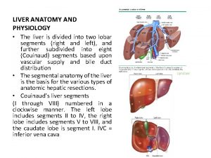
 Liver hilus
 Difference between anatomy and physiology
 Iliac regions
 Google com
 The central sulcus divides which two lobes? (figure 14-13)
 Endomysium
 Http://anatomy and physiology
 Chapter 1 introduction to human anatomy and physiology
 Appendectomy anatomy and physiology
 Aohs foundations of anatomy and physiology 1
 Aohs foundations of anatomy and physiology 1
 Anatomical planes
 Anatomy and physiology chapter 8 special senses
 Chapter 13 anatomy and physiology of pregnancy
 Teks anatomy and physiology
 Science olympiad anatomy and physiology 2020 cheat sheet
 Anatomy and physiology chapter 2
 Stomach anatomy and physiology ppt
 Physiology
 Chapter 7:9 lymphatic system
 Chapter 14 the digestive system and body metabolism
 Chapter 10 blood anatomy and physiology
 Aohs foundations of anatomy and physiology 1
 Aohs foundations of anatomy and physiology 1
 What produces bile
 Anatomy and physiology chapter 15
 Cornell notes for anatomy and physiology
 Human anatomy & physiology edition 9
 Necessary life functions anatomy and physiology
 Holes anatomy and physiology chapter 1
 Holes essential of human anatomy and physiology
 Anatomy and physiology unit 7 cardiovascular system
 Gi tract histology
 Anatomy and physiology
 Medial and lateral
 The speed at which the body consumes energy
 Aohs foundations of anatomy and physiology 1
 Olecranal region
 Human physiology exam 1
