ACUTE APPENDICITIS Rahaf Jereisat Introduction Inflammation of the


 ANAEROBIC B. fragilis 80 B.")
")


")
 -")

 - Intestinal obstruction - Ca colon")
 2. Any")


 scan - Appendix dilated and the wall is thickened. Periappendiceal phlegmon,")
")


- Slides: 65
ACUTE APPENDICITIS Rahaf Jereisat
Introduction *Inflammation of the appendix. *The most common medical abdominal emergency.
Epidemiology Highest Incidence in 2 nd and 3 rd decade of life. Incidence is equal among males and females before puberty In teenagers and young adults M: F ratio is 3: 2 at age 25 Thereafter the greater incidence in males decline Seasonal variation, more cases between may and august.
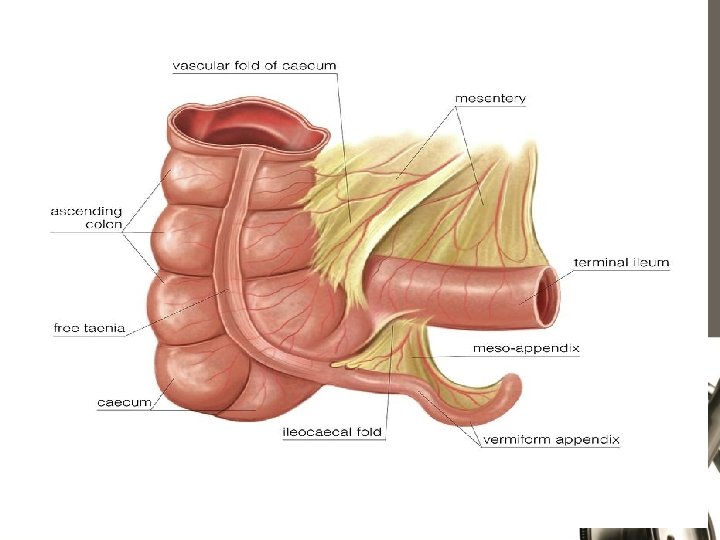
Anatomy The appendix is a narrow blind-ended tube that is attached to the posteromedial end of the cecum. Average length of 6 – 9 cm, Outer diameter 38 mm and luminal diameter 1 -3 mm. Histologically the appendix consist of three layers The serosa, muscularis layer and submucosa Formed by lymphoid aggregates and neuroendocrine complexes.
• Position: constant at the confluence of the 3 taenia coli of caecum • Mesoappendix: Arises from the lower surface of the mesentery of terminal ileum • Appendicular Artery: Branch of Ileo-colic artery – End Artery (pass through mesoappendix) • 04 -06 Lymphatic channels traverse Mesoappendix ----> Ileo-caecal LNs • Innervation : Superior mesenteric plexuses T 10 -L 1 and vagus nerve
Innervation: Superior mesenteric plexuses T 10 -L 1 and vagus nerve
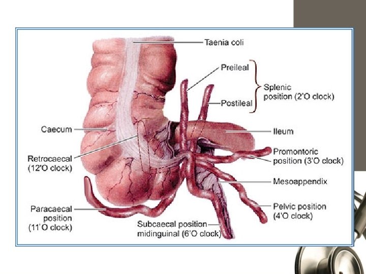
Anatomical Positions RETROCAECAL 74% PELVIC 21% PARACAECAL 2% SUBCAECAL 1. 5% PREILEAL 1% POSTILEAL 0. 5%
The function of the appendix: Used to be thought of as a vestigial remenant. Immunological organ that secrete immunoglobulin particularly immunoglobulin A and May function as a reservoir for healthy bacteria. In the base of the crypts of the mucus membrane lies the argentaffin cells which secrete serotonin (most frequent site for carcinoid tumor)
Etiology Fecolith: inspissated fecal material, calcium phosphate, bacteria, epithelial debris • Low dietary fibre and increased consumption of refined carbs • Improved hygiene and change in normal flora • Obstruction: Faecoliths/ stricture/ Foreign body Worm infestations: Oxyuris vermicularis Neoplasms: Ca caecum, carcinoids Viral
Obstruction of the lumen due to fecaliths or hypertrophy of lymphoid tissue is proposed as the main etiologic factor in acute appendicitis. The proximal obstruction of the appendiceal lumen produces a closed-loop obstruction, and continuing normal secretion by the appendiceal mucosa rapidly produces distension. Distension increases from continued mucosal secretion and from rapid multiplication of the resident bacteria of the appendix. This causes reflex nausea and vomiting, and the visceral pain increases. Capillaries and venules are occluded but arterial inflow continues, resulting in engorgement and vascular congestion. process soon involves the serosa of the appendix and in turn the parietal peritoneum. This produces the characteristic shift in pain to the right lower quadrant. As distension, bacterial invasion, compromise of the vascular supply, and infarction progress, perforation occurs
Pathology Lymphatic hyperplasia Luminal obstruction Increased intra-luminal pressure Edema, mucosal ulceration Bacterial translocation to submucosa
Resolution Venous obstruction Ischaemia of appendix wall Invasion of muscularis propria, submucosa
Ischemic necrosis of appendix wall Gangrenous appendicitis Peritonitis Appendix filled with pus Paracecal abscess Microperforations Greater omentum and loops of SB become adherant to inflammed appendix walling off the spread of contamination Phlegmonous mass/ appendicular mass Inflammation resolve with mucus distended organ Mucocele of appendix
Microbiology : • The flora of the inflamed appendix differs from that of the normal appendix. About 60% of aspirates of inflamed appendices have anaerobes compared to 25% of aspirates from normal appendices. • Virtually all grow Escherichia coli and Bacteroides species on culture. • 62% Fusobacterium nucleatum/necrophorum • Other usual species (Peptostreptococcus, Pseudomonas, Bacteroides splanchnicus, Bacteroides intermedius, Lactobacillus). • Patients with gangrene or perforated appendicitis appear to have more tissue invasion by Bacteroides.
Bacteriology of perforated appendicitis TYPE OF BACTERIA PATIENTS (%) ANAEROBIC B. fragilis 80 B. thetiaotaomicron 61 Bilophila wadsworthia 55 Peptostrptococcus spp 46 AEROBIC E. coli 77 S. viridans 43 Group D streptococcus 27 P. aeruginosa 18
Complications 1. Diffuse peritonitis 2. Perforation of gangerous appendix (mc in tip and base) 3. Delayed perforation of appendicular abscess. 4. 20% of perforated appendix: adhesions and infertility.
• Risk factors for perforation: Children: underdeveloped omentum 1. Extremes of age. Elderly: comorbidities and atrophic 2. Immunosuppression 3. Diabetes mellitus (adhesions)Limit the ability of omentum to 4. Previous abdominal surgery wall off the spread of infection 5. Use of analgesics 6. Pelvic appendix, subhepatic 7. Situs inversus, Midgut malrotation
Clinical Presentation Symptoms • Loss of appetite • Appendicitis usually starts with periumbilical and diffuse pain that eventually localizes to the right lower quadrant. • Nausea (compression in nerves) and vomiting (usually in obstructive appendicitis and children)
• Atypical symptoms include • Indigestion • Flatulence and inability to defecate (middle age) • Constipation (middle age and elderly) • Diarrhea and retching (pre and post ileal) • suprapubic pain, dysuria and urinary frequency (pelvic)
Clinical features • Signs: 1. Pyrexia 2. Localized tenderness in RIF 3. Muscle guarding 4. Rebound tenderness 5. Rovsing’s sign 6. Dunphy sign 7. Psoas sign 8. Obturator sign
Area of skin hyperaesthesia bounded by lines joining anterior superior iliac spine, the pubic symphysis and umbili cus.
Special clinical scenarios • According to position: 1. Retro-caecal - Silent appendix - Quadratus lumborum rigidity - Psoas sign - Loin tenderness
Special clinical scenarios 2. Pelvic - Early diarhoea - Increased urinary frequency - Deep tenderness over symphysis pubis - DRE: Rectovesical pouch/POD tenderness - Obturator/Psoas sign +ve
Special clinical scenarios 3. Post-ileal - Diarrhoea - Marked retching - Ill defined tenderness to rt of umbilicus
Special clinical scenarios • As per age: 1. Infants - Uncommon <36 mths - Difficult to diagnose - Diffuse peritonitis common - High incidence of perforation
Special clinical scenarios 2. Children - Vomiting - Marked anorexia 3. Elderly - High incidence of gangrene & perforation - Features of SAIO 4. Obese - Diminished signs/ delayed dignosis - Midline/ Laparoscopic approach
Special clinical scenarios 5. Pregnancy - Most common extra-uterine cause of acute abdomen Highest rate of - Delayed presentation appendicitis to cause abortion is - Fetal loss in the second trimester • 10% • Upto 20% : Perforation
Differential Diagnosis 1. Children - Mesenteric adenitis (pain moves as you move the patient) - Meckel’s diverticulitis - Gastroenteritis - Intussusception - HS purpura - Lobar pneumonia
2. Adults - Perforated peptic ulcer - Torsion of testis (always check genitalia) - Regional enteritis Valentino's syndrome is pain presenting in - Ureteric colic the RLQ caused by perforated duodenal ulcer. - Pancreatitis - Pyelonephritis - Crohn’s (mc site is ileocecal vale) - Medical causes: FMF, DKA, PNH, acute porphyria, herpes zoster
Differential Diagnosis 3. Adult female Ask about: mestrual cycle, vaginal discharge and back pain - Mittelschmerz important in relation to PID - Pyelonephritis - Ectopic pregnancy - Torsion/ rupture of ovarian cyst - Endometriosis
Differential Diagnosis 4. Elderly - Diverticulitis (redundant sigmoid) - Intestinal obstruction - Ca colon - Mesenteric infarction (vascular problems / SLE)
Differential Diagnosis for Appendicular Mass 1. Ectopic kidney (may be in RIF) 2. Any retroperitoneal pathology or tumor
Laboratory Findings • CBC : Mild leukocytosis accompanied by a polymorphonuclear prominence (white blood cell count >18, 000 cells/mm 3 raise the possibility of a perforated appendix with or without an abscess And the opposite could be due to lymphopenia or septic reaction) • C-reactive protein : elevated but can have up to a 12 -hour delay • Urine analysis : to rule out the urinary tract infection. however, several white or red blood cells can be present from irritation of the ureter or bladder. (RBC in retrocecal) • Pregnancy test BHCG A decreasing inflammatory response may indicate spontaneous resolution.
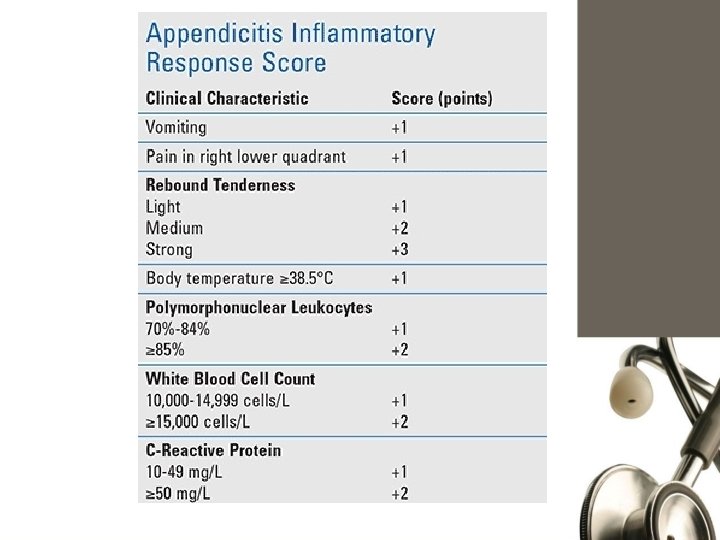
Clinical Scoring Systems
Imaging Studies 1. Plain Abdominal X-Rays 2. Graded Compression U/S 3. CT
Abdominal XRay • Fecalith • loss of psoas shadow • distention of SB loop or cecum • air under diaphragm
Plain Abdominal X-Rays • Appendicoliths picked up in only 1015% cases • Can be combined with Barium enema • Low sensitivity • Low specificity 20% of normal Appendices do not fill up
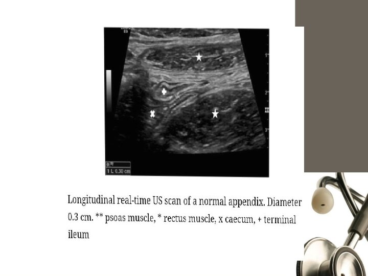
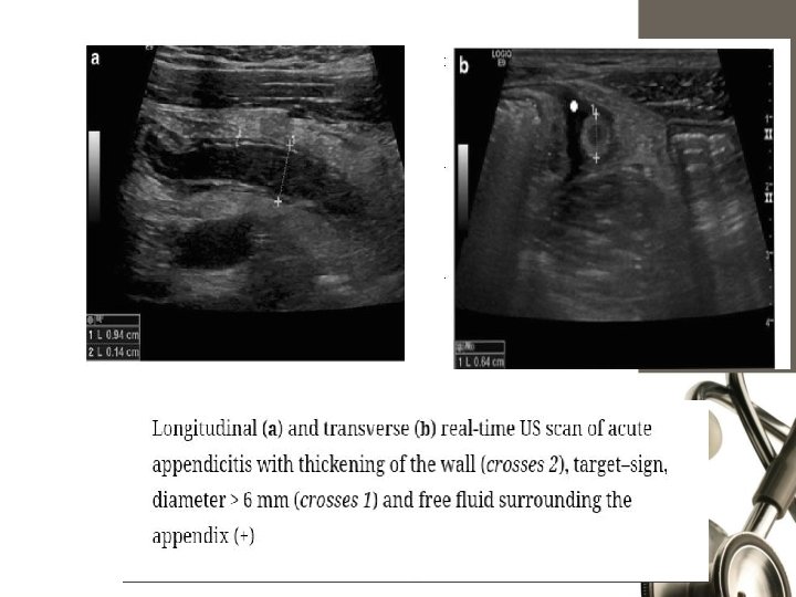
Graded Compression U/S Blind ending nonperistaltic bowel loop. Non compressible. Thickened wall>3 mm, lumen >6 mm. Presence of periappendicular fluid. Target sign. Appendicolith.
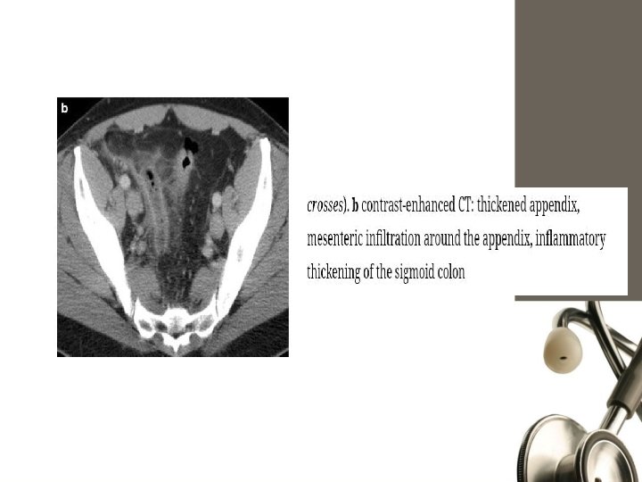
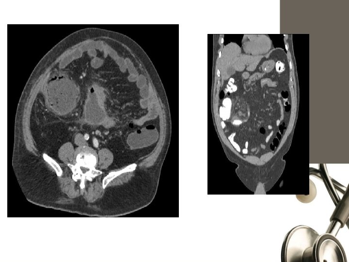
computed tomography (CT) scan - Appendix dilated and the wall is thickened. Periappendiceal phlegmon, and free fluid Periappendiceal fat stranding Thickened mesoappendix Fecaliths but their presence is not pathognomonic of appendicitis.
Ultrasonography. sensitivity of 55% to 96% and a specificity of 85% to 98%. (CT) scan 92% to 97% sensitivity, 85% to 94% specificity
Computed Tomography • Rational use: - Elderly - Atypical presentations - Neoplasms - Acute diverticulits - Intestinal obstruction • MRI: ? ?
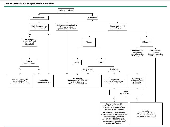
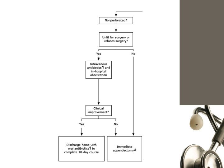
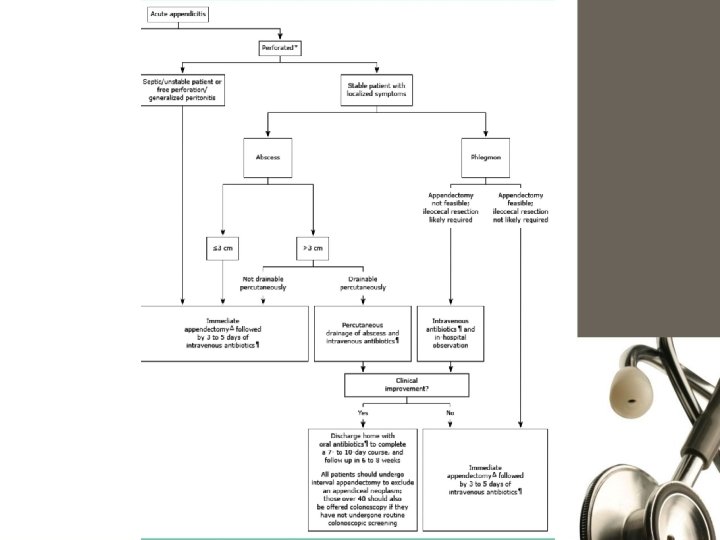
Management of Appendicular mass • Late presentation • Clinically mass & fever, Confirm by CT or US • Admit, Labs, NPO if not vomiting, NG if vomiting, Fluids Antibiotics (Broad spectrum: cefriaxone and metronidazole) Monitor Vitals q 4 Hrs • Tx: Conservative mgt(Oschner Sherren’s Regime) After 24 -48 Hrs: Improvement (less pain, no vomiting no fever, bowel motion)
No improvement: Vomiting, tachycardia, fibrile, incrase size of mass 2 possibilities: 1. Bowel obstruction by adhesions 2. Mass became abscess, confirm by CT, tx by CT guided percutaneous drainage and antibiotics
Mass same size: 1. Mass resulting from Crohn’s disease 2. Cecum CA Do lower GI endoscopy
Mucocele Deal with it carefully to avoid rupture Ruptue: pseudomyxoma peritonei (rare, diffuse gelaninous material in the peritoneum, more in females, can also be caused by neoplastic mucus secreting cells, can obstruct ureter –cause renal failure- or IVC. Tx with open surgery If it reached cecum: rt hemicolonectomy Another cause of pseudomyxoma peritonei is cystadenocarcinoma of the ovaries
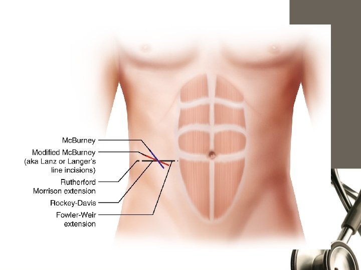
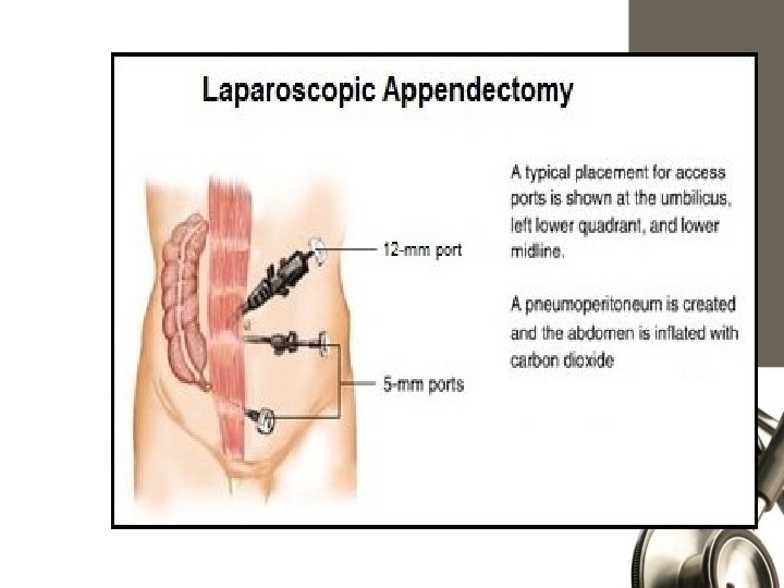
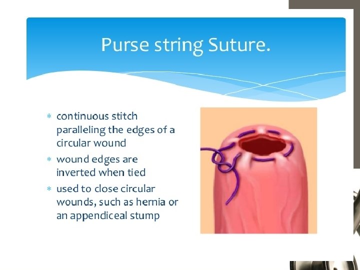
OPERATIVE INTERVENTIONS FOR THE APPENDIx Open Appendectomy Laparoscopic Appendectomy General Anesthesia A right lower quadrant incision at Mc. Burney’s point with A Mc. Burney (oblique) or Rocky-Davis (transverse) right lower quadrant muscle splitting incision If perforated appendicitis is suspected or the diagnosis is in doubt, a lower midline laparotomy can be considered Typically uses three ports. Typically, a 10 - or 12 -mm port is placed at the umbilicus, whereas two 5 -mm ports are placed suprapubic and in the left lower quadrant. Inc risk for surgical site infection Inc risk Of intra abdominal abscess Decreased operative duration less pain, shorter length of stay, and quicker return to normal activity
Problems Intra-operatively 1. Appendix not found: - Mobilise caecum - Taenia coli to be traced to their confluence 2. Appendicular tumour (carcinoid): - <2 cms: Appendicectomy - >2 cms: Rt hemicolectomy 3. Appendix normal (higher in females, more ddx) tx: Remove it. 4. Abscess and can’t be removed easily
Incidental appendectomy is defined as the removal of a clinically normal appendix during non-appendiceal surgery. In oncology patients who undergo laprotomy for any cause Prophylactic removal in: AIDS, handicaps AIDS patients have a higher incidence of appendicitis and its complications with no leukocytosis.
POSTOPERATIVE CARE AND COMPLICATIONS Non perforated. After either open or laparoscopic appendectomy for nonperforated appendicitis, patients may be started on a clear liquid diet and advanced as tolerated to a regular diet. Antibiotics are not required postoperatively. Most patients are discharged within 24 to 48 hours of surgery. Same-day discharge is feasible, most commonly following a laparoscopic appendectomy perforated appendicitis often develop an ileus postoperatively regardless of the surgical approach (open versus laparoscopic). Thus, diet should only be advanced as the clinical situation warrants. Patients may be discharged once they tolerate a regular diet, usually in five to seven days. Three to five days of intravenous antibiotics is recommended for perforated appendicitis after appendectomy
Post OP Complications 1. 2. 3. 4. 5. 6. 7. Wound infection Intra-abdominal abscess Ileus Portal pyaemia( pyelephlebitis) Adhesive intestinal obstruction Damage to ileoinguinal nerve Recurrent ot stump appendicitis
Notes Appendictomy in crohn’s if base is intact Amyand hernia is a rare form of an inguinal hernia in which the vermiform appendix is located within the hernial sac. It is seen in less than 1% of inguinal hernia. It should not be confused with an appendixcontaining femoral hernia, known as De Garengeot hernia.
Appendiceal Tumors • Carcinoid m. c, neuroendocrine that secrete 5 HT= serotonin, Mets to liver • Adenocarcinoma • Malignant mucoid adenocarcinoma (pseudomyxoma peritonei if it rupture) • Lymphoma Carcinoid tumor either intraabdominal or extra abdominal In lungs it’ll give flushing, wheezing