The Heart Valves Mohammad Saifur Rohman MD Ph


- Slides: 81
The Heart Valves Mohammad Saifur Rohman MD. Ph. D. Consultant of Interventional cardiology Department of Cardiology and Vascular Medicine Faculty of Medicine Brawijaya University 1
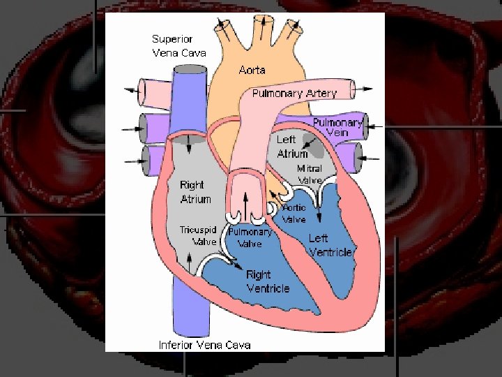
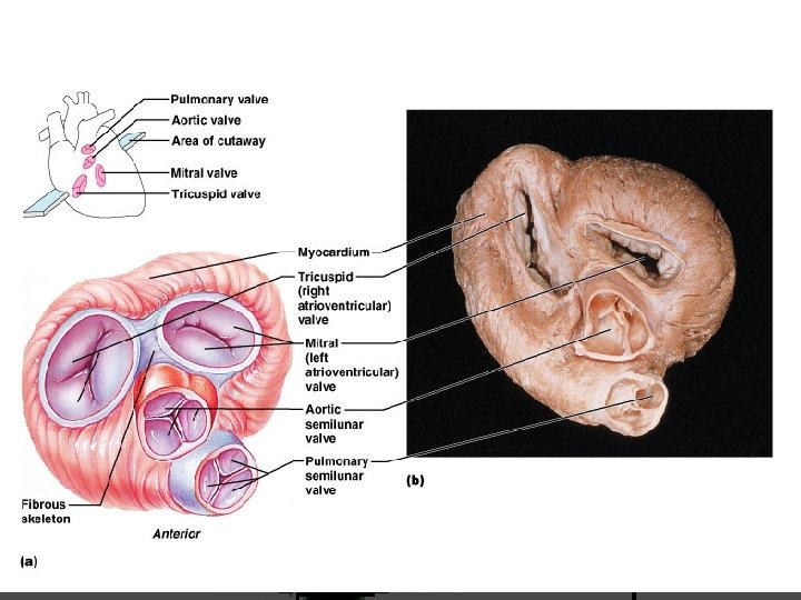
more on valves 3
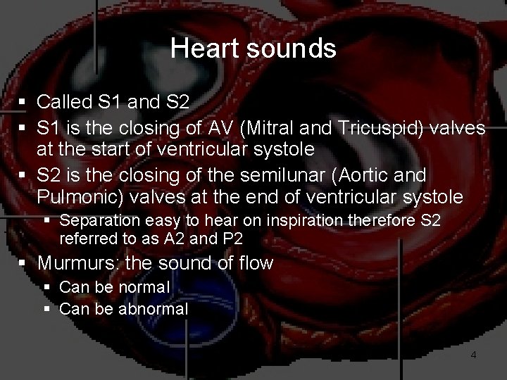
Heart sounds § Called S 1 and S 2 § S 1 is the closing of AV (Mitral and Tricuspid) valves at the start of ventricular systole § S 2 is the closing of the semilunar (Aortic and Pulmonic) valves at the end of ventricular systole § Separation easy to hear on inspiration therefore S 2 referred to as A 2 and P 2 § Murmurs: the sound of flow § Can be normal § Can be abnormal 4
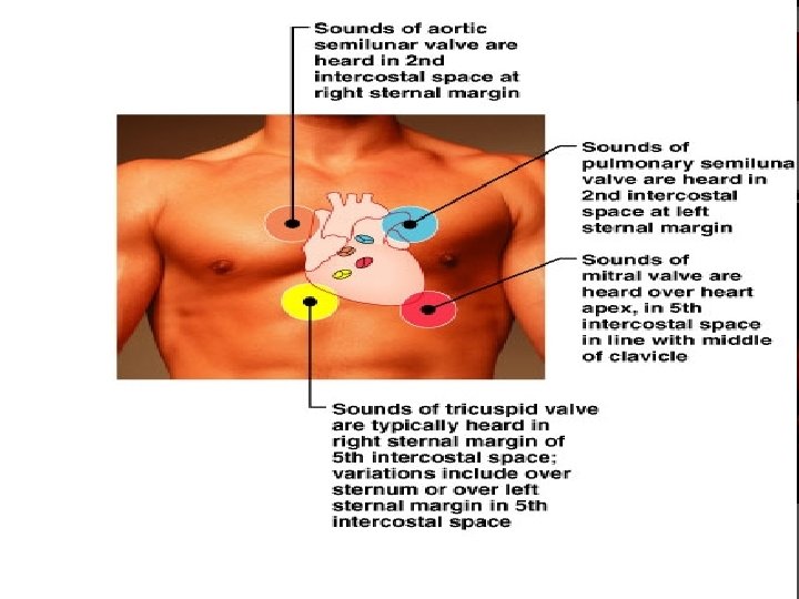
Places to auscultate 5
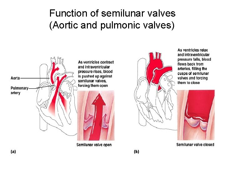
Function of semilunar valves (Aortic and pulmonic valves) 6
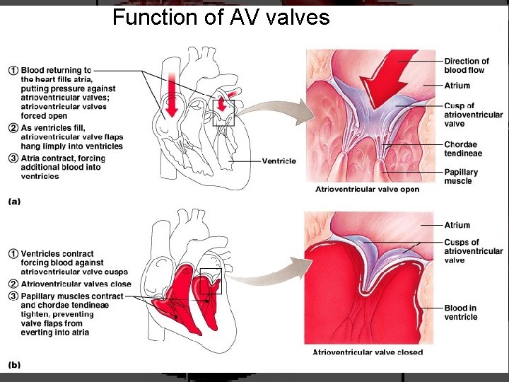
Function of AV valves 7
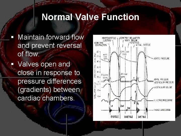
Normal Valve Function § Maintain forward flow and prevent reversal of flow. § Valves open and close in response to pressure differences (gradients) between cardiac chambers.
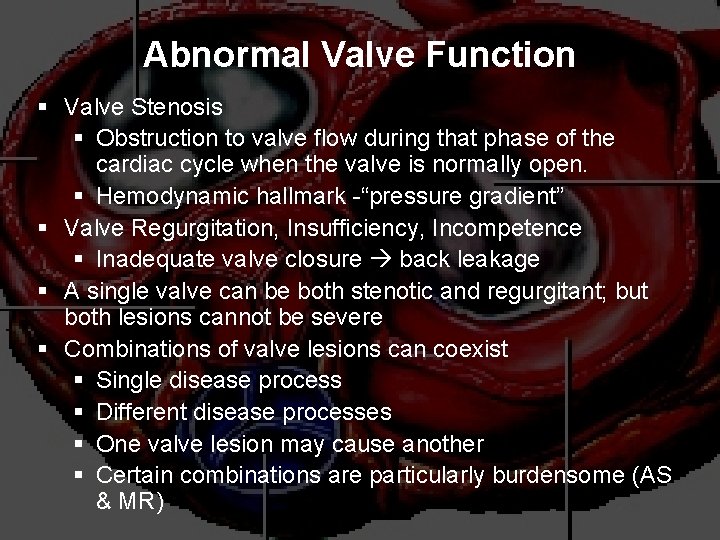
Abnormal Valve Function § Valve Stenosis § Obstruction to valve flow during that phase of the cardiac cycle when the valve is normally open. § Hemodynamic hallmark -“pressure gradient” § Valve Regurgitation, Insufficiency, Incompetence § Inadequate valve closure back leakage § A single valve can be both stenotic and regurgitant; but both lesions cannot be severe § Combinations of valve lesions can coexist § Single disease process § Different disease processes § One valve lesion may cause another § Certain combinations are particularly burdensome (AS & MR)
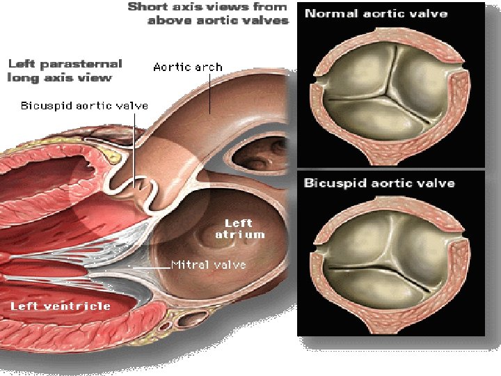
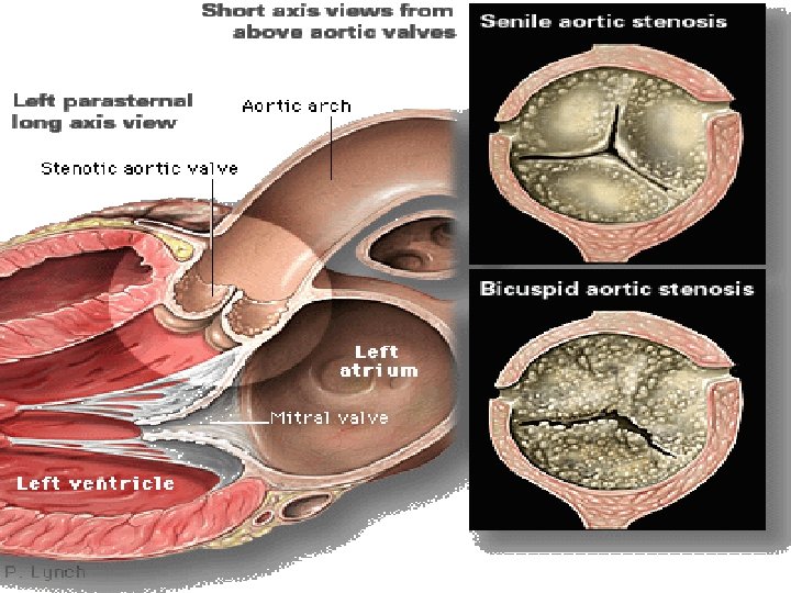
Aortic Valve Disease: Etiology § Aortic Stenosis § Degenerative calcific (senile) § Congenital – Uni or bicuspid § Rheumatic § Prosthetic § Acute Aortic Insufficiency § Infective endocarditis § Acute Aortic Dissection § Marfan’s Syndrome § Chest trauma § Chronic Aortic Insufficiency § Aortic leaflet disease § Infective endocarditis § Rheumatic § Bicuspid Aortic valve § Prolapse & congenital VSD § Prosthetic § Aortic root disease § Aortic aneurysm/dissection § Marfan’s syndrome § Connective tissue disorders § Syphilis § HTN § Annulo-aortic ectasia
Aortic Stenosis Overview: § § Normal Aortic Valve Area: 3 -4 cm 2 Symptoms: Occur when valve area is 1/4 th of normal area. § Types: § Supravalvular § Subvalvular § Valvular
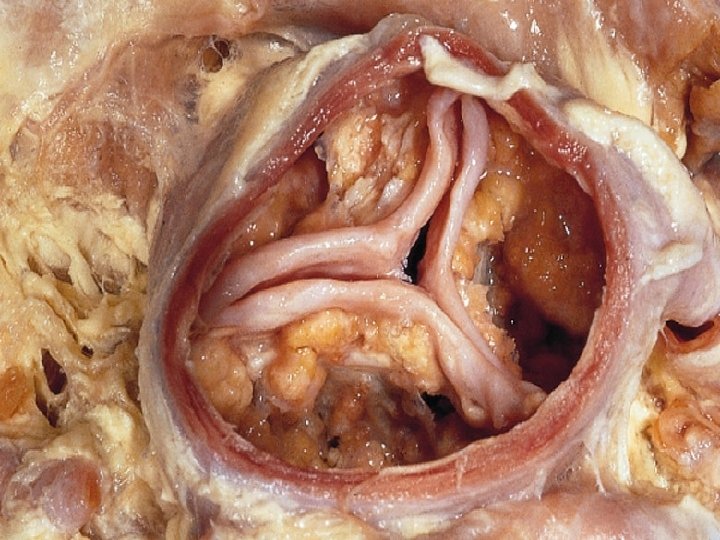
Etiology of Aortic Stenosis § Congenital § Rheumatic § Degenerative/Calcification Patients under 70: >50% have a congenital cause Patients over 70: 50% due to degenerative
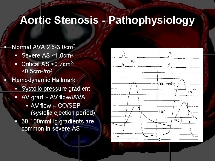
Aortic Stenosis - Pathophysiology § Normal AVA 2. 5 -3. 0 cm 2 § Severe AS <1. 0 cm 2 § Critical AS <0. 7 cm 2; <0. 5 cm 2/m 2 § Hemodynamic Hallmark § Systolic pressure gradient § AV grad ~ AV flow//AVA § AV flow = CO/SEP (systolic ejection period) § 50 -100 mm. Hg gradients are common in severe AS
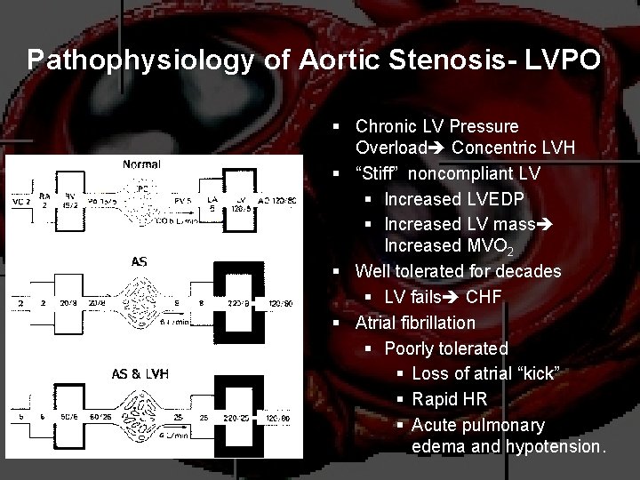
Pathophysiology of Aortic Stenosis- LVPO § Chronic LV Pressure Overload Concentric LVH § “Stiff” noncompliant LV § Increased LVEDP § Increased LV mass Increased MVO 2 § Well tolerated for decades § LV fails CHF § Atrial fibrillation § Poorly tolerated § Loss of atrial “kick” § Rapid HR § Acute pulmonary edema and hypotension.
Pathophysiology of Aortic Stenosis § A pressure gradient develops between the left ventricle and the aorta. (increased afterload) § LV function initially maintained by compensatory pressure hypertrophy § When compensatory mechanisms exhausted, LV function declines.
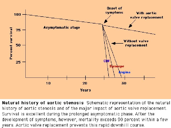
Aortic Stenosis: Natural History & Clinical Symptoms § Asymptomatic for many years § Symptoms develop when valve is critically narrowed and LV function deteriorates § Bicuspid AV 5 th - 6 th decade § Senile AS 7 th-8 th decades § Classic Symptom Triad § Angina pectoris – 5 years § CHF 1 -2 years § Syncope 2 -3 years § Sudden Death § Natural History Studies§ Pts grad 25 mm. Hg – 20% chance of intervention in 15 years § Pts with asymptomatic severe AS require close f/u § Gradient progression § 6 -10 mm. Hg/yr § Risk Factors § Age > 70 § CAD, hyperlipidemia § Chronic renal failure
Natural History § Mild AS to Severe AS: § 8% in 10 years § 22% in 22 years § 38% in 25 years § The onset of symptoms is a poor prognostic indicator.
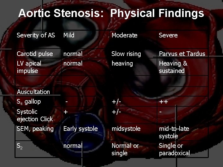
Aortic Stenosis: Physical Findings Severity of AS Mild Moderate Severe Carotid pulse normal Slow rising Parvus et Tardus LV apical impulse normal heaving Heaving & sustained - +/- ++ Systolic ejection Click + +/- - SEM, peaking Early systole mid-to-late systole S 2 normal Normal or single Single or paradoxical Auscultation S 4 gallop
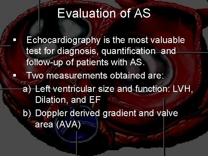
Evaluation of AS § Echocardiography is the most valuable test for diagnosis, quantification and follow-up of patients with AS. § Two measurements obtained are: a) Left ventricular size and function: LVH, Dilation, and EF b) Doppler derived gradient and valve area (AVA)
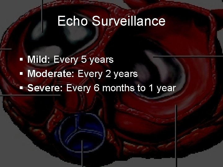
Echo Surveillance § Mild: Every 5 years § Moderate: Every 2 years § Severe: Every 6 months to 1 year
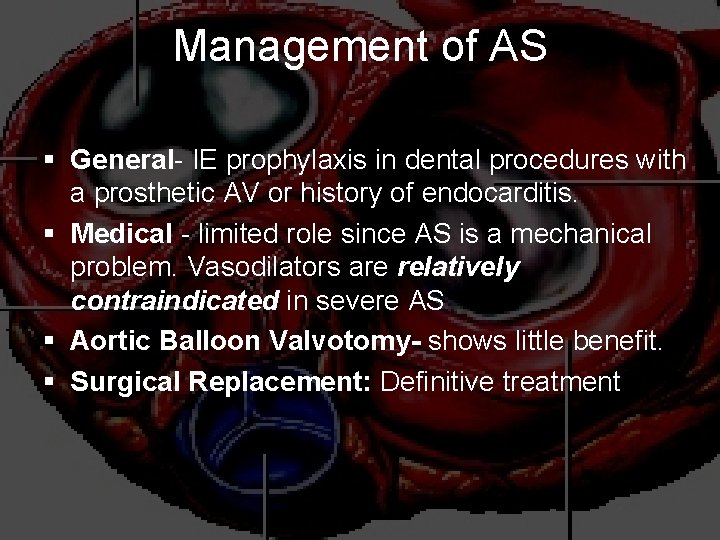
Management of AS § General- IE prophylaxis in dental procedures with a prosthetic AV or history of endocarditis. § Medical - limited role since AS is a mechanical problem. Vasodilators are relatively contraindicated in severe AS § Aortic Balloon Valvotomy- shows little benefit. § Surgical Replacement: Definitive treatment
Balloon Aortic Valvuloplasty § Indications for BAV in critical Aortic Stenosis § Younger patients with congenital AS and predominant commissural fusion § Bridge to eventual AVR § Moderate to severe heart failure/cardiogenic shock § Extremely high risk for AVR § Urgent/emergent need for noncardiac surgery § Patient with limited lifespan – cardiac or noncardiac § Patient refuses surgery
Simplified Indications for Surgery in Aortic Stenosis § Any SYMPTOMATIC patient with severe AS (includes symptoms with exercise) § Any patient with decreasing EF § Any patient undergoing CABG with moderate or severe AS
Aortic Regurgitation Overview § Definition: Leakage of blood into LV during diastole due to ineffective coaptation of the aortic cusps
Etiology of Acute AR § Endocarditis § Aortic Dissection § Physical Findings: § Wide pulse pressure § Diastolic murmur § Florid pulmonary edema
Etiology of Chronic AR § Bicuspid aortic valve § Rheumatic § Infective endocarditis
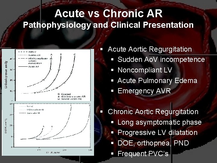
Acute vs Chronic AR Pathophysiology and Clinical Presentation § Acute Aortic Regurgitation § Sudden Ao. V incompetence § Noncompliant LV § Acute Pulmonary Edema § Emergency AVR § Chronic Aortic Regurgitation § Long asymptomatic phase § Progressive LV dilatation § DOE, orthopnea, PND § Frequent PVC’s
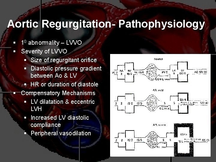
Aortic Regurgitation- Pathophysiology § 10 abnormality – LVVO § Severity of LVVO § Size of regurgitant orifice § Diastolic pressure gradient between Ao & LV § HR or duration of diastole § Compensatory Mechanisms § LV dilatation & eccentric LVH § Increased LV diastolic compliance § Peripheral vasodilation
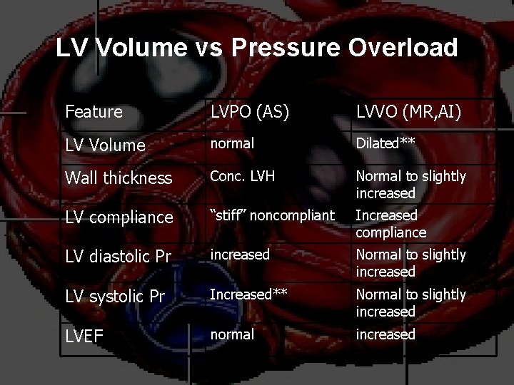
LV Volume vs Pressure Overload Feature LVPO (AS) LVVO (MR, AI) LV Volume normal Dilated** Wall thickness Conc. LVH Normal to slightly increased LV compliance “stiff” noncompliant Increased compliance LV diastolic Pr increased Normal to slightly increased LV systolic Pr Increased** Normal to slightly increased LVEF normal increased
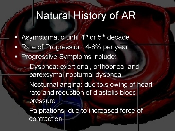
Natural History of AR § Asymptomatic until 4 th or 5 th decade § Rate of Progression: 4 -6% per year § Progressive Symptoms include: - Dyspnea: exertional, orthopnea, and paroxsymal nocturnal dyspnea - Nocturnal angina: due to slowing of heart rate and reduction of diastolic blood pressure - Palpitations: due to increased force of contraction
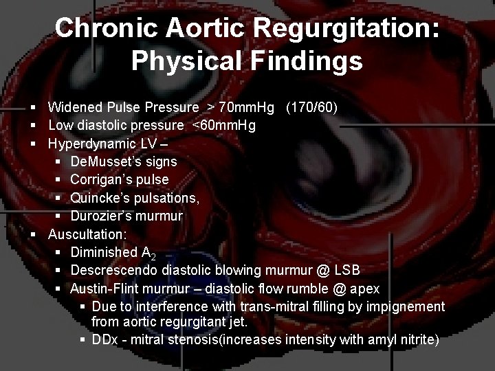
Chronic Aortic Regurgitation: Physical Findings § Widened Pulse Pressure > 70 mm. Hg (170/60) § Low diastolic pressure <60 mm. Hg § Hyperdynamic LV – § De. Musset’s signs § Corrigan’s pulse § Quincke’s pulsations, § Durozier’s murmur § Auscultation: § Diminished A 2 § Descrescendo diastolic blowing murmur @ LSB § Austin-Flint murmur – diastolic flow rumble @ apex § Due to interference with trans-mitral filling by impignement from aortic regurgitant jet. § DDx - mitral stenosis(increases intensity with amyl nitrite)
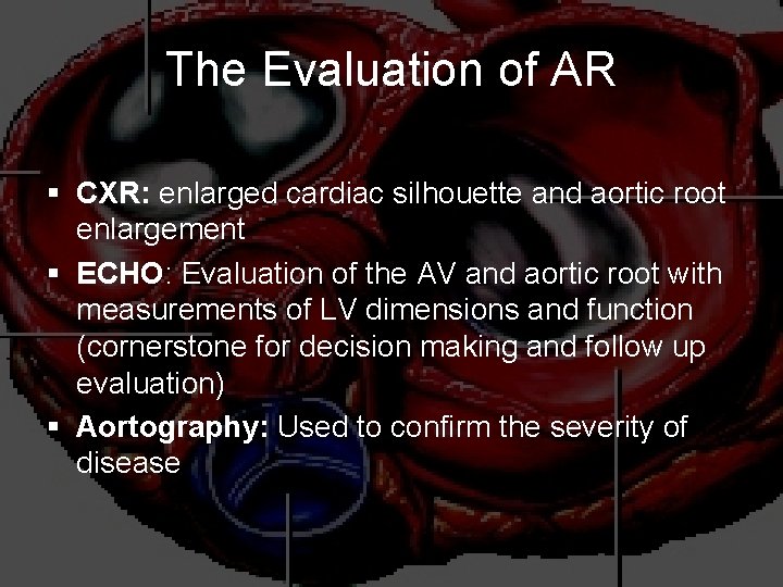
The Evaluation of AR § CXR: enlarged cardiac silhouette and aortic root enlargement § ECHO: Evaluation of the AV and aortic root with measurements of LV dimensions and function (cornerstone for decision making and follow up evaluation) § Aortography: Used to confirm the severity of disease
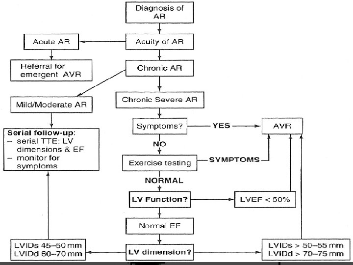
Management of AR § § General: IE prophylaxis in dental procedures with a prosthetic AV or history of endocarditis. Medical: Vasodilators (ACEI’s), Nifedipine improve stroke volume and reduce regurgitation only if pt symptomatic or HTN. Serial Echocardiograms: to monitor progression. Surgical Treatment: Definitive Tx
Treatment of Acute AR § § § True Surgical Emergency: Positive inotrope: (eg, dopamine, dobutamine) Vasodilators: (eg, nitroprusside) Avoid beta-blockers Do not even consider a balloon pump
Simplified Indications for Surgical Treatment of AR § ANY Symptoms at rest or exercise § Asymptomatic treatment if: § EF drops below 50% or LV becomes dilated
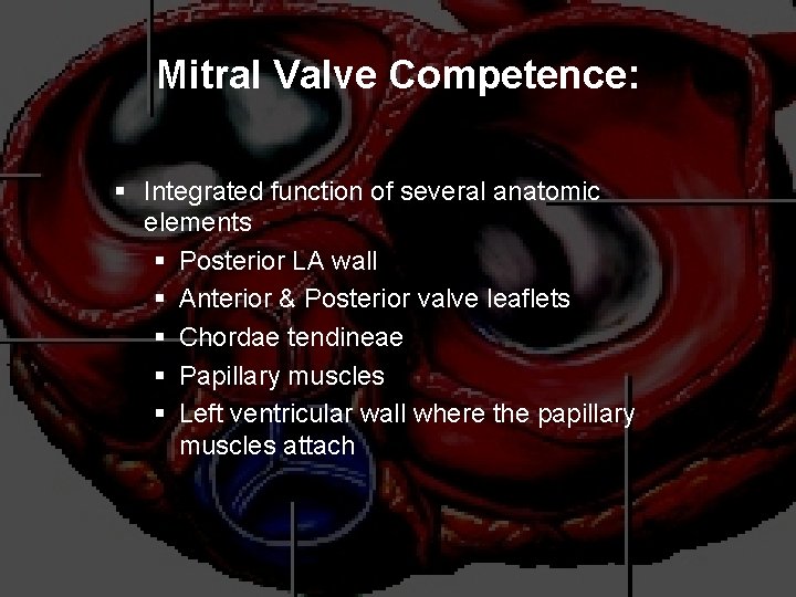
Mitral Valve Competence: § Integrated function of several anatomic elements § Posterior LA wall § Anterior & Posterior valve leaflets § Chordae tendineae § Papillary muscles § Left ventricular wall where the papillary muscles attach
Mitral Valve Disease: Etiology v Mitral Stenosis v Rheumatic - 99. 9%!!! v Congenital v Prosthetic valve stenosis v Mitral Annular Calcification v Left Atrial Myxoma v Acute Mitral Regurgitation v Infective endocarditis v Ischemic Heart disease v. Papillary ms rupture v Mitral valve prolapse v. Chordal rupture v Chest trauma v Chronic Mitral Regurgitation v Ischemic Heart disease v. Papillary ms dysfunction v. Inferior & posterior MI v Mitral Valve prolapse v Infective endocarditis v Rheumatic v Prosthetic v Mitral annular calcification v Cardiomyopathy v. LV dilatation v. IHSS
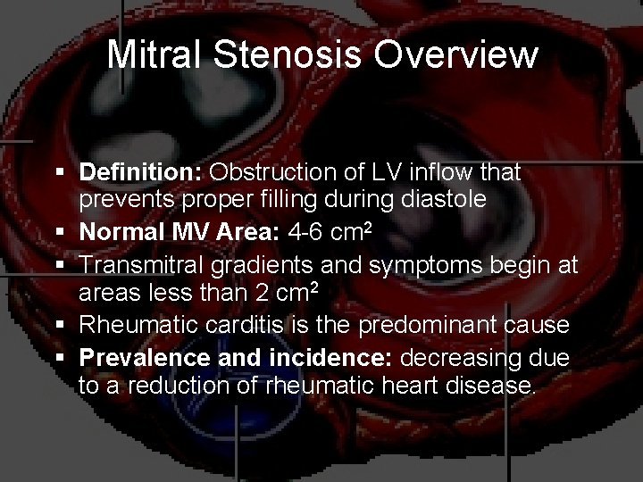
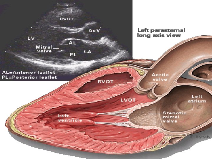
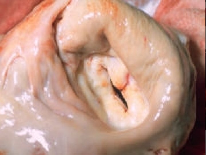
Mitral Stenosis Overview § Definition: Obstruction of LV inflow that prevents proper filling during diastole § Normal MV Area: 4 -6 cm 2 § Transmitral gradients and symptoms begin at areas less than 2 cm 2 § Rheumatic carditis is the predominant cause § Prevalence and incidence: decreasing due to a reduction of rheumatic heart disease.
Etiology of Mitral Stenosis § Rheumatic heart disease: 77 -99% of all cases § Infective endocarditis: 3. 3% § Mitral annular calcification: 2. 7%
MS Pathophysiology § Progressive Dyspnea (70%): LA dilation pulmonary congestion (reduced emptying) § § worse with exercise, fever, tachycardia, and pregnancy Increased Transmitral Pressures: Leads to left atrial enlargement and atrial fibrillation. Right heart failure symptoms: due to Pulmonary venous HTN Hemoptysis: due to rupture of bronchial vessels due to elevated pulmonary pressure
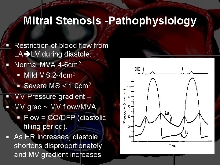
Mitral Stenosis -Pathophysiology § Restriction of blood flow from LA LV during diastole. § Normal MVA 4 -6 cm 2. § Mild MS 2 -4 cm 2. § Severe MS < 1. 0 cm 2. § MV Pressure gradient – § MV grad ~ MV flow//MVA. § Flow = CO/DFP (diastolic filling period). § As HR increases, diastole shortens disproportionately and MV gradient increases.
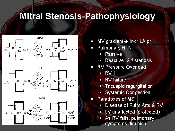
Mitral Stenosis-Pathophysiology § MV gradient Incr LA pr § Pulmonary HTN § Passive § Reactive- 2 nd stenosis § RV Pressure Overload § RVH § RV failure § Tricuspid regurgitation § Systemic Congestion § Paradoxes of MS § Disease of Pulm Arts & RV § LV unaffected (protected) § As RV fails, pulmonary symptoms diminish
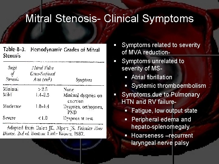
Mitral Stenosis- Clinical Symptoms § Symptoms related to severity of MVA reduction§ Symptoms unrelated to severity of MS§ Atrial fibrillation § Systemic thromboembolism § Symptoms due to Pulmonary HTN and RV failure§ Fatigue, low output state § Peripheral edema and hepato-splenomegaly § Hoarseness –recurrent laryngeal nerve palsy
Natural History of MS § Disease of plateaus: § Mild MS: 10 years after initial RHD insult § Moderate: 10 years later § Severe: 10 years later § Mortality: Due to progressive pulmonary congestion, infection, and thromboembolism.
Mitral Stenosis: Physical Findings § Auscultatory findings § S 1 – variable intensity; increased early, progressively decreases § OS –opening snap, variable intensity § A 2 -OS interval – varies inversely with severity of MS; shortens as MVA diminishes § Low-pitched diastolic rumble @ apex § Duration of murmur correlates with severity of MS § Pre-systolic accentuation § Increased P 2 § Body habitus – thin, asthenic, female § Low BP § LA lift & RV tap
Heart Sounds in MS § Diastolic murmur: § Low-pitched diastolic rumble most prominent at the apex. § Heard best with the patient lying on the left side in held expiration § Intensity of the diastolic murmur does not correlate with the severity of the stenosis
Heart Sounds in MS § Loud Opening S 1 snap: heard at the apex when leaflets are still mobile § Due to the abrupt halt in leaflet motion in early diastole, after rapid initial rapid opening, due to fusion at the leaflet tips. § A shorter S 2 to opening snap interval indicates more severe disease.
Evaluation of MS § ECG: may show atrial fibrillation and LA enlargement § CXR: LA enlargement and pulmonary congestion. Occasionally calcified MV § ECHO: The GOLD STANDARD for diagnosis. Asses mitral valve mobility, gradient and mitral valve area
Management of MS Serial echocardiography: § Mild: 3 -5 years § Moderate: 1 -2 years § Severe: yearly § Medications: MS like AS is a mechanical problem and medical therapy does not prevent progression § -blockers, CCBs, Digoxin which control heart rate and hence prolong diastole for improved diastolic filling § Diuretics for fluid overload
Management of MS § Identify patient early who might benefit from percutaneous mitral balloon valvotomy. § IE prophylaxis: Patients with prosthetic valves or a Hx of IE for dental procedures.
Simplified Indications for Mitral valve replacement § ANY SYMPTOMATIC Patient with NYHA Class III or IV Symptoms § Asymptomatic moderate or Severe MS with a pliable valve suitable for PMBV
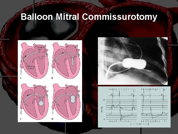
Balloon Mitral Commissurotomy
Chronic Mitral Regurgitation Overview § § Definition: Backflow of blood from the LV to the LA during systole Mild (physiological) MR is seen in 80% of normal individuals.
Etiologies of Chronic Mitral Regurgitation § § Myxomatous degeneration (MVP) Ischemic MR Rheumatic heart disease Infective Endocarditis
Acute MR § Endocarditis § Acute MI: § Malfunction or disruption of prosthetic valve
Pathophysiology of MR § Pure Volume Overload § Compensatory Mechanisms: Left atrial enlargement, LVH and increased contractility § Progressive left atrial dilation and right ventricular dysfunction due to pulmonary hypertension. § Progressive left ventricular volume overload leads to dilation and progressive heart failure.
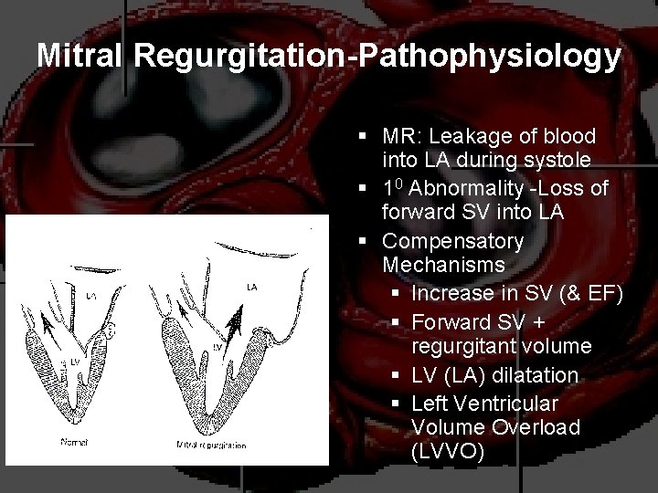
Mitral Regurgitation-Pathophysiology § MR: Leakage of blood into LA during systole § 10 Abnormality -Loss of forward SV into LA § Compensatory Mechanisms § Increase in SV (& EF) § Forward SV + regurgitant volume § LV (LA) dilatation § Left Ventricular Volume Overload (LVVO)
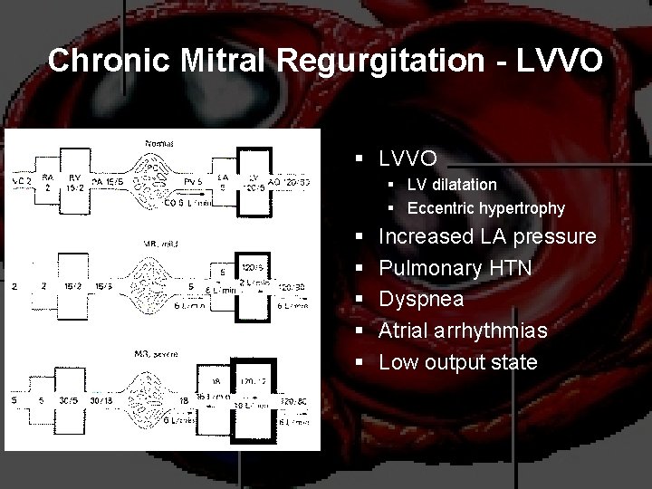
Chronic Mitral Regurgitation - LVVO § LV dilatation § Eccentric hypertrophy § § § Increased LA pressure Pulmonary HTN Dyspnea Atrial arrhythmias Low output state
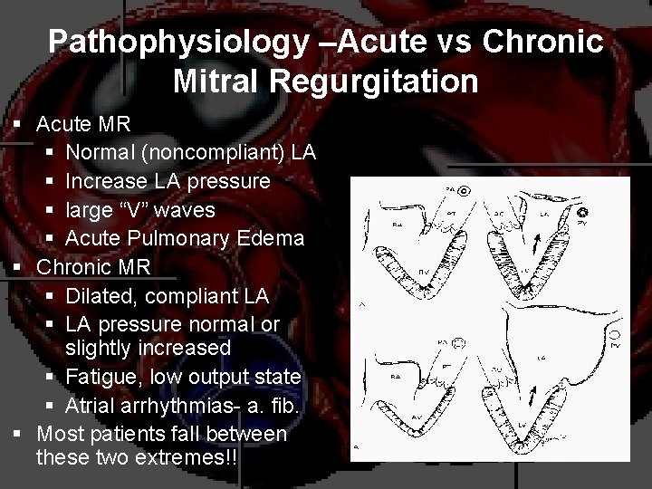
Pathophysiology –Acute vs Chronic Mitral Regurgitation § Acute MR § Normal (noncompliant) LA § Increase LA pressure § large “V” waves § Acute Pulmonary Edema § Chronic MR § Dilated, compliant LA § LA pressure normal or slightly increased § Fatigue, low output state § Atrial arrhythmias- a. fib. § Most patients fall between these two extremes!!
Physical Exam findings in MR § Auscultation: soft S 1 and a holosystolic murmur at the apex radiating to the axilla § S 3 (CHF/LA overload) & diastolic flow rumble § In chronic MR, the intensity of the murmur does correlate with the severity. § Exertion Dyspnea: ( exercise intolerance) § Heart Failure: May coincide with increased hemodynamic burden e. g. , pregnancy, infection or atrial fibrillation
Mitral Regurgitation: Physical Findings § Auscultatory Findings § P 2 – increased § Holosystolic blowing murmur @ apex § MVP – mid-systolic click § IHSS – murmur increases with Valsalva § Acute MR – descrescendo systolic murmur § Hyperdynamic Left Ventricle § Brisk carotid upstrokes § Hyperdynamic LV apical impulse § LA lift; RV tap
The Natural History of MR § Compensatory phase: 10 -15 years § Patients with asymptomatic severe MR have a 5%/year mortality rate § Once the patient’s EF becomes <60% and/or becomes symptomatic, mortality rises sharply § Mortality: From progressive dyspnea and heart failure
Imaging studies in MR § ECG: May show, LA enlargement, atrial fibrillation and LV hypertrophy with severe MR § CXR: LA enlargement, central pulmonary artery enlargement. § ECHO: Estimation of LA, LV size and function. Valve structure assessment § TEE if transthoracic echo is inconclusive
Management of Acute MR § Myocardial infarction: Cardiac cath or thrombolytics § Most other cases of mitral regurgitation is afterload reduction: § Diuretics and nitrates § nitroprusside, even in the setting of a normal blood pressure.
Management of Acute MR § Do not attempt to alleviate tachycardia with betablockers. Mild-to-moderate tachycardia is beneficial in these patients because it allows less time for the heart to have backfill, which lowers regurgitant volume.
Treatment of Acute MR § Balloon Pump § Nitroprusside even if hypotensive § Emergent Surgery
Management of MR § Medications a) Vasodilator such as hydralazine b) Rate control for atrial fibrillation with blockers, CCB, digoxin c) Anticoagulation in atrial fibrillation and flutter d) Diuretics for fluid overload
Management of MR § § Serial Echocardiography: § Mild: 2 -3 years § Moderate: 1 -2 years § Severe: 6 -12 months IE prophylaxis: Patients with prosthetic valves or a Hx of IE for dental procedures.
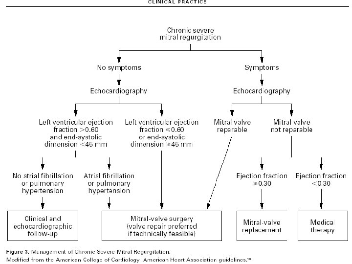
Simplified Indications for MV Replacement in Severe MR § ANY Symptoms at rest or exercise with (repair if feasible) § Asymptomatic: § If EF <60% § If new onset atrial fibrillation
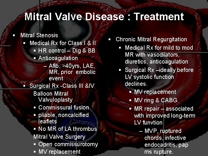
Mitral Valve Disease : Treatment § Mitral Stenosis § Medical Rx for Class I & II § HR control – Dig & BB § Anticoagulation – Afib, >40 yrs, LAE, MR, prior embolic event § Surgical Rx -Class III &IV Balloon Mitral Valvuloplasty § Commissural fusion § pliable, noncalcified leaflets § No MR of LA thrombus Mitral Valve Surgery § Open commissurotomy § MV replacement § Chronic Mitral Regurgitation § Medical Rx for mild to mod MR with vasodilators, diuretics, anticoagulation § Surgical Rx –ideally before LV systolic function declines. § MV replacement § MV ring & CABG § MR repair – associated with improved long-term LV funvtion – MVP, ruptured chords, infective endocadritis, pap ms rupture.
THANK YOU