
Urologic Disorders Mohammad Alomar Consultant Urologist Assistant professor






- Slides: 47
Urologic Disorders Mohammad Alomar Consultant Urologist Assistant professor of Surgery September 2015
Urologic Disorders Urinary tract infections Urolithiasis Benign Prostatic Hyperplasia and voiding dysfunction
Urinary tract infections Urethritis Acute Pyelonephritis Epididymitis/orchitis Chronic Pyelonephritis Prostatitis Renal Abscess cystitis
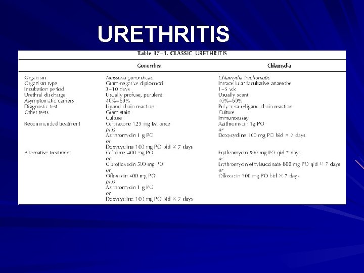
URETHRITIS S&S – urethral discharge – burning on urination – Asymptomatic Gonococcal vs. Nongonococcal DX: – incubation period(3 -10 days vs. 1 -5 wks) – Urethral swab – Serum: Chlamydia-specific ribosomal RNA
URETHRITIS
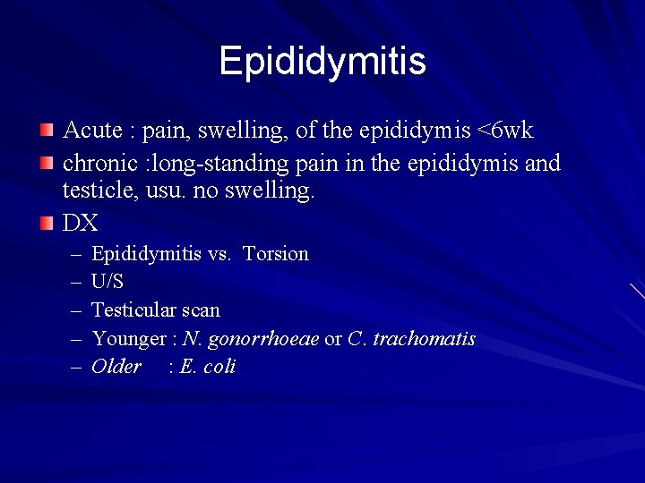
Epididymitis Acute : pain, swelling, of the epididymis <6 wk chronic : long-standing pain in the epididymis and testicle, usu. no swelling. DX – – – Epididymitis vs. Torsion U/S Testicular scan Younger : N. gonorrhoeae or C. trachomatis Older : E. coli
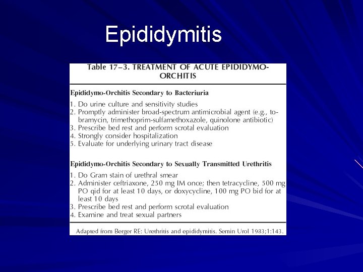
Epididymitis
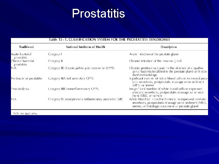
Prostatitis Syndrome that presents with inflammation± infection of the prostate gland including: – – Dysuria, frequency dysfunctional voiding Perineal pain Painful ejaculation
Prostatitis
Prostatitis Acute Bacterial Prostatitis : – Rare – Acute pain – Storage and voiding urinary symptoms – Fever, chills, malaise, N/V – Perineal and suprapubic pain – Tender swollen hot prostate. – Rx : Abx and urinary drainage
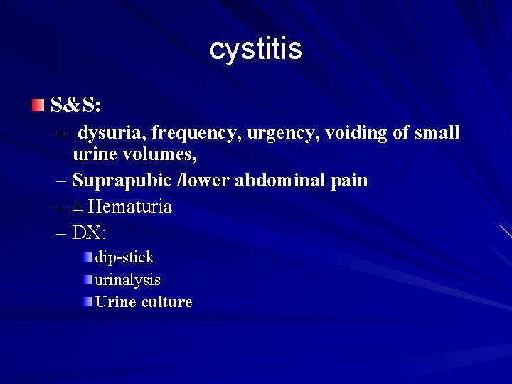
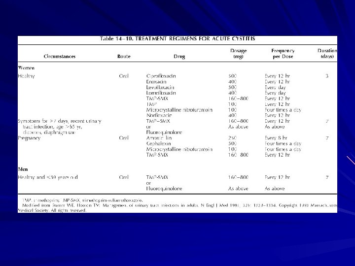
cystitis S&S: – dysuria, frequency, urgency, voiding of small urine volumes, – Suprapubic /lower abdominal pain – ± Hematuria – DX: dip-stick urinalysis Urine culture
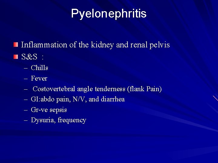
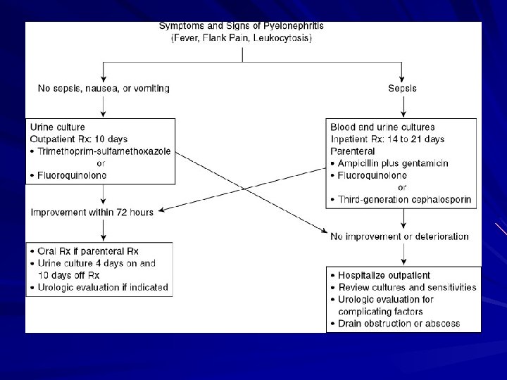
Pyelonephritis Inflammation of the kidney and renal pelvis S&S : – – – Chills Fever Costovertebral angle tenderness (flank Pain) GI: abdo pain, N/V, and diarrhea Gr-ve sepsis Dysuria, frequency
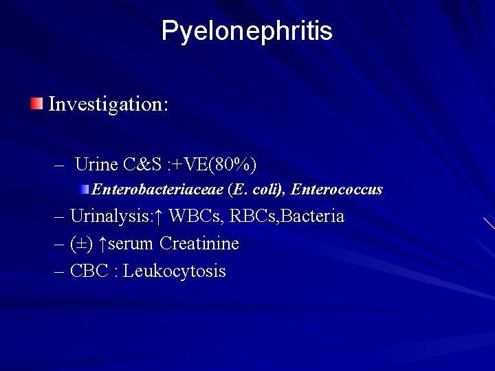
Pyelonephritis Investigation: – Urine C&S : +VE(80%) Enterobacteriaceae (E. coli), Enterococcus – Urinalysis: ↑ WBCs, RBCs, Bacteria – (±) ↑serum Creatinine – CBC : Leukocytosis
Pyelonephritis Imaging: – IVP – U/S – CT
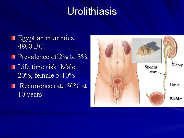
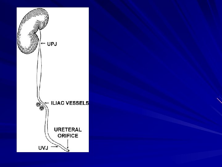
Urolithiasis Egyptian mummies 4800 BC Prevalence of 2% to 3%, Life time risk: Male : 20%, female 5 -10% Recurrence rate 50% at 10 years
Urolithiasis Risk factors: – Intrinsic Factors Genetics Age (2 Os-4 Os) Sex M>F
Urolithiasis Extrinsic Factors – Geography (mountainous, desert, tropics ) – Climate (July - October) – Water Intake – Diet (purines , oxalates, Na ) – Occupation (sedentary occupations )
Urolithiasis How do stones form – supersaturated→ Crystal Growth – Aggregation of crystals →stone
Urolithiasis Most people have crystals in their urine, so why not everyone gets stones? – Anatomic abnormalities – Modifiers of crystal formation: Inhibitors/promoters Citrate Mg, urinary proteins(nephrocalcin) oxalate
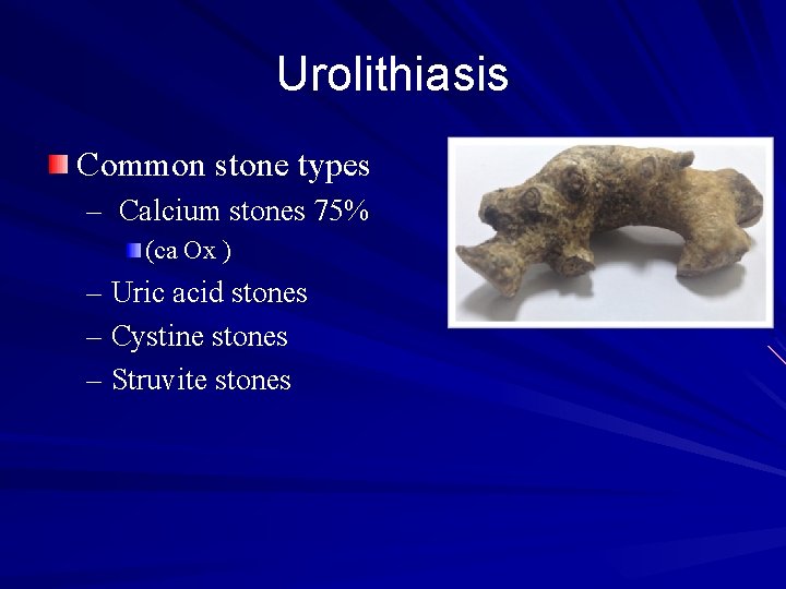
Urolithiasis Common stone types – Calcium stones 75% (ca Ox ) – Uric acid stones – Cystine stones – Struvite stones
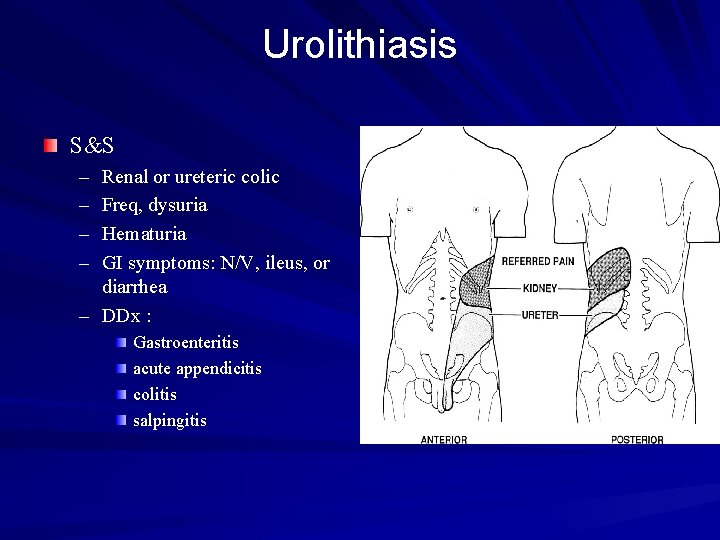
Urolithiasis S&S – – Renal or ureteric colic Freq, dysuria Hematuria GI symptoms: N/V, ileus, or diarrhea – DDx : Gastroenteritis acute appendicitis colitis salpingitis
Urolithiasis Cont. S&S – Restless ↑HR, ↑ BP fever (If UTI) Tender CVA
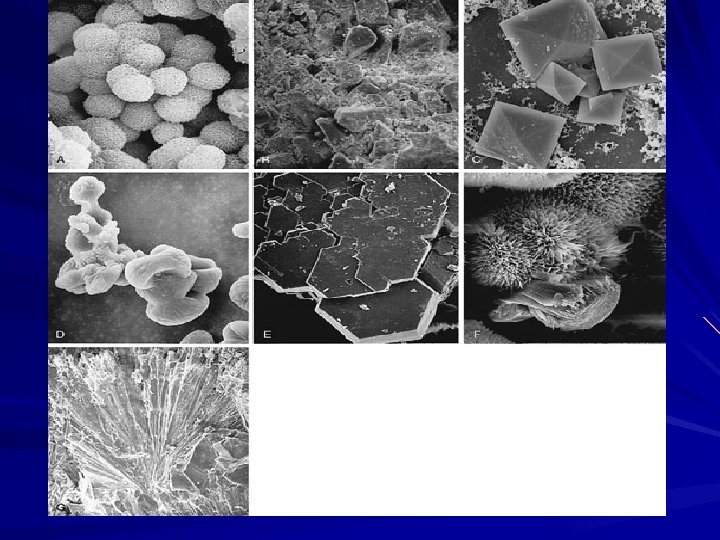
Urolithiasis Investigation Urinalysis : – RBC – WBC – Bacteria – Crystals
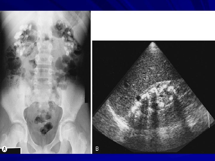
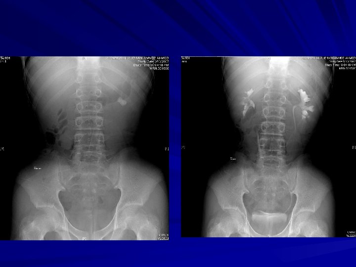
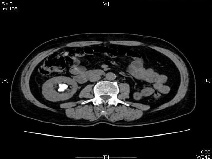
Urolithiasis Investigation Imaging – Plain Abdominal Films (KUB) – Intravenous Urography (IVP) – Ultrasonography (U/S) – Computed Tomography (CT)
Urolithiasis Management Conservative – – Hydration Analgesia Antiemetic Stones (<5 mm ) >90% spontaneous Passage Indication for admission – – Renal impairment Refractory pain Pyelonephritis intractable N/V
Urolithiasis Management Extracorporeal Shock Wave lithotripsy (SWL) Ureteroscopy Percutaneous Nephrolithotripsy (PNL) Open Sx
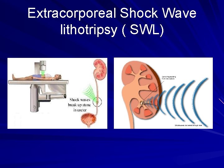
Extracorporeal Shock Wave lithotripsy ( SWL)
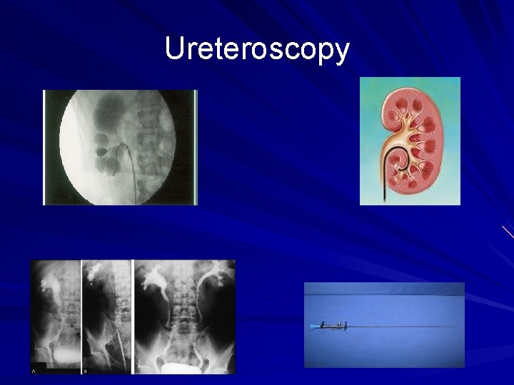
Ureteroscopy
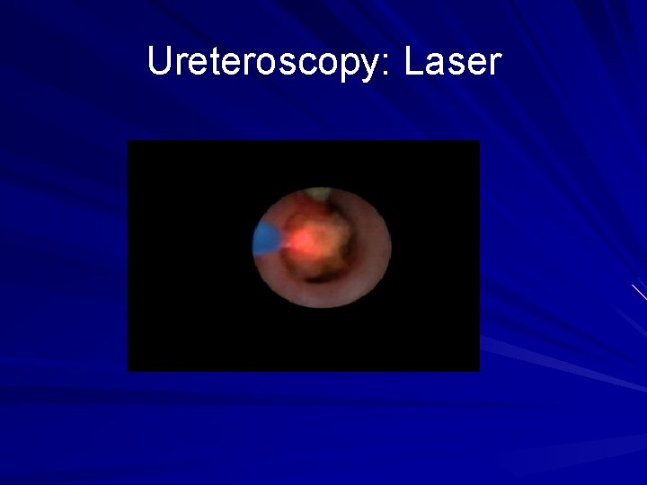
Ureteroscopy: Laser
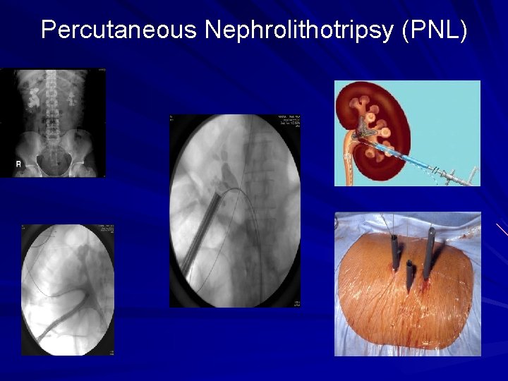
Percutaneous Nephrolithotripsy (PNL)
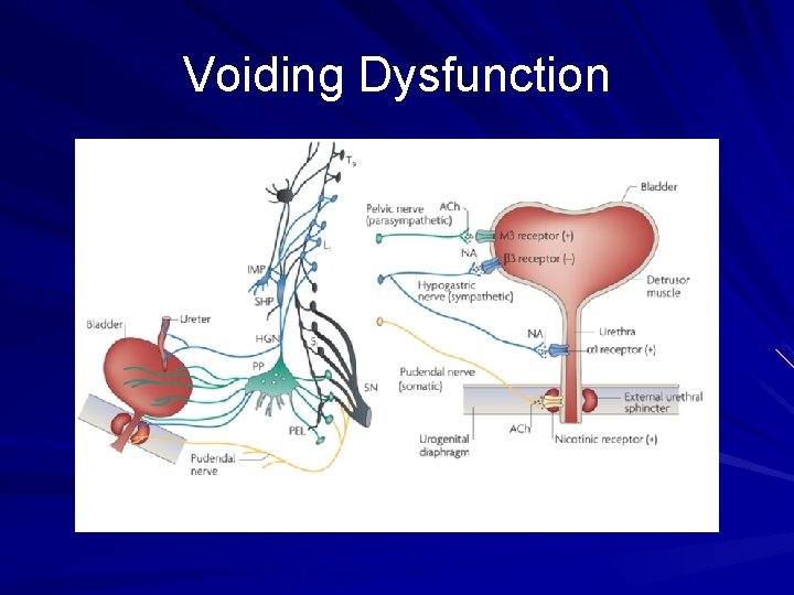
Voiding Dysfunction
Voiding Dysfunction Failure to store – Bladder problems overactivity Hypersensitivity – Outlet problem Stress incontinence Sphincter deficiency – combination Failure to Empty – Bladder problems Neurologic Myogenic idiopathic – Outlet problem BPH Urethral stricture Sphincter dyssynergia – combination
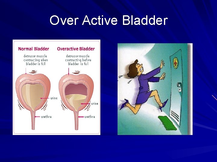
Over Active Bladder
Benign Prostatic Hyperplasia BPH Clinically: – – – LUTS poor bladder emptying urinary retention urinary tract infection Hematuria Renal insufficiency
Benign Prostatic Hyperplasia Physical Examination – 1 -DRE 2 - Focused neurologic exam Prostate Ca rectal Ca anal tone neurologic problems – Abdomen: distended bladder
Benign Prostatic Hyperplasia Urinalysis , culture – UTI – Hematuria Serum Creatinine Serum Prostate-Specific Antigen Flow rate Ultrasound (Kidney, Bladder And Prostate)
Benign Prostatic Hyperplasia Treatment options – medical therapy α-Adrenergic Blockers – – – Tamsulocin Alfuzocin Terazocin Androgen Suppression – Finasteride
Benign Prostatic Hyperplasia Surgical Rx – Endoscopic – Transurethral Resection of the Prostate TURP – Laser ablation – prostatic stents
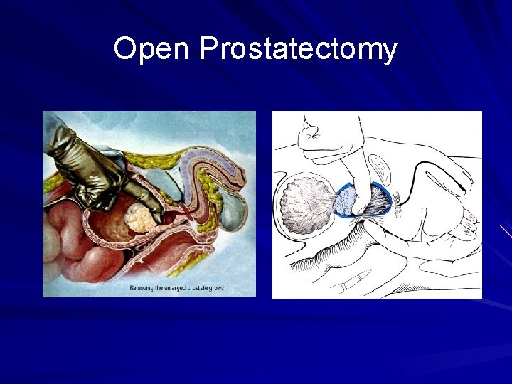
Open Prostatectomy