
Heart Structure Function 16 17 March 2015 What



- Slides: 17
Heart Structure & Function 16 -17 March 2015 What questions do you have about the
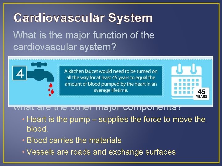
Cardiovascular System What is the major function of the cardiovascular system? • To transport materials (oxygen, nutrients, wastes, hormones, immune components, heat, etc. ) throughout the body What is the heart’s role in this system, and what are the other major components? • Heart is the pump – supplies the force to move the blood. • Blood carries the materials • Vessels are roads and exchange surfaces
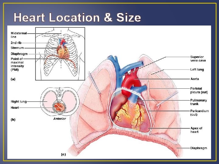
Heart Location & Size
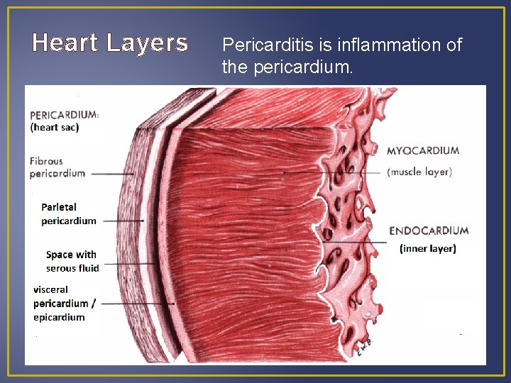
Heart Layers Pericarditis is inflammation of the pericardium.
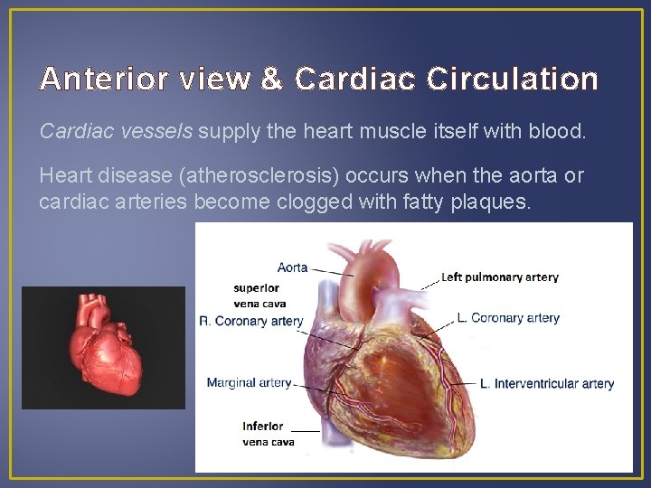
Anterior view & Cardiac Circulation Cardiac vessels supply the heart muscle itself with blood. Heart disease (atherosclerosis) occurs when the aorta or cardiac arteries become clogged with fatty plaques.
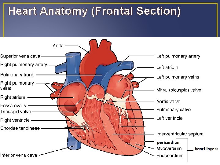
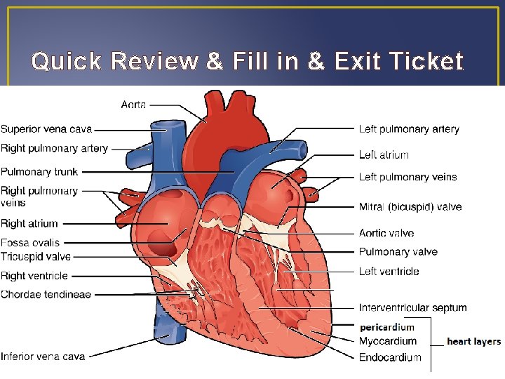
Heart Anatomy (Frontal Section)
The Heart is a Double Pump • The right side of the heart receives oxygen-poor blood from the body and pumps it to the lungs pulmonary circulation • The left side of the heart receives oxygen-rich blood from the lungs and pumps it to the body systemic circulation
The heart has four chambers Each side has an atrium for collecting blood and a ventricle for pumping blood to the lungs or body Blood collects in atria while ventricles contract. Then, as ventricles relax, atria contract slightly and ventricles refill. Two sides of heart contract and relax simultaneously.
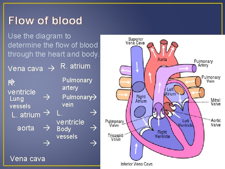
Flow of blood Use the diagram to determine the flow of blood through the heart and body. Vena cava R. atrium R. ventricle Lung vessels Pulmonary artery Pulmonary vein L. atrium L. ventricle aorta Body Vena cava vessels
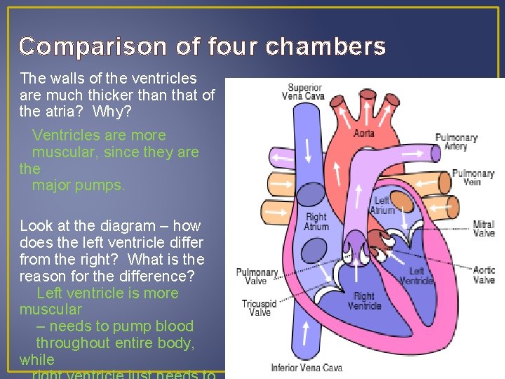
Comparison of four chambers The walls of the ventricles are much thicker than that of the atria? Why? Ventricles are more muscular, since they are the major pumps. Look at the diagram – how does the left ventricle differ from the right? What is the reason for the difference? Left ventricle is more muscular – needs to pump blood throughout entire body, while
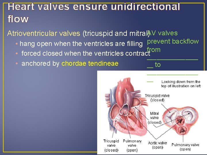
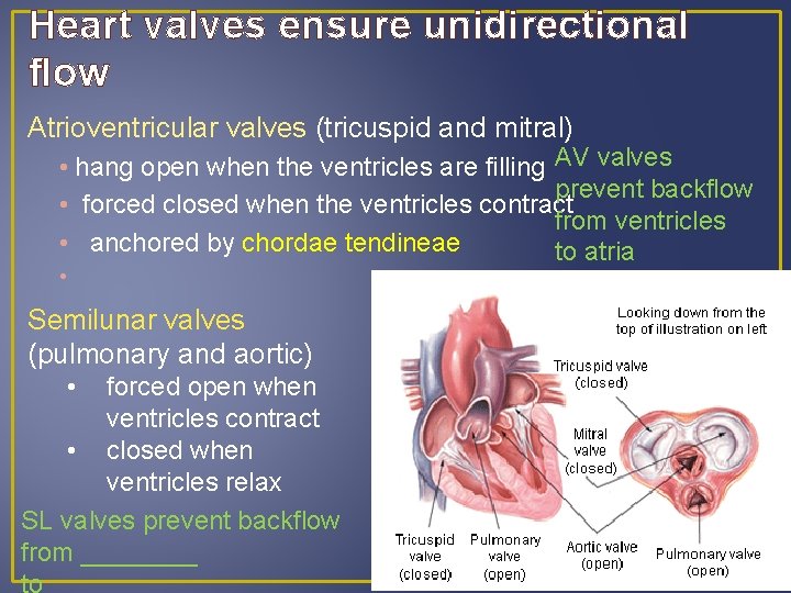
Heart valves ensure unidirectional flow Atrioventricular valves (tricuspid and mitral)AV valves • hang open when the ventricles are filling prevent backflow from • forced closed when the ventricles contract________ • anchored by chordae tendineae __ to ________ __
Heart valves ensure unidirectional flow Atrioventricular valves (tricuspid and mitral) • hang open when the ventricles are filling AV valves prevent backflow • forced closed when the ventricles contract from ventricles • anchored by chordae tendineae to atria • Semilunar valves (pulmonary and aortic) • forced open when ventricles contract • closed when ventricles relax SL valves prevent backflow from ____ to_____
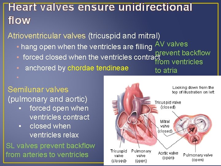
Heart valves ensure unidirectional flow Atrioventricular valves (tricuspid and mitral) • hang open when the ventricles are filling AV valves prevent backflow • forced closed when the ventricles contract from ventricles • anchored by chordae tendineae to atria • Semilunar valves (pulmonary and aortic) • forced open when ventricles contract • closed when ventricles relax SL valves prevent backflow from arteries to ventricles
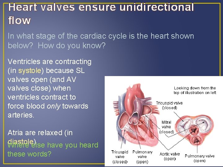
Heart valves ensure unidirectional flow In what stage of the cardiac cycle is the heart shown below? How do you know? Ventricles are contracting (in systole) because SL valves open (and AV valves close) when ventricles contract to force blood only towards arteries. Atria are relaxed (in diastole) Where else have you heard these words?
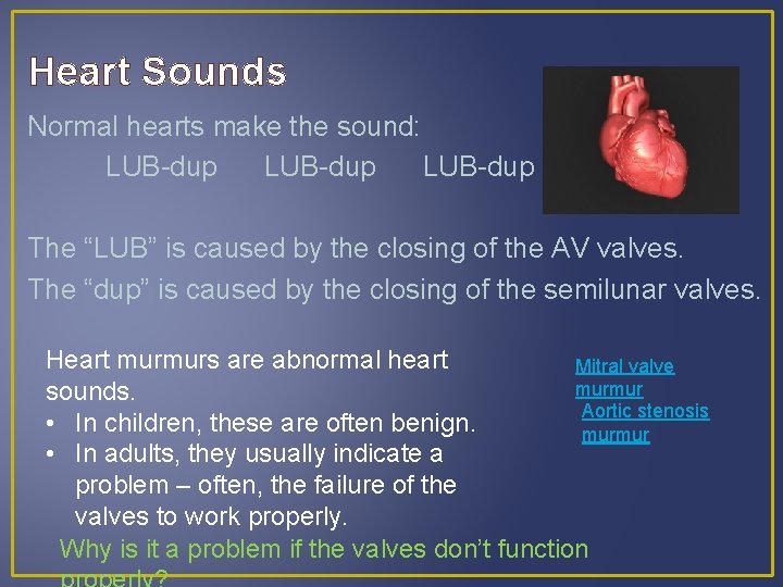
Heart Sounds Normal hearts make the sound: LUB-dup The “LUB” is caused by the closing of the AV valves. The “dup” is caused by the closing of the semilunar valves. Heart murmurs are abnormal heart Mitral valve murmur sounds. Aortic stenosis • In children, these are often benign. murmur • In adults, they usually indicate a problem – often, the failure of the valves to work properly. Why is it a problem if the valves don’t function
Quick Review & Fill in & Exit Ticket
Closure & HW What questions did we answer today? What haven’t we answered? What new questions do you have? HOMEWORK Wed / Thursday – Present answers to questions Friday / Monday – Review Exercise 20 Quiz next Wednesday or Friday