Pulmonary embolism Mohammad Jomaa Refrances Davidson Medscape Pulmonary

Pulmonary embolism Mohammad Jomaa Refrances • Davidson • Medscape
 is when a blood clot (thrombus) becomes lodged in an artery")
�Pulmonary embolism (PE) is when a blood clot (thrombus) becomes lodged in an artery in the lung and blocks blood flow to the lung. � 1: 1000 � Increase CT > increase diagnosis � Not desease? ! � complication

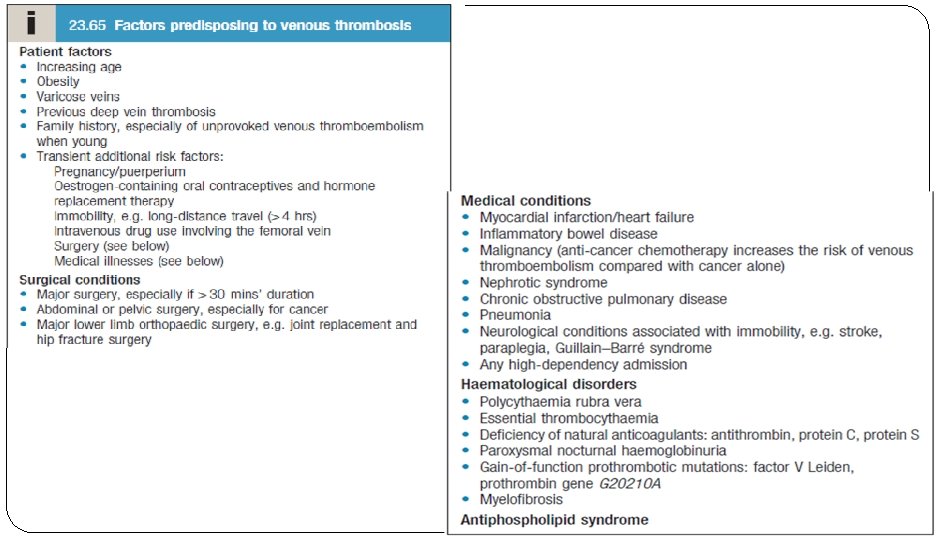
causes �The majority of pulmonary emboli arise from the propagation of lower limb deep vein thrombosis. �Rare causes include �septic emboli (from endocarditis affecting the tricuspid or pulmonary valves) � tumour (especially choriocarcinoma) � fat following fracture of long bones such as the femur �air, and amniotic fluid, which may enter the mother’s circulation following delivery
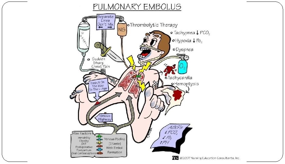

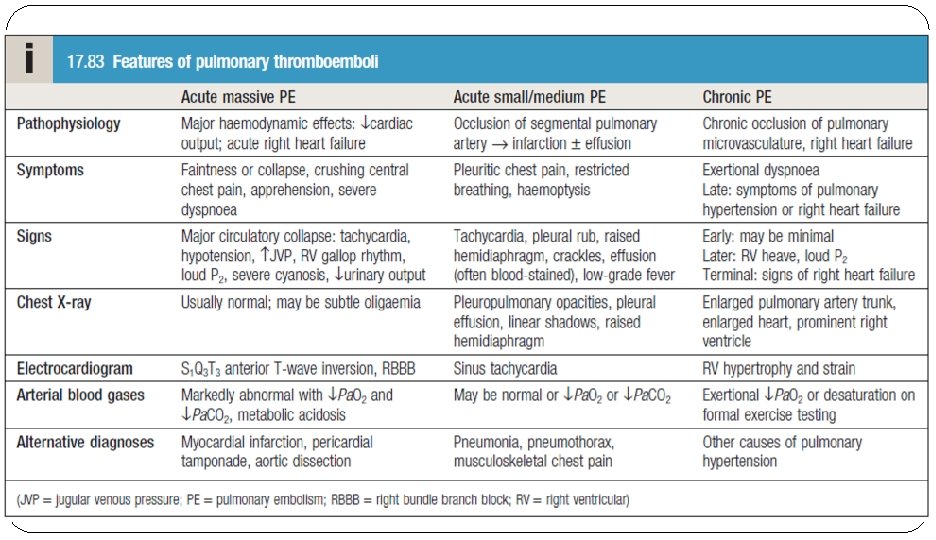
Clinical features �Clinical presentation varies, depending on number, size and distribution of emboli and on underlying cardiorespiratory reserve �A recognised risk factor is present in 80– 90% �There are both respiratory and hemodynamic consequences associated with pulmonary embolism. �Respiratory consequences �Increased alveolar dead space �Hypoxemia �Hyperventilation �Hemodynamic consequences �Pulmonary embolism reduces the cross-sectional area of the pulmonary vascular bed, resulting in an increment in pulmonary vascular resistance, which, in turn, increases the right ventricular afterload. If the afterload is increased severely, right ventricular failure may ensue. In addition, the humoral and reflex mechanisms contribute to the pulmonary arterial constriction. Following the initiation of anticoagulant therapy, the resolution of emboli usually occurs rapidly during the first 2 weeks of therapy; however, it can persist on chest imaging studies for months to years. Chronic pulmonary hypertension may occur with failure of the initial embolus to undergo lyses or in the setting of recurrent thromboemboli.
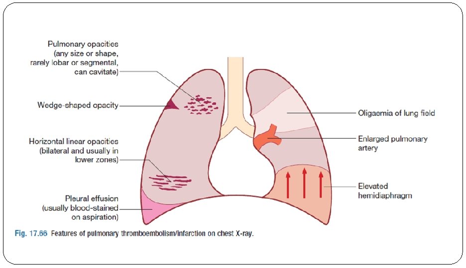
 � only found in 8%-14% of confirmed pulmonary embolism. However, it")
Oligemia (Westermark sign) � only found in 8%-14% of confirmed pulmonary embolism. However, it is highly specific and should raise one's suspicion of pulmonary embolism if present � Westermark's sign refers to a focal area of enhanced or increased translucency due to oligaemia, which occurs due to impaired vascularisation of the lung due to primary mechanical obstruction or reflex vasoconstriction. The sign is formed by dilatation of the pulmonary arteries proximal to the site of emboli followed by a sharp and demarcated collapse of the distal

S 1 Q 3 T 3 pattern �The ECG showed the finding of sinus tachycardia along with S wave in lead I, Q wave and inverted T wave in lead III which has been associated with acute massive PE causing cor pulmonale

modified wells criteria

Investigations �chest X-ray � is most useful in excluding key differential diagnoses, e. g. pneumonia or pneumothorax. � Normal appearances in an acutely breathless and hypoxaemic patient should raise the suspicion of PE, as should bilateral changes in anyone presenting with unilateral pleuritic chest pain. �The ECG �often normal but is useful in excluding other important differential diagnoses, such as acute myocardial infarction and pericarditis. �The most common findings in PE include sinus tachycardia and anterior Twave inversion but these are non-specific; larger emboli may cause right heart strain revealed by an S Q T pattern, ST-segment and T-wave changes, or the appearance of right bundle branch block. 1 3 3

Cont… � Arterial blood gases �typically show a reduced Pa. O 2 and a normal or low Pa. CO 2, and an increased alveolar– arterial oxygen gradient, but may be normal in a significant minority. A metabolic acidosis may be seen in acute massive PE with cardiovascular collapse. � D-dimer �An elevated D-dimer is of limited value, as it may be raised in a variety of other conditions, including myocardial infarction, pneumonia and sepsis. �However, low levels, particularly in the context of a low clinical risk, have a high negative predictive value and further investigation is usually unnecessary. �The result of the D-dimer assay should be disregarded in high-risk patients, as further investigation is mandatory even when normal. � The serum troponin I �may be elevated, reflecting right heart strain. � White Blood Cell Count �Increase � Brain Natriuretic Peptide � Increase
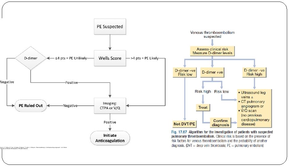

� CTPA � is the first-line diagnostic test �It has the advantage of visualising the distribution and extent of the emboli or highlighting an alternative diagnosis, such as consolidation, pneumothorax or aortic dissection. �The sensitivity of CT scanning may be increased by simultaneous visualisation of the femoral �and popliteal veins, although this is not widely practised. As the contrast media may be nephrotoxic, care should be taken in patients with renal impairment, and CTPA avoided in those with a history of allergy to iodinated contrast media. �In these cases, either V/Q scanning or ventilation/perfusion single photon emission computed tomography (V/Q SPECT) may be

Cont… �Colour Doppler ultrasound of the leg veins �may be used in patients with suspected PE, particularly if there are clinical signs in a limb, as many will have identifiable proximal thrombus in the leg veins. �Bedside echocardiography �is extremely helpful in the differential diagnosis and assessment of acute circulatory collapse. �Acute dilatation of the right heart is usually present in massive PE, and thrombus (embolism in transit) may be visible. Important differential diagnoses, including left ventricular failure, aortic dissection and pericardial tamponade, can also be identified. �Conventional pulmonary angiography �still useful in selected settings or for the delivery of catheter-based therapies.

Management �General measures �Prompt recognition and treatment are potentially life-saving. �Sufficient oxygen should be given to hypoxaemic patients to maintain arterial oxygen saturation above 90%. �Circulatory shock should be treated with intravenous fluids or plasma expander, but inotropic agents are of limited value as the hypoxic dilated right ventricle is already close to maximally stimulated by endogenous catecholamines. �Diuretics and vasodilators should also be avoided, as they will reduce cardiac output. �Opiates may be necessary to relieve pain and distress but should be used with caution in the hypotensive patient. �External cardiac massage may be successful in the moribund patient by dislodging and breaking up a large central embolus.

� Anticoagulation �The mainstay of treatment for all forms of VTE is anticoagulation. �This can be achieved in several ways. 1. One option is to use LMWH followed by warfarin. Treatment of acute VTE with LMWH should continue for a minimum of 5 days. Patients treated with warfarin should achieve a target INR of 2. 5 (range 2– 3) with LMWH continuing until the INR is above 2. Alternatively, patients may be treated with a DOAC. Rivaroxaban and apixaban may be used immediately from diagnosis without the need for LMWH, while the licences for dabigatran and edoxaban include initial treatment with LMWH for a minimum of 5 days before commencing the DOAC. 3. In patients with active cancer and VTE, there is evidence that maintenance anticoagulation with LMWH is associated with a lower recurrence rate than warfarin. 4. Patients who have had VTE and have a strong contraindication to anticoagulation and those who continue to have new pulmonary emboli despite therapeutic anticoagulation should have an inferior vena cava (IVC) filter inserted to prevent life-threatening PE. �The optimal initial period of anticoagulation is between 6 weeks and 6 months. �Patients with a provoked VTE in the presence of a temporary risk factor, which is then removed, can usually be treated for short periods (e. g. 3 months), and indeed anticoagulation for more than 6 months does not alter the rate of recurrence following discontinuation of therapy. �If there are ongoing risk factors that cannot be alleviated, such as active cancer, long-term

� For patients with unprovoked VTE, the optimum duration of anticoagulation can be difficult to establish. � Recurrence of VTE is about 2– 3% per annum in patients who have a temporary medical risk factor at presentation and about 7– 10% per annum in those with apparently unprovoked VTE. This plateaus at around 30– 40% recurrence at 5 years. � As such, many patients who have had unprovoked episodes of VTE will benefit from long-term anticoagulation. � Several factors predict risk of recurrence following an episode of unprovoked VTE. The strongest predictors of recurrence are male sex and a positive D-dimer assay measured 1 month after stopping anticoagulant therapy. These factors are

�Thrombolytic and surgical therapy �Thrombolysis is indicated in any patient presenting with acute massive PE accompanied by cardiogenic shock. �In the absence of shock, the benefits are less clear but thrombolysis may be considered in those presenting with right ventricular dilatation and hypokinesis or severe hypoxaemia. �Patients must be screened carefully for haemorrhagic risk, as there is a high risk of intracranial haemorrhage. �Surgical pulmonary embolectomy may be considered in selected patients but carries a high mortality.

�Caval filters �A patient in whom anticoagulation is contraindicated, who has suffered massive haemorrhage on anticoagulation, or recurrent VTE despite anticoagulation, should be considered for an inferior vena caval filter. �Retrievable caval filters are particularly useful in individuals with temporary risk factors. The caval filter should be used only until anticoagulation can be safely initiated, at which time the filter should be removed if possible. �Risk: �increasing risk for long-term complications such as �DVT “insertion site thrombosis” �filter migration �IVC occlusion �insertion site thrombosis

Complications of PE � Complications of pulmonary embolism include the following: � Sudden cardiac death � Obstructive shock � Pulseless electrical activity � Atrial or ventricular arrhythmias � Secondary pulmonary arterial hypertension � Cor pulmonale � Severe hypoxemia � Right-to-left intracardiac shunt � Lung infarction � Pleural effusion � Paradoxical embolism � Heparin-induced thrombocytopenia

Prognosis �Immediate mortality is greatest in those with echocardiographic evidence of right ventricular dysfunction or cardiogenic shock. �Once anticoagulation is commenced, however, the risk of mortality rapidly falls. �The risk of recurrence is highest in the first 6– 12 months after the initial event, and at 10 years around one-third of individuals will have suffered a further event.

- Slides: 24