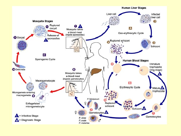
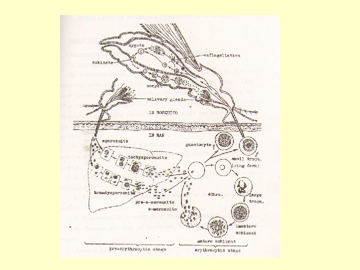
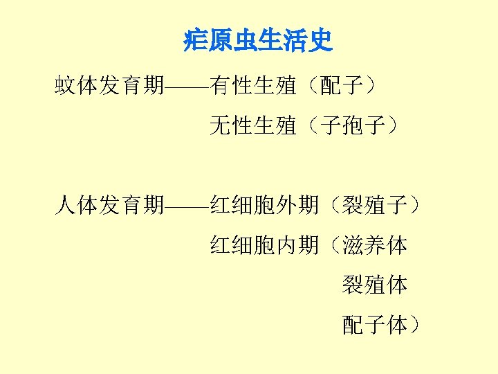
Sporozoites Preerythrocytic stage in liver P vivax young



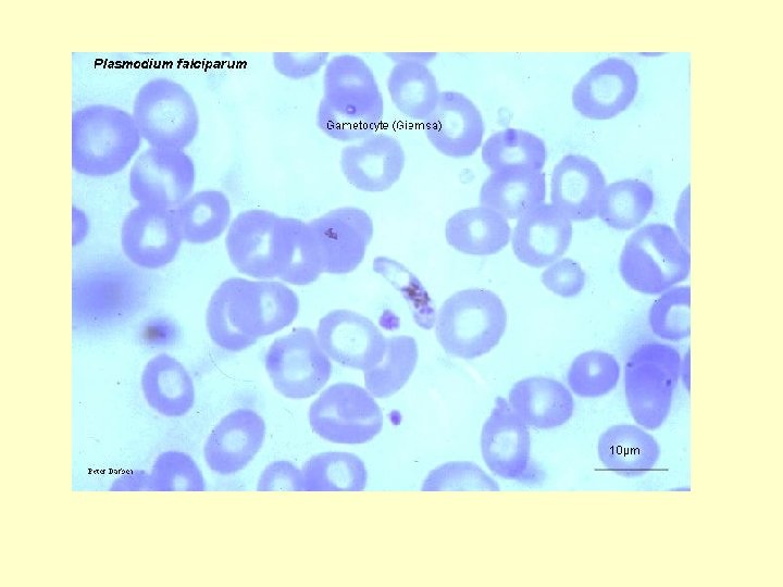
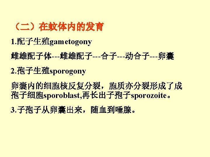


























- Slides: 87
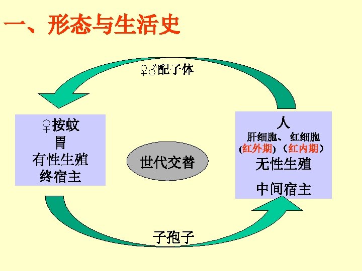
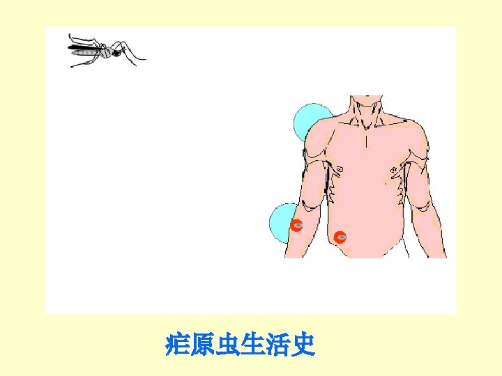
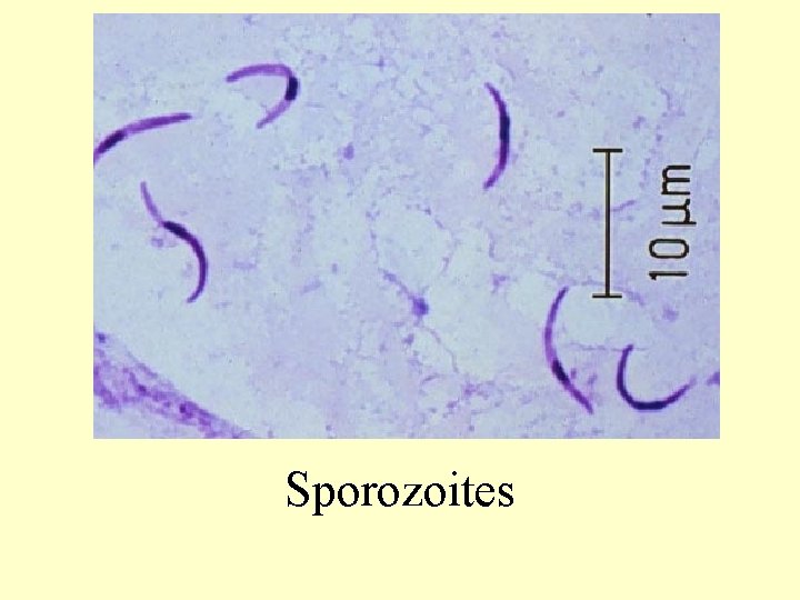
Sporozoites
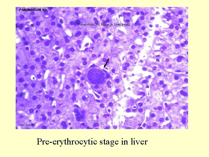
Pre-erythrocytic stage in liver
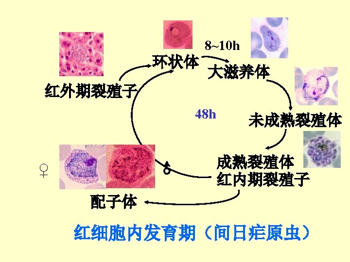
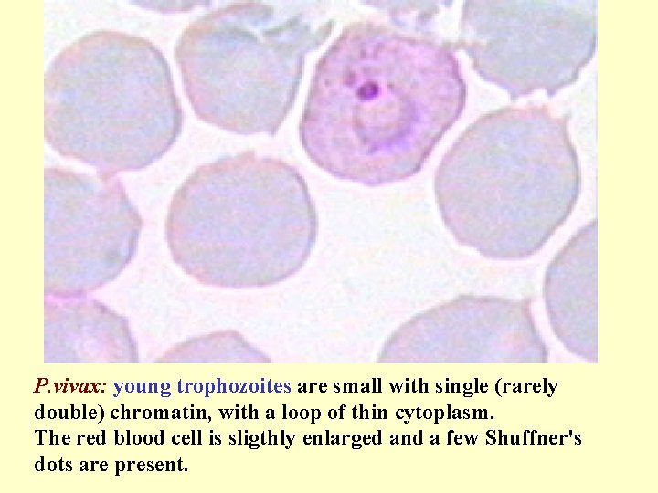
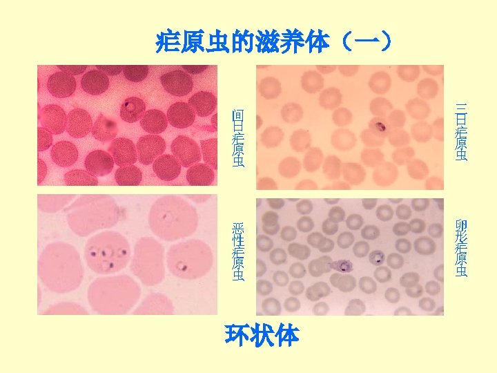
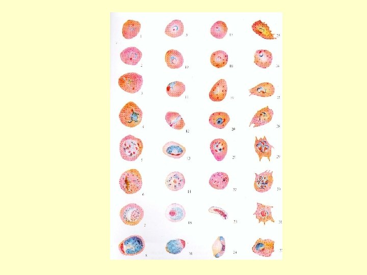
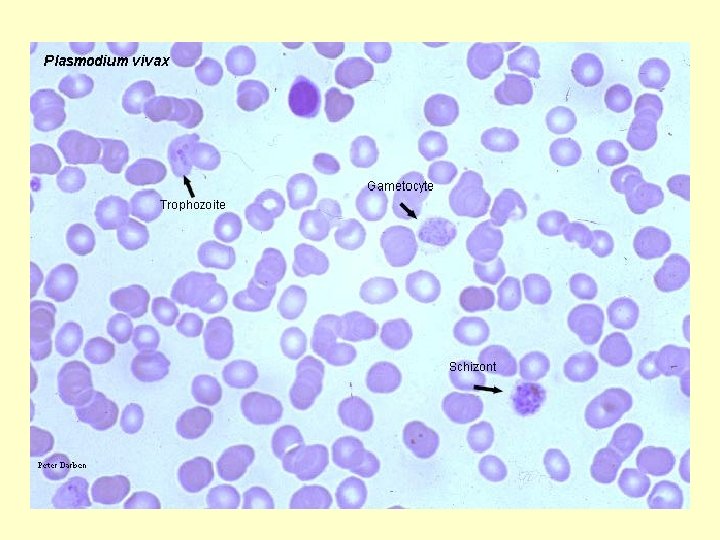
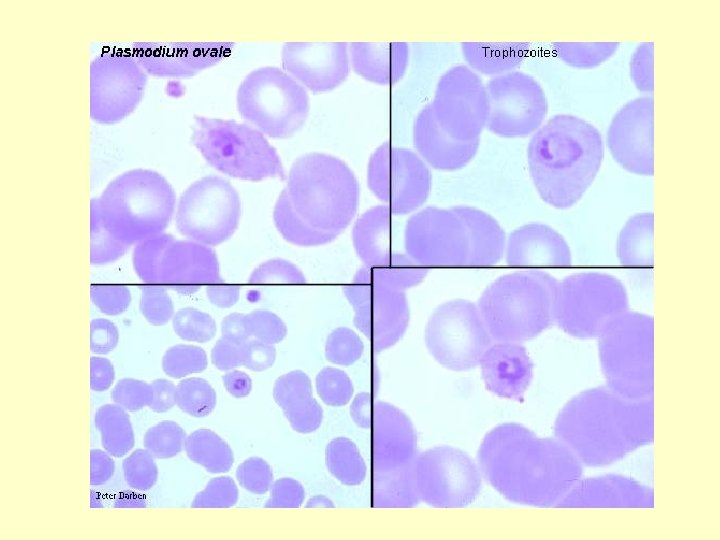
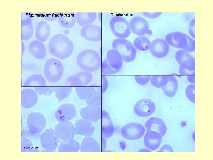
P. vivax: young trophozoites are small with single (rarely double) chromatin, with a loop of thin cytoplasm. The red blood cell is sligthly enlarged and a few Shuffner's dots are present.
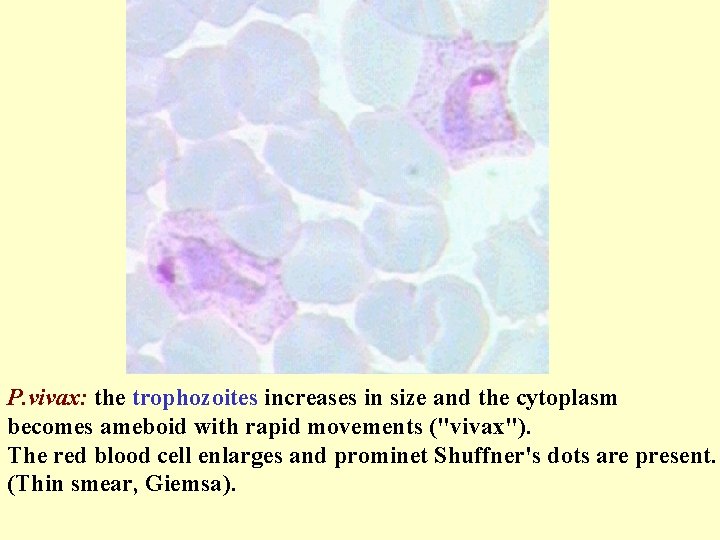
P. vivax: the trophozoites increases in size and the cytoplasm becomes ameboid with rapid movements ("vivax"). The red blood cell enlarges and prominet Shuffner's dots are present. (Thin smear, Giemsa).
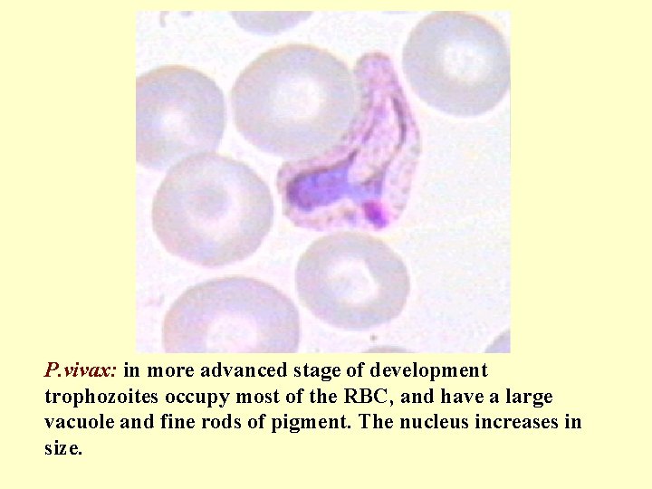
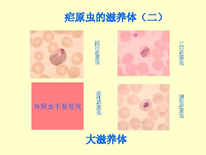
P. vivax: in more advanced stage of development trophozoites occupy most of the RBC, and have a large vacuole and fine rods of pigment. The nucleus increases in size.
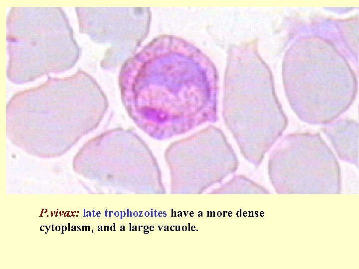
P. vivax: late trophozoites have a more dense cytoplasm, and a large vacuole.
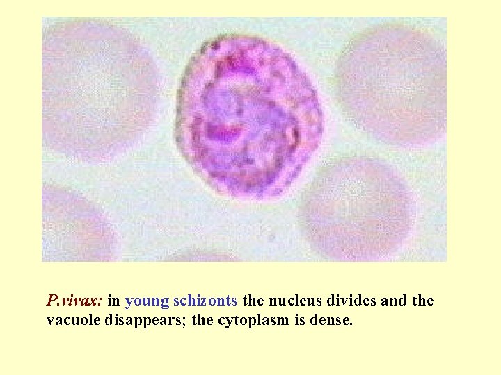
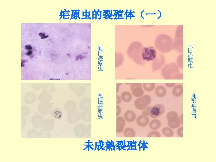
P. vivax: in young schizonts the nucleus divides and the vacuole disappears; the cytoplasm is dense.
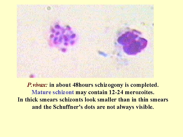
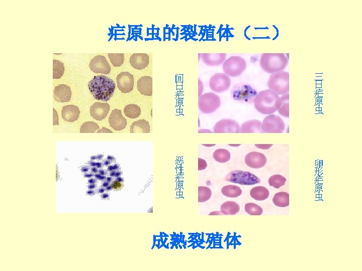
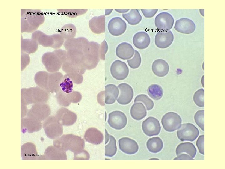
P. vivax: in about 48 hours schizogony is completed. Mature schizont may contain 12 -24 merozoites. In thick smears schizonts look smaller than in thin smears and the Schuffner's dots are not always visible.
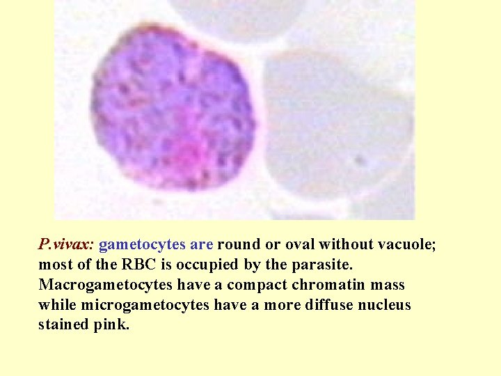
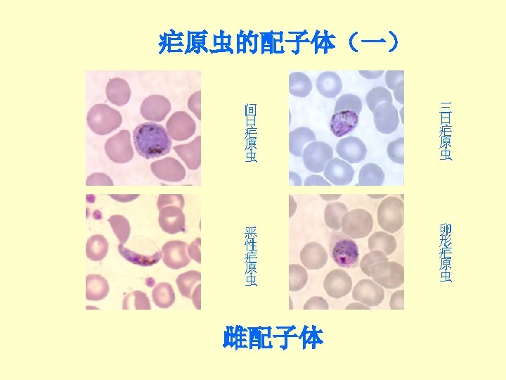
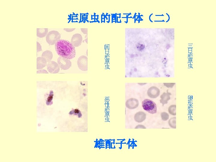
P. vivax: gametocytes are round or oval without vacuole; most of the RBC is occupied by the parasite. Macrogametocytes have a compact chromatin mass while microgametocytes have a more diffuse nucleus stained pink.
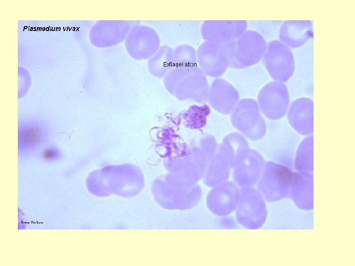
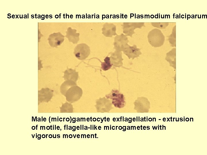
Sexual stages of the malaria parasite Plasmodium falciparum Male (micro)gametocyte exflagellation - extrusion of motile, flagella-like microgametes with vigorous movement.
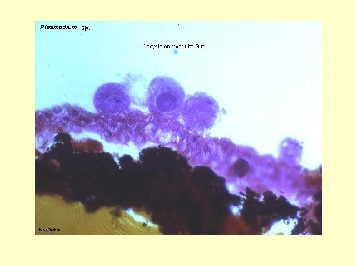
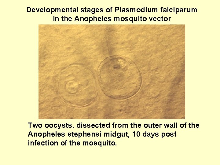
Developmental stages of Plasmodium falciparum in the Anopheles mosquito vector Two oocysts, dissected from the outer wall of the Anopheles stephensi midgut, 10 days post infection of the mosquito.
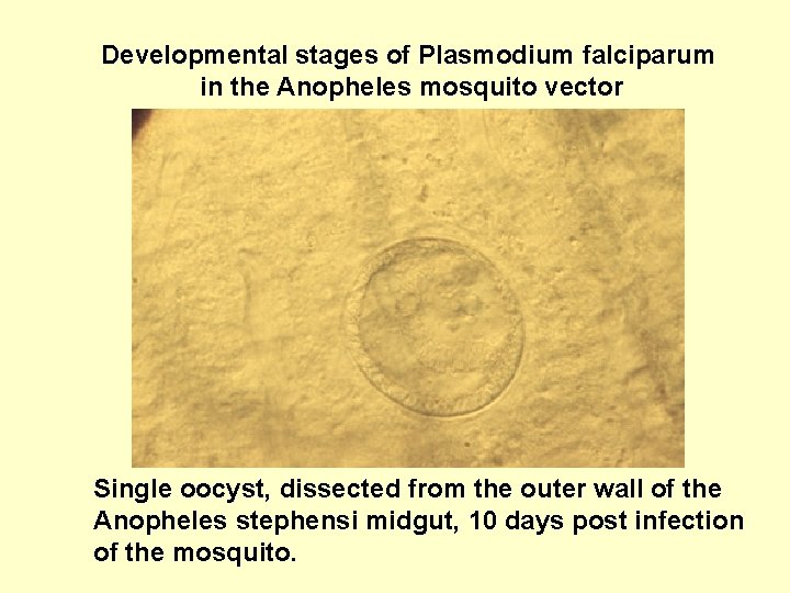
Developmental stages of Plasmodium falciparum in the Anopheles mosquito vector Single oocyst, dissected from the outer wall of the Anopheles stephensi midgut, 10 days post infection of the mosquito.
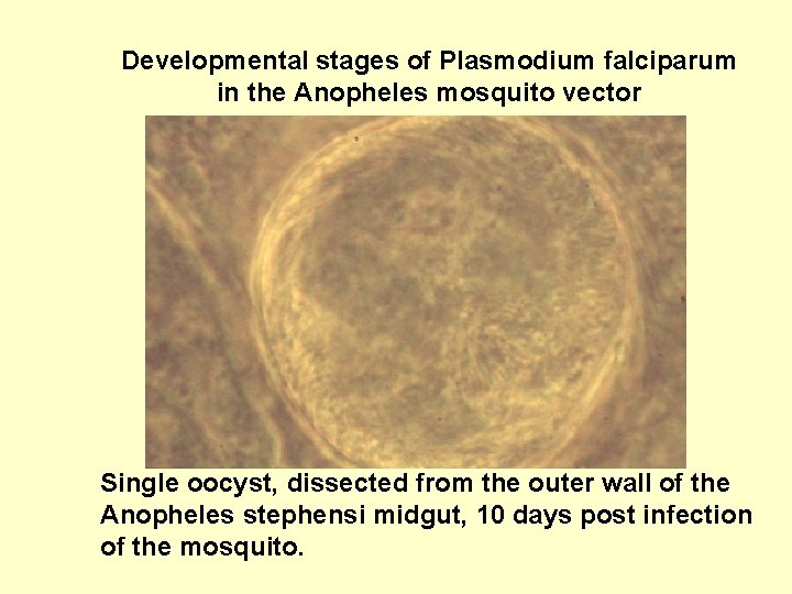
Developmental stages of Plasmodium falciparum in the Anopheles mosquito vector Single oocyst, dissected from the outer wall of the Anopheles stephensi midgut, 10 days post infection of the mosquito.
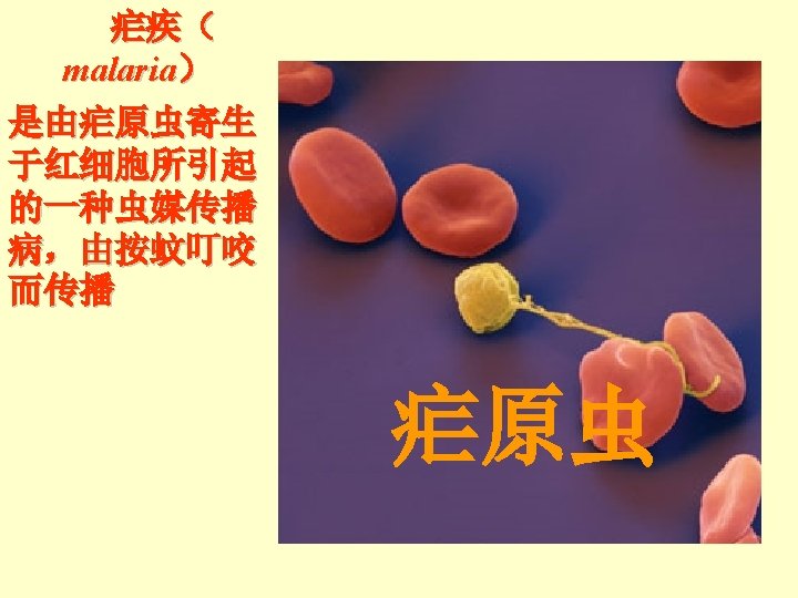
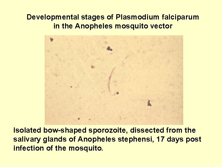
Developmental stages of Plasmodium falciparum in the Anopheles mosquito vector Isolated bow-shaped sporozoite, dissected from the salivary glands of Anopheles stephensi, 17 days post infection of the mosquito.
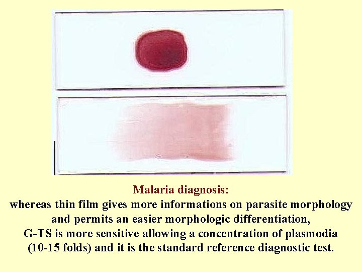
Malaria diagnosis: whereas thin film gives more informations on parasite morphology and permits an easier morphologic differentiation, G-TS is more sensitive allowing a concentration of plasmodia (10 -15 folds) and it is the standard reference diagnostic test.
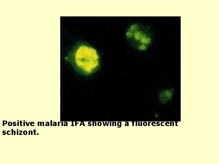
Positive malaria IFA showing a fluorescent schizont.
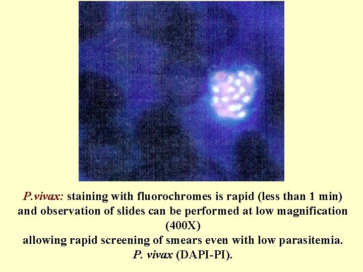
P. vivax: staining with fluorochromes is rapid (less than 1 min) and observation of slides can be performed at low magnification (400 X) allowing rapid screening of smears even with low parasitemia. P. vivax (DAPI-PI).
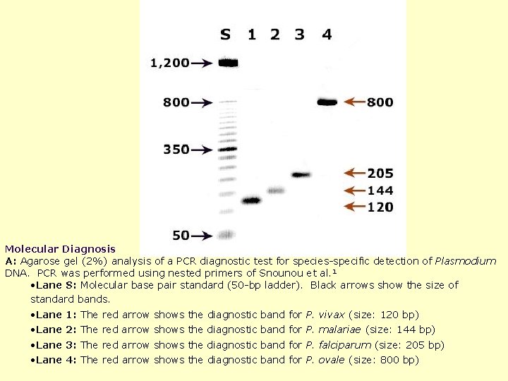
Molecular Diagnosis A: Agarose gel (2%) analysis of a PCR diagnostic test for species-specific detection of Plasmodium DNA. PCR was performed using nested primers of Snounou et al. 1 • Lane S: Molecular base pair standard (50 -bp ladder). Black arrows show the size of standard bands. • Lane 1: The red arrow shows the diagnostic band for P. vivax (size: 120 bp) 2: The red arrow shows the diagnostic band for P. malariae (size: 144 bp) 3: The red arrow shows the diagnostic band for P. falciparum (size: 205 bp) 4: The red arrow shows the diagnostic band for P. ovale (size: 800 bp)