
RealTime Harmonic Contrast Material specific US of Focal

- Slides: 35
Real-Time Harmonic Contrast Material – specific US of Focal Liver Lesions Radio. Graphics 2005; 25: 333– 349 Presented by intern 李世瑜 August 24 th , 2005 MRI會議室
Introduction n “first-generation” US contrast media (Air) q q n Microbubble rupture needed to obtain an adequate echo signal. only high-mechanical index technologies “Second-generation” US contrast media q q Gases other than air continuous real-time assessment by low acoustic pressure (low mechanical index)
“II” contrast media n Vascular phases: q q q n arterial (early) phase (15– 35 seconds) portal (venous) phase (35– 90 seconds) sinusoidal (parenchymal or late vascular) phase (90– 240 seconds) Lesion echogenicity defined : q Compare to surrounding parenchymal echo levels at the same imaging time and depth.
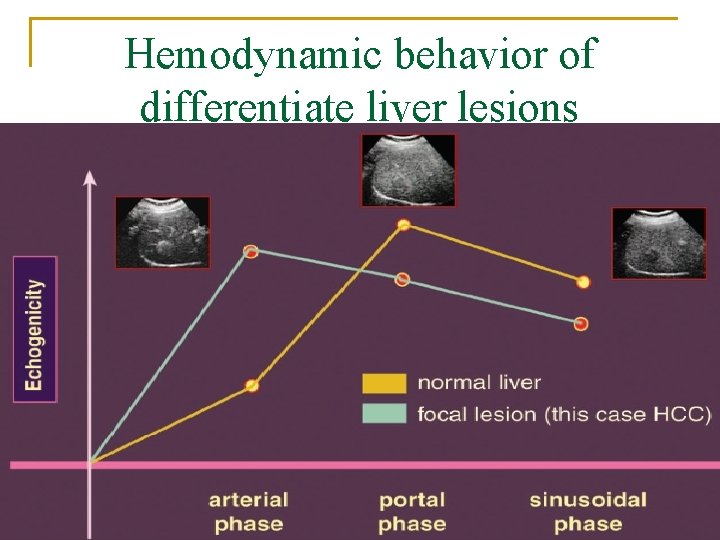
Hemodynamic behavior of differentiate liver lesions
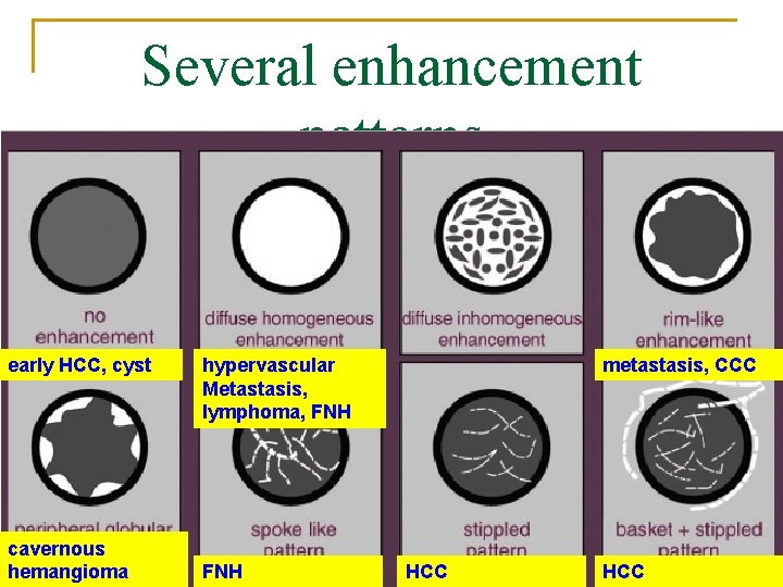
Several enhancement patterns early HCC, cyst cavernous hemangioma hypervascular Metastasis, lymphoma, FNH metastasis, CCC HCC
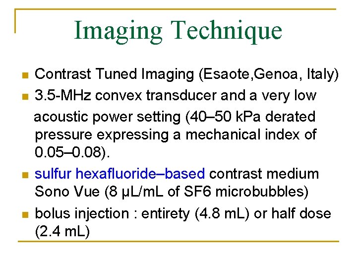
Imaging Technique n n Contrast Tuned Imaging (Esaote, Genoa, Italy) 3. 5 -MHz convex transducer and a very low acoustic power setting (40– 50 k. Pa derated pressure expressing a mechanical index of 0. 05– 0. 08). sulfur hexafluoride–based contrast medium Sono Vue (8 μL/m. L of SF 6 microbubbles) bolus injection : entirety (4. 8 m. L) or half dose (2. 4 m. L)
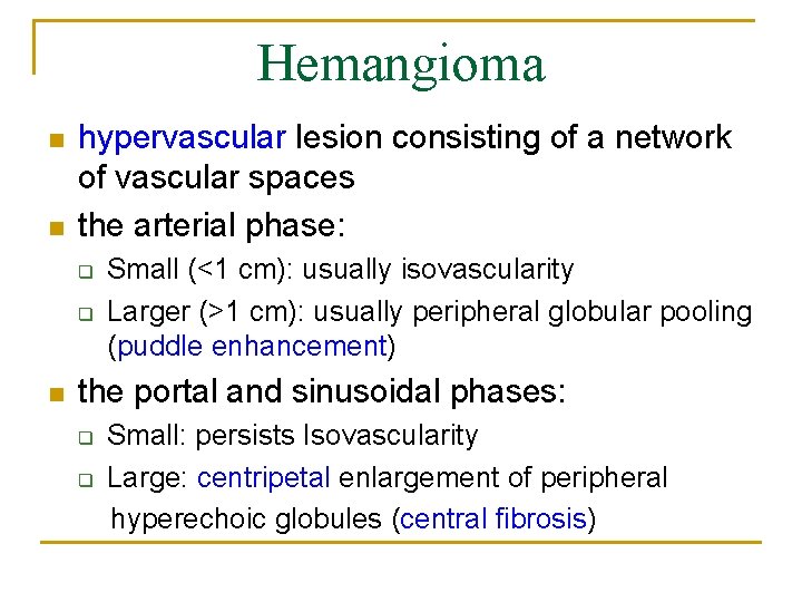
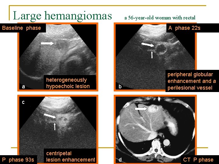
Hemangioma n n hypervascular lesion consisting of a network of vascular spaces the arterial phase: q q n Small (<1 cm): usually isovascularity Larger (>1 cm): usually peripheral globular pooling (puddle enhancement) the portal and sinusoidal phases: q q Small: persists Isovascularity Large: centripetal enlargement of peripheral hyperechoic globules (central fibrosis)
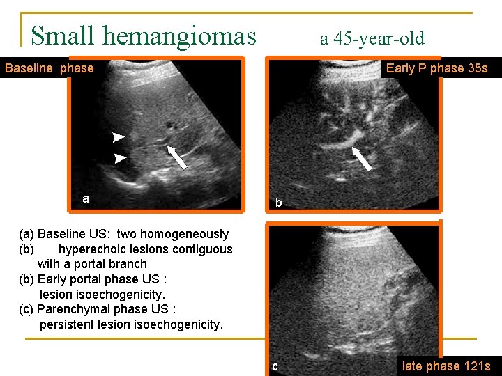
Small hemangiomas a 45 -year-old woman Baseline phase a Early P phase 35 s b (a) Baseline US: two homogeneously (b) hyperechoic lesions contiguous with a portal branch (b) Early portal phase US : lesion isoechogenicity. (c) Parenchymal phase US : persistent lesion isoechogenicity. c late phase 121 s
Large hemangiomas a 56 -year-old woman with rectal cancerphase Baseline a A phase 22 s heterogeneously hypoechoic lesion b centripetal lesion enhancement d peripheral globular enhancement and a perilesional vessel c P phase 93 s CT P phase
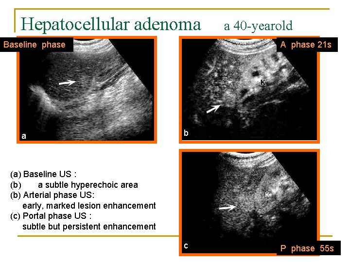
Hepatocellular Adenoma n n n hypervascular lesion without portal tracts Typical: Large, subcapsular tributary arteries the arterial phase: q q n Discrete, perilesional feeding arteries manifest as enhancement around the tumor capsule never seen in HCC portal and sinusoidal phases: q q Hypervascularity followed by isovascularity Hypoechoic appearance is never seen
Hepatocellular adenoma woman Baseline phase a a 40 -yearold A phase 21 s b (a) Baseline US : (b) a subtle hyperechoic area (b) Arterial phase US: early, marked lesion enhancement (c) Portal phase US : subtle but persistent enhancement c P phase 55 s
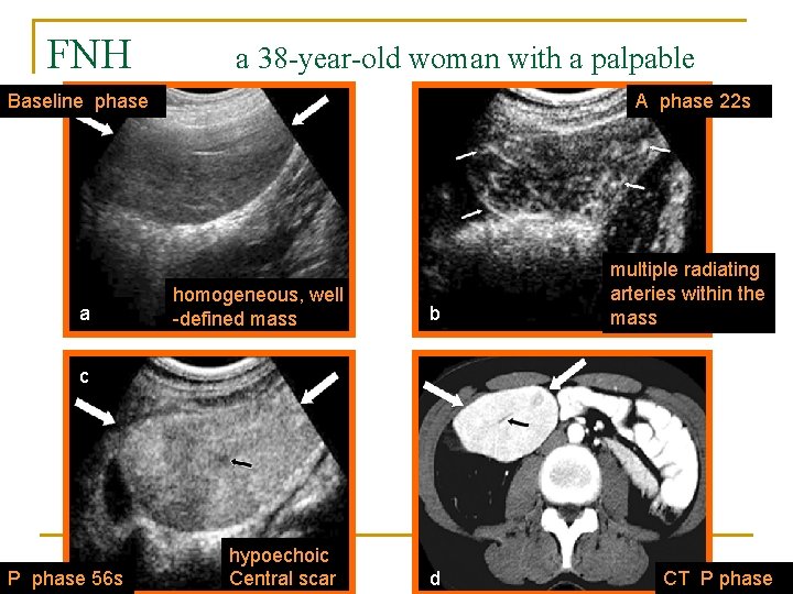
Focal Nodular Hyperplasia n n n hypervascular, hyperplastic lesion caused by preexisting vascular malformation peripheral pseudocapsule and a central or eccentric fibrous scar the arterial phase q q q n tortuous feeding artery and a central artery “wheel spoke” or “central spider” pattern hypoechoic central scar (full arterial phase) the portal and sinusoidal phases q isoechoic or hyperechoic appearance
FNH a 38 -year-old woman with a palpable massphase Baseline a A phase 22 s homogeneous, well -defined mass b multiple radiating arteries within the mass c P phase 56 s hypoechoic Central scar d CT P phase
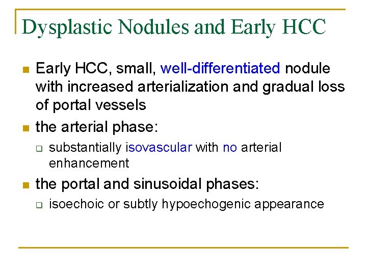
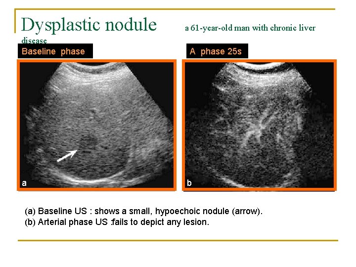
Dysplastic Nodules and Early HCC n n Early HCC, small, well-differentiated nodule with increased arterialization and gradual loss of portal vessels the arterial phase: q n substantially isovascular with no arterial enhancement the portal and sinusoidal phases: q isoechoic or subtly hypoechogenic appearance
Dysplastic nodule disease Baseline phase a a 61 -year-old man with chronic liver A phase 25 s b (a) Baseline US : shows a small, hypoechoic nodule (arrow). (b) Arterial phase US : fails to depict any lesion.
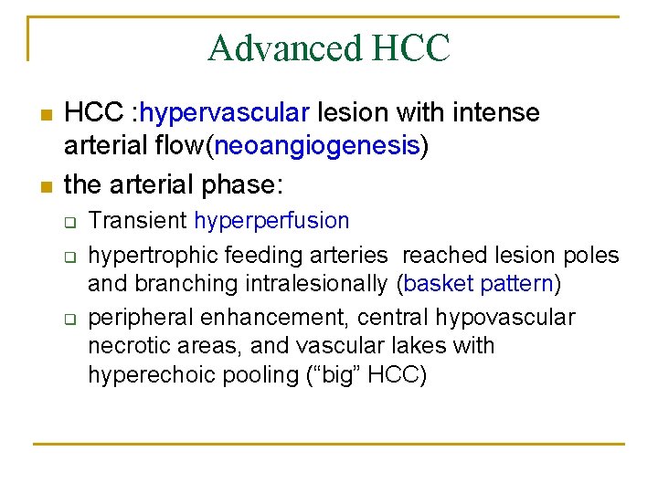
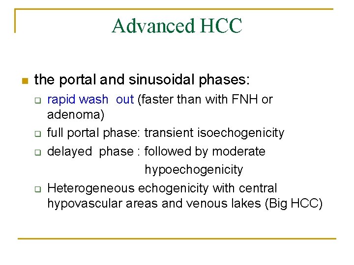
Advanced HCC n n HCC : hypervascular lesion with intense arterial flow(neoangiogenesis) the arterial phase: q q q Transient hyperperfusion hypertrophic feeding arteries reached lesion poles and branching intralesionally (basket pattern) peripheral enhancement, central hypovascular necrotic areas, and vascular lakes with hyperechoic pooling (“big” HCC)
Advanced HCC n the portal and sinusoidal phases: q q rapid wash out (faster than with FNH or adenoma) full portal phase: transient isoechogenicity delayed phase : followed by moderate hypoechogenicity Heterogeneous echogenicity with central hypovascular areas and venous lakes (Big HCC)
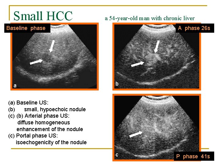
Small HCC a 54 -year-old man with chronic liver diseasephase Baseline a A phase 26 s b (a) Baseline US: (b) small, hypoechoic nodule (c) (b) Arterial phase US: diffuse homogeneous enhancement of the nodule (c) Portal phase US: isoechogenicity of the nodule c P phase 41 s
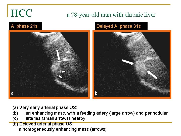
HCC disease A phase 21 s a a 78 -year-old man with chronic liver Delayed A phase 31 s b (a) Very early arterial phase US: (b) an enhancing mass, with a feeding artery (large arrow) and perinodular (c) arteries (small arrows) nearby. (b) Delayed arterial phase US: a homogeneously enhancing mass (arrows)
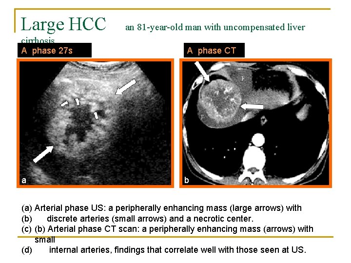
Large HCC cirrhosis A phase 27 s a an 81 -year-old man with uncompensated liver A phase CT b (a) Arterial phase US: a peripherally enhancing mass (large arrows) with (b) discrete arteries (small arrows) and a necrotic center. (c) (b) Arterial phase CT scan: a peripherally enhancing mass (arrows) with small (d) internal arteries, findings that correlate well with those seen at US.
Cholangiocellular Carcinoma n the arterial phase: q n peripheral rim-like enhancement or heterogeneous enhancement the portal and sinusoidal phase: q q q hypoechoic appearance Persistent subtle rim enhancement is possible Satellite lesions may also appeared
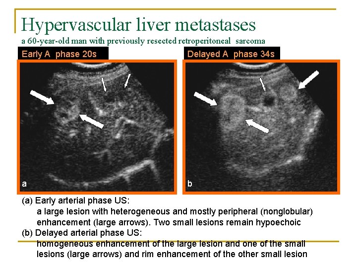
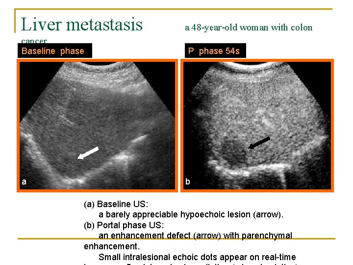
Metastasis n n Some degree of arterial neoangiogenesis (peripheral macrovessels and central microvessels) Hypo- or hypervascular on arterial phase images depending on the organ of origin
Metastasis n the arterial phase: q n peripheral rim-like enhancement (target like appearance) the portal and sinusoidal phases q q q no centripetal filling Washout is rapid black holes on a bright background
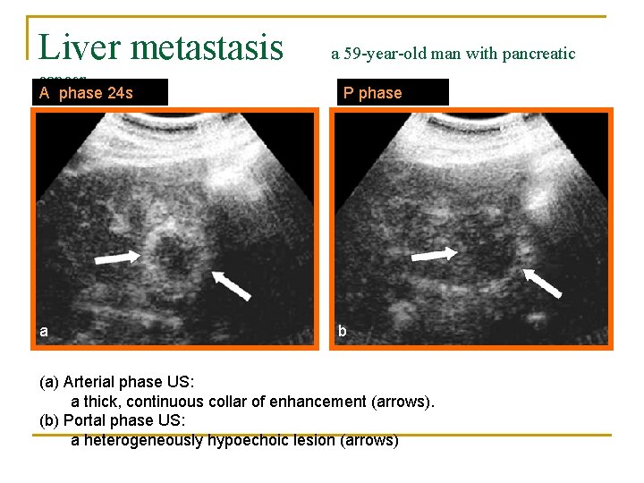
Liver metastasis cancer A phase 24 s a a 59 -year-old man with pancreatic P phase b (a) Arterial phase US: a thick, continuous collar of enhancement (arrows). (b) Portal phase US: a heterogeneously hypoechoic lesion (arrows)
Hypervascular liver metastases a 60 -year-old man with previously resected retroperitoneal sarcoma Early A phase 20 s Delayed A phase 34 s a b (a) Early arterial phase US: a large lesion with heterogeneous and mostly peripheral (nonglobular) enhancement (large arrows). Two small lesions remain hypoechoic (b) Delayed arterial phase US: homogeneous enhancement of the large lesion and one of the small lesions (large arrows) and rim enhancement of the other small lesion
Liver metastasis cancer Baseline phase a a 48 -year-old woman with colon P phase 54 s b (a) Baseline US: a barely appreciable hypoechoic lesion (arrow). (b) Portal phase US: an enhancement defect (arrow) with parenchymal enhancement. Small intralesional echoic dots appear on real-time
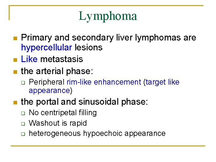
Lymphoma n n n Primary and secondary liver lymphomas are hypercellular lesions Like metastasis the arterial phase: q n Peripheral rim-like enhancement (target like appearance) the portal and sinusoidal phase: q q q No centripetal filling Washout is rapid heterogeneous hypoechoic appearance
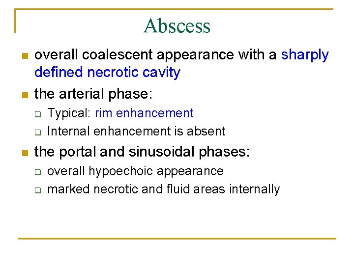
Abscess n n overall coalescent appearance with a sharply defined necrotic cavity the arterial phase: q q n Typical: rim enhancement Internal enhancement is absent the portal and sinusoidal phases: q q overall hypoechoic appearance marked necrotic and fluid areas internally
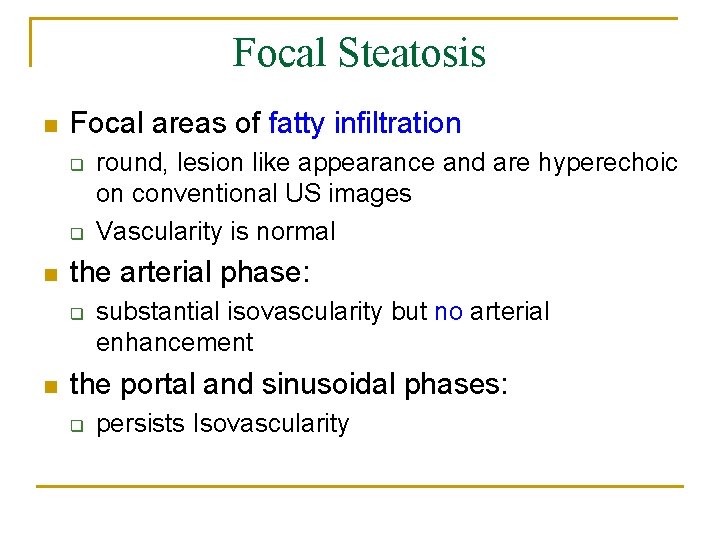
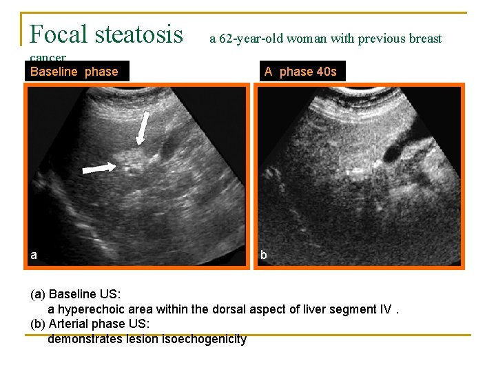
Focal Steatosis n Focal areas of fatty infiltration q q n the arterial phase: q n round, lesion like appearance and are hyperechoic on conventional US images Vascularity is normal substantial isovascularity but no arterial enhancement the portal and sinusoidal phases: q persists Isovascularity
Focal steatosis cancer Baseline phase a a 62 -year-old woman with previous breast A phase 40 s b (a) Baseline US: a hyperechoic area within the dorsal aspect of liver segment IV. (b) Arterial phase US: demonstrates lesion isoechogenicity
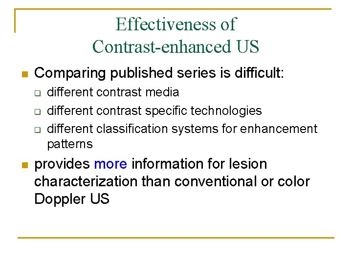
Effectiveness of Contrast-enhanced US n Comparing published series is difficult: q q q n different contrast media different contrast specific technologies different classification systems for enhancement patterns provides more information for lesion characterization than conventional or color Doppler US
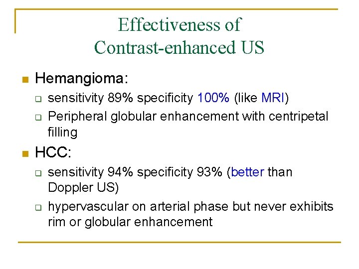
Effectiveness of Contrast-enhanced US n Hemangioma: q q n sensitivity 89% specificity 100% (like MRI) Peripheral globular enhancement with centripetal filling HCC: q q sensitivity 94% specificity 93% (better than Doppler US) hypervascular on arterial phase but never exhibits rim or globular enhancement
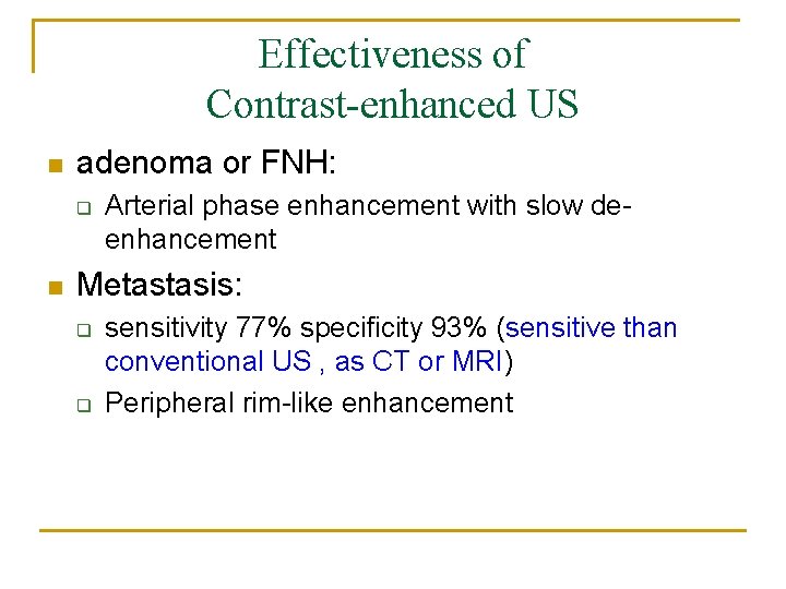
Effectiveness of Contrast-enhanced US n adenoma or FNH: q n Arterial phase enhancement with slow deenhancement Metastasis: q q sensitivity 77% specificity 93% (sensitive than conventional US , as CT or MRI) Peripheral rim-like enhancement
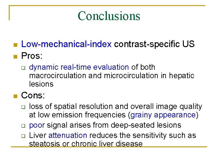
Conclusions n n Low-mechanical-index contrast-specific US Pros: q n dynamic real-time evaluation of both macrocirculation and microcirculation in hepatic lesions Cons: q q q loss of spatial resolution and overall image quality at low emission frequencies (grainy appearance) poor signal arises from deep-seated lesions Liver attenuation reduces the sensitivity such as steatosis or chronic liver disease
Thanks for your attention!!