
Neglected Tropical Diseases Keeley Morris Greg Leos Inger




- Slides: 42
Neglected Tropical Diseases Keeley Morris Greg Leos Inger Vilcins
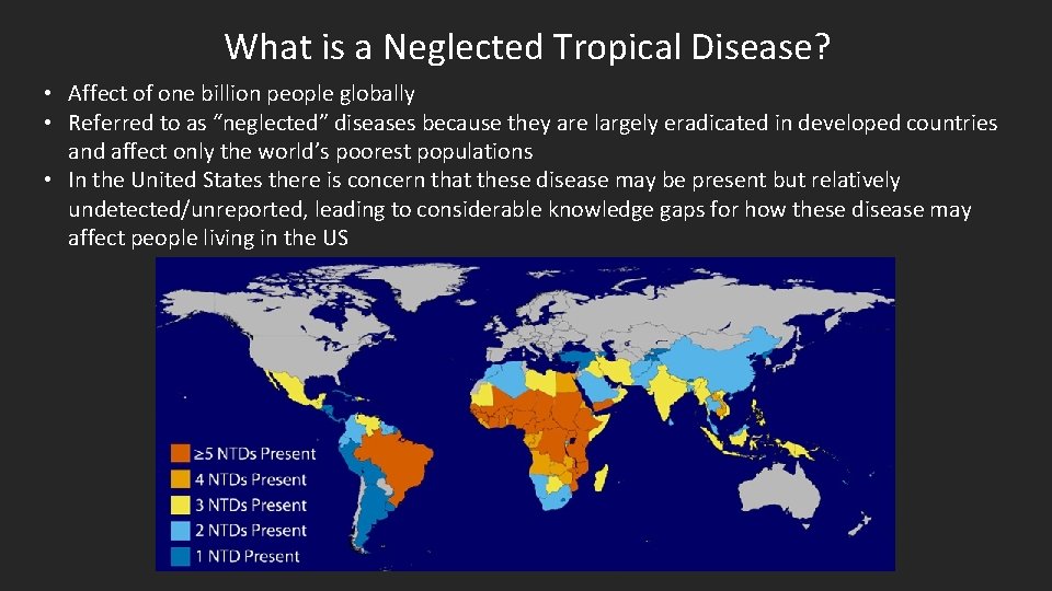
What is a Neglected Tropical Disease? • Affect of one billion people globally • Referred to as “neglected” diseases because they are largely eradicated in developed countries and affect only the world’s poorest populations • In the United States there is concern that these disease may be present but relatively undetected/unreported, leading to considerable knowledge gaps for how these disease may affect people living in the US
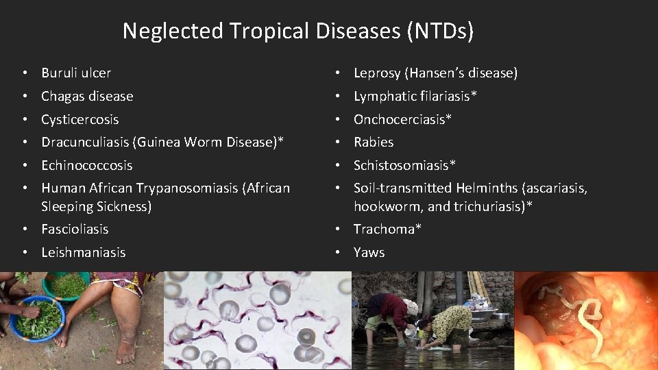
Neglected Tropical Diseases (NTDs) • • • Buruli ulcer Chagas disease Cysticercosis Dracunculiasis (Guinea Worm Disease)* Echinococcosis Human African Trypanosomiasis (African Sleeping Sickness) • Fascioliasis • Leishmaniasis • • • Leprosy (Hansen’s disease) Lymphatic filariasis* Onchocerciasis* Rabies Schistosomiasis* Soil-transmitted Helminths (ascariasis, hookworm, and trichuriasis)* • Trachoma* • Yaws
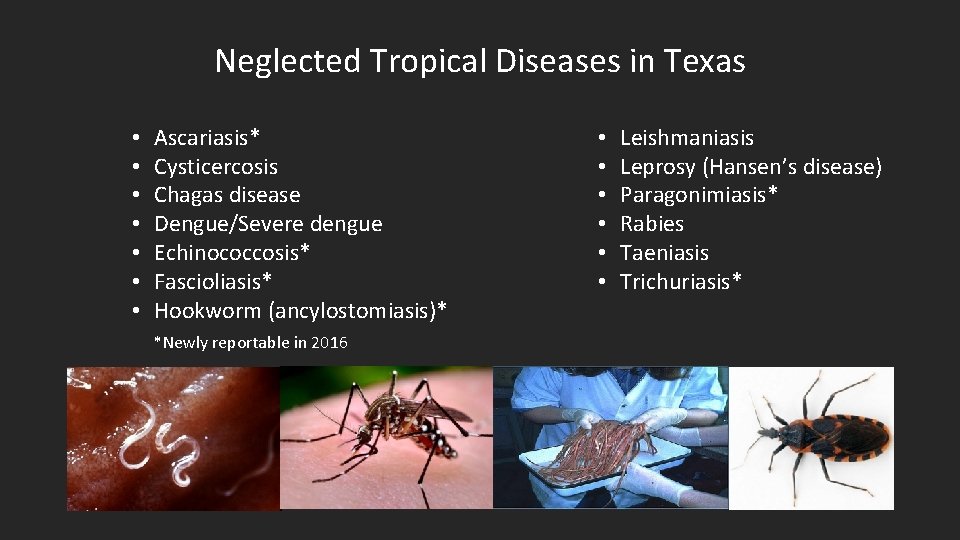
Neglected Tropical Diseases in Texas • • Ascariasis* Cysticercosis Chagas disease Dengue/Severe dengue Echinococcosis* Fascioliasis* Hookworm (ancylostomiasis)* *Newly reportable in 2016 • • • Leishmaniasis Leprosy (Hansen’s disease) Paragonimiasis* Rabies Taeniasis Trichuriasis*
Newly Reportable NTDs HCID Team • Ascariasis • Hookworm (ancylostomiasis) Foodborne Team • Fascioliasis Zoonosis • Echinococcosis • Paragonimiasis • Trichuriasis Main Contact: Keeley Morris Main Contact: Greg Leos For HCID and Foodborne Teams fax to EAIDB at 512 -776 -7616 For Zoonosis Control Branch fax to 512 -776 -7454 Main Contact: Inger Vilcins
HCID Team NTDs – Soil Transmitted Helminths Keeley Morris, MPH
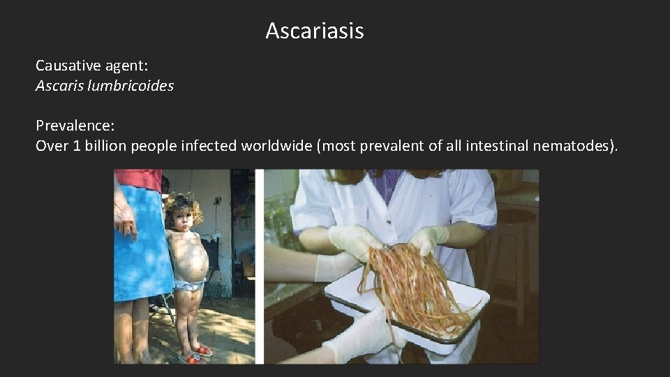
Ascariasis Causative agent: Ascaris lumbricoides Prevalence: Over 1 billion people infected worldwide (most prevalent of all intestinal nematodes).
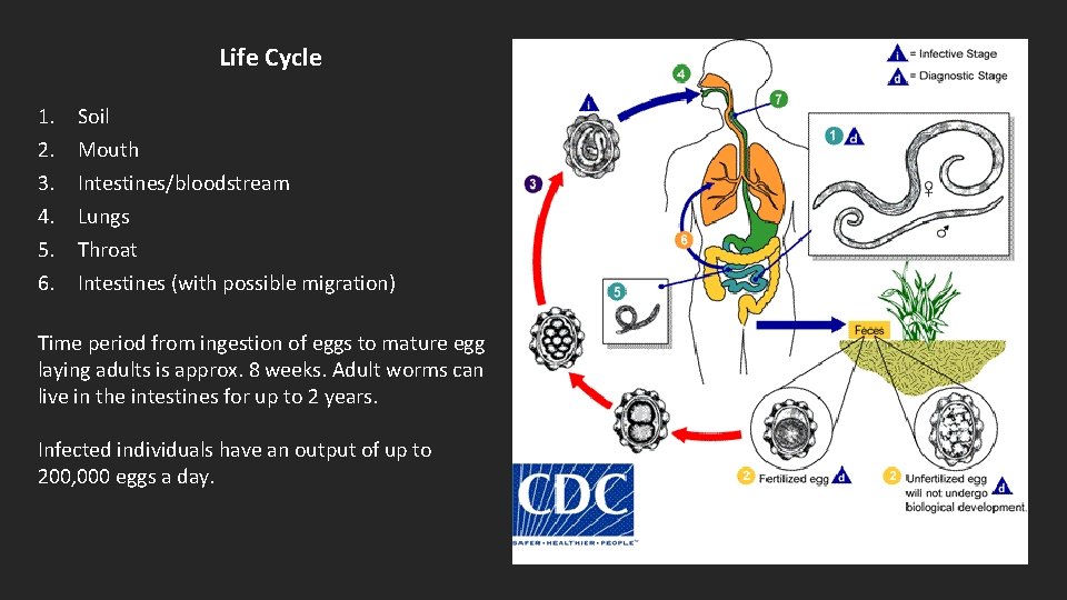
Life Cycle 1. 2. 3. 4. 5. 6. Soil Mouth Intestines/bloodstream Lungs Throat Intestines (with possible migration) Time period from ingestion of eggs to mature egg laying adults is approx. 8 weeks. Adult worms can live in the intestines for up to 2 years. Infected individuals have an output of up to 200, 000 eggs a day.
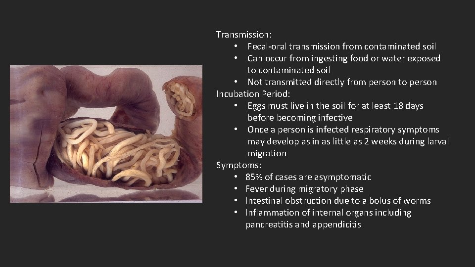
Transmission: • Fecal-oral transmission from contaminated soil • Can occur from ingesting food or water exposed to contaminated soil • Not transmitted directly from person to person Incubation Period: • Eggs must live in the soil for at least 18 days before becoming infective • Once a person is infected respiratory symptoms may develop as in as little as 2 weeks during larval migration Symptoms: • 85% of cases are asymptomatic • Fever during migratory phase • Intestinal obstruction due to a bolus of worms • Inflammation of internal organs including pancreatitis and appendicitis
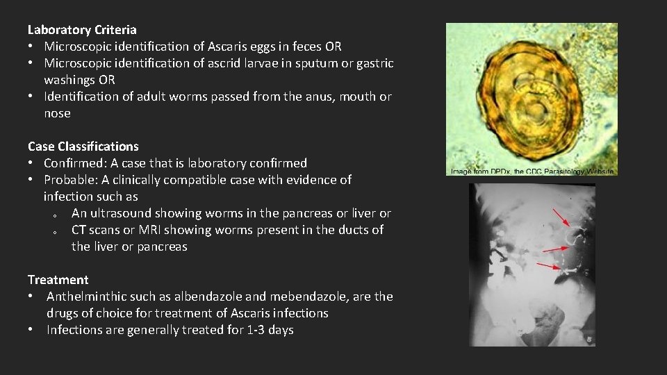
Laboratory Criteria • Microscopic identification of Ascaris eggs in feces OR • Microscopic identification of ascrid larvae in sputum or gastric washings OR • Identification of adult worms passed from the anus, mouth or nose Case Classifications • Confirmed: A case that is laboratory confirmed • Probable: A clinically compatible case with evidence of infection such as o An ultrasound showing worms in the pancreas or liver or o CT scans or MRI showing worms present in the ducts of the liver or pancreas Treatment • Anthelminthic such as albendazole and mebendazole, are the drugs of choice for treatment of Ascaris infections • Infections are generally treated for 1 -3 days
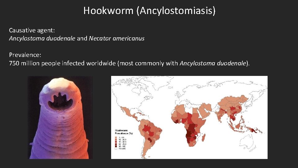
Hookworm (Ancylostomiasis) Causative agent: Ancylostoma duodenale and Necator americanus Prevalence: 750 million people infected worldwide (most commonly with Ancylostoma duodenale).
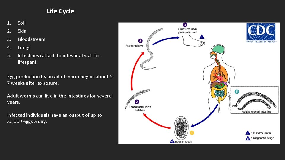
Life Cycle 1. 2. 3. 4. 5. Soil Skin Bloodstream Lungs Intestines (attach to intestinal wall for lifespan) Egg production by an adult worm begins about 57 weeks after exposure. Adult worms can live in the intestines for several years. Infected individuals have an output of up to 30, 000 eggs a day.
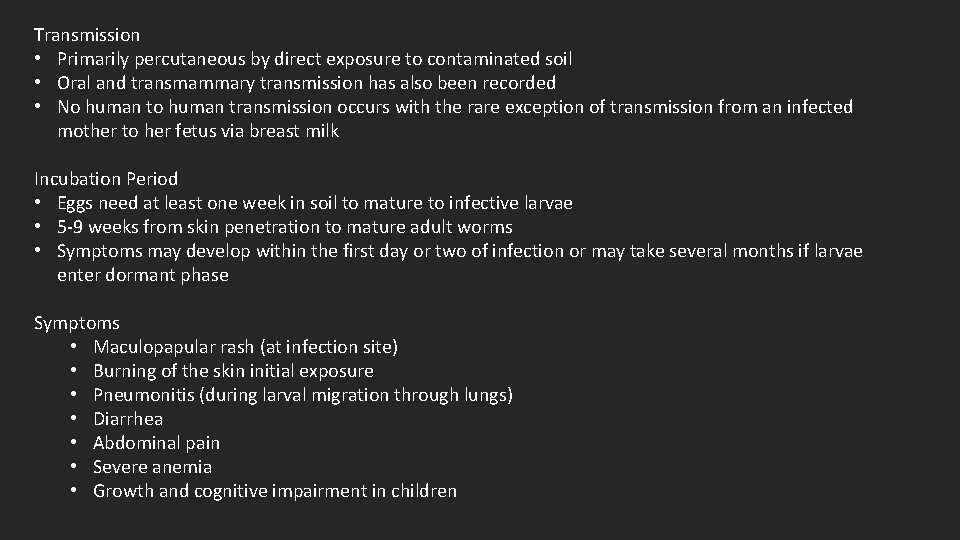
Transmission • Primarily percutaneous by direct exposure to contaminated soil • Oral and transmammary transmission has also been recorded • No human transmission occurs with the rare exception of transmission from an infected mother to her fetus via breast milk Incubation Period • Eggs need at least one week in soil to mature to infective larvae • 5 -9 weeks from skin penetration to mature adult worms • Symptoms may develop within the first day or two of infection or may take several months if larvae enter dormant phase Symptoms • Maculopapular rash (at infection site) • Burning of the skin initial exposure • Pneumonitis (during larval migration through lungs) • Diarrhea • Abdominal pain • Severe anemia • Growth and cognitive impairment in children
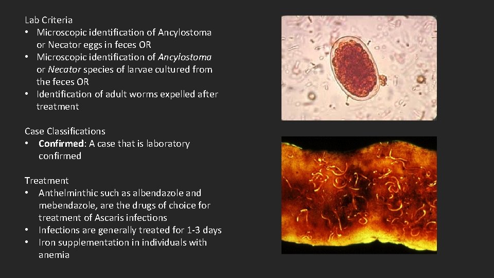
Lab Criteria • Microscopic identification of Ancylostoma or Necator eggs in feces OR • Microscopic identification of Ancylostoma or Necator species of larvae cultured from the feces OR • Identification of adult worms expelled after treatment Case Classifications • Confirmed: A case that is laboratory confirmed Treatment • Anthelminthic such as albendazole and mebendazole, are the drugs of choice for treatment of Ascaris infections • Infections are generally treated for 1 -3 days • Iron supplementation in individuals with anemia
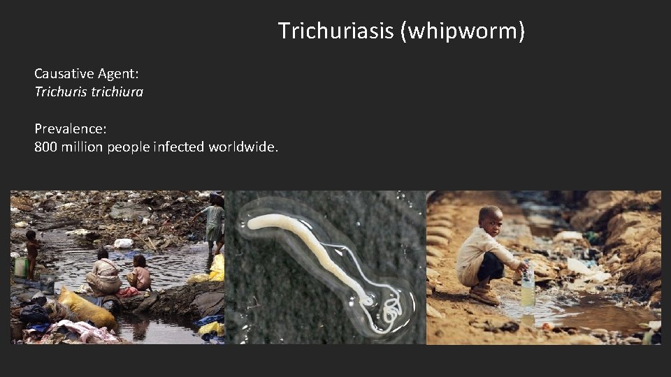
Trichuriasis (whipworm) Causative Agent: Trichuris trichiura Prevalence: 800 million people infected worldwide.
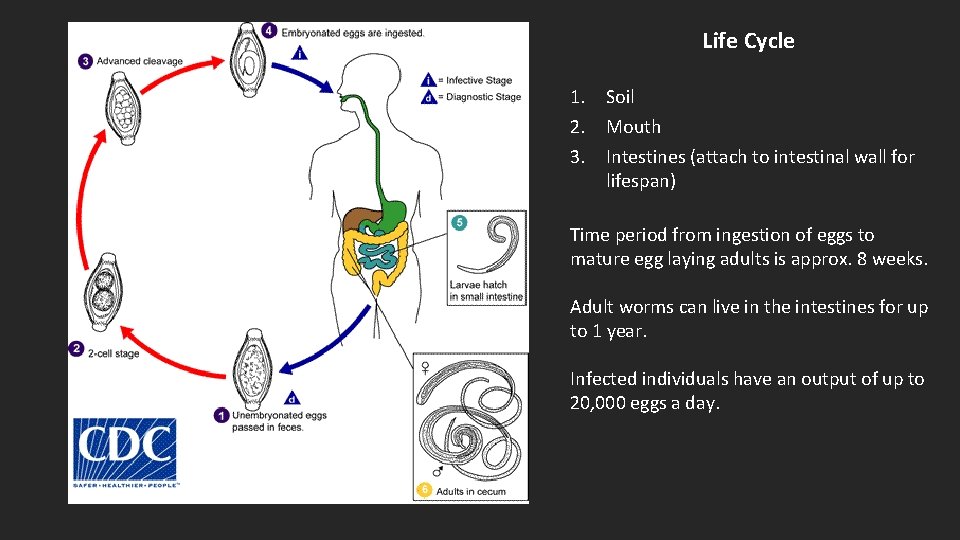
Life Cycle 1. Soil 2. Mouth 3. Intestines (attach to intestinal wall for lifespan) Time period from ingestion of eggs to mature egg laying adults is approx. 8 weeks. Adult worms can live in the intestines for up to 1 year. Infected individuals have an output of up to 20, 000 eggs a day.
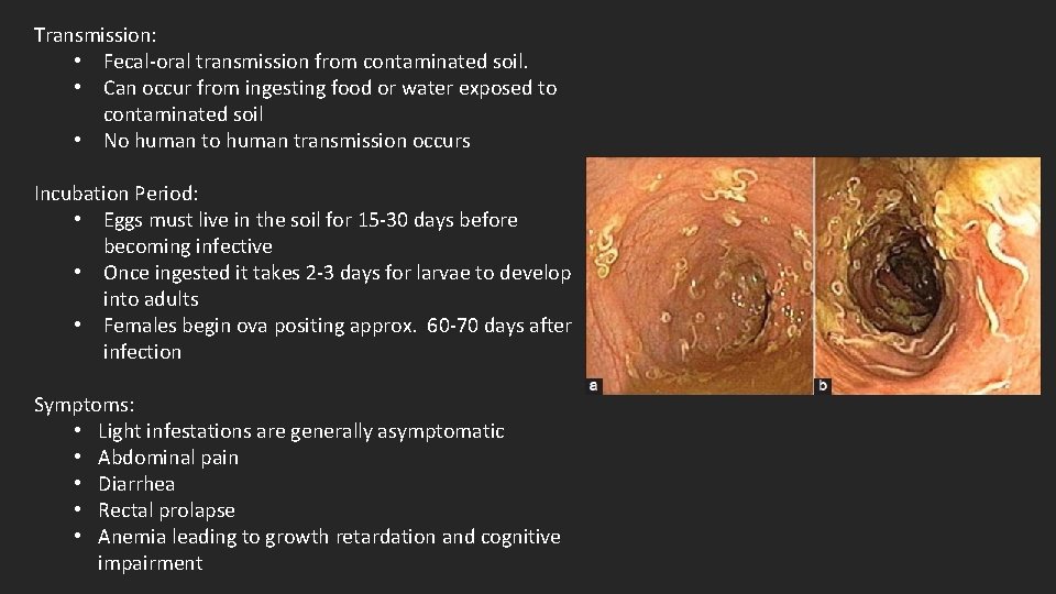
Transmission: • Fecal-oral transmission from contaminated soil. • Can occur from ingesting food or water exposed to contaminated soil • No human transmission occurs Incubation Period: • Eggs must live in the soil for 15 -30 days before becoming infective • Once ingested it takes 2 -3 days for larvae to develop into adults • Females begin ova positing approx. 60 -70 days after infection Symptoms: • Light infestations are generally asymptomatic • Abdominal pain • Diarrhea • Rectal prolapse • Anemia leading to growth retardation and cognitive impairment
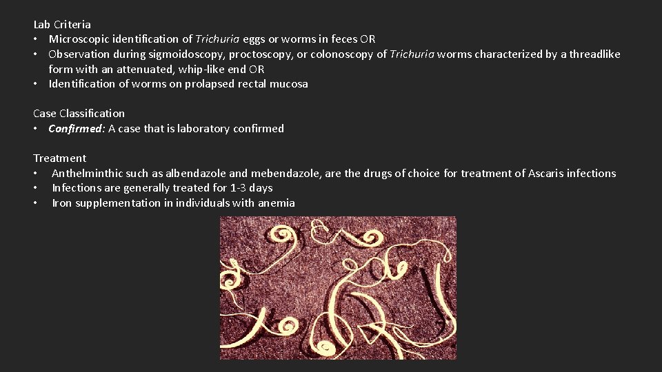
Lab Criteria • Microscopic identification of Trichuria eggs or worms in feces OR • Observation during sigmoidoscopy, proctoscopy, or colonoscopy of Trichuria worms characterized by a threadlike form with an attenuated, whip-like end OR • Identification of worms on prolapsed rectal mucosa Case Classification • Confirmed: A case that is laboratory confirmed Treatment • Anthelminthic such as albendazole and mebendazole, are the drugs of choice for treatment of Ascaris infections • Infections are generally treated for 1 -3 days • Iron supplementation in individuals with anemia
Foodborne Team NTDs Greg Leos, MPH, CPH
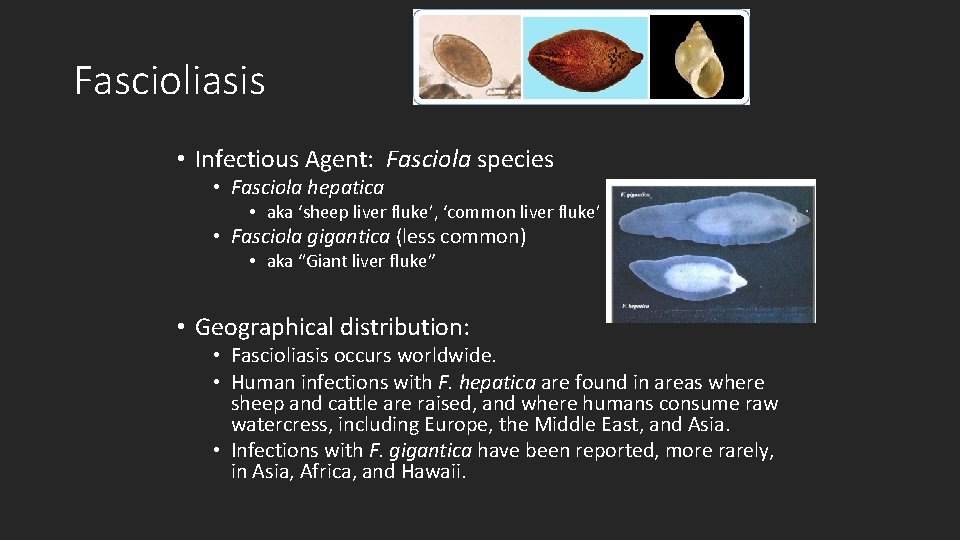
Fascioliasis • Infectious Agent: Fasciola species • Fasciola hepatica • aka ‘sheep liver fluke’, ‘common liver fluke’ • Fasciola gigantica (less common) • aka “Giant liver fluke” • Geographical distribution: • Fascioliasis occurs worldwide. • Human infections with F. hepatica are found in areas where sheep and cattle are raised, and where humans consume raw watercress, including Europe, the Middle East, and Asia. • Infections with F. gigantica have been reported, more rarely, in Asia, Africa, and Hawaii.
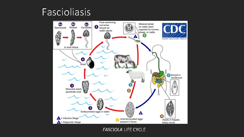
Fascioliasis FASCIOLA LIFE CYCLE
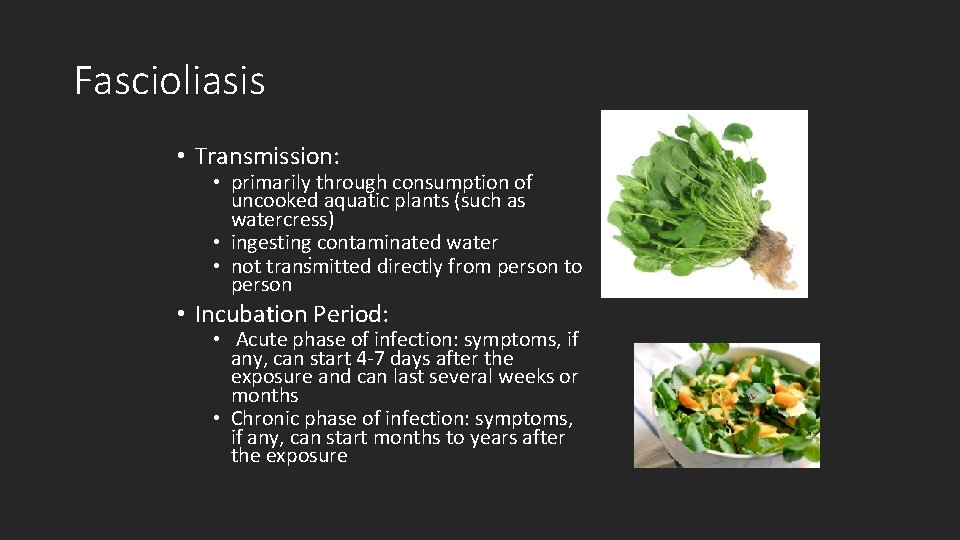
Fascioliasis • Transmission: • primarily through consumption of uncooked aquatic plants (such as watercress) • ingesting contaminated water : • not transmitted directly from person to person • Incubation Period: • Acute phase of infection: symptoms, if any, can start 4 -7 days after the exposure and can last several weeks or months • Chronic phase of infection: symptoms, if any, can start months to years after the exposure
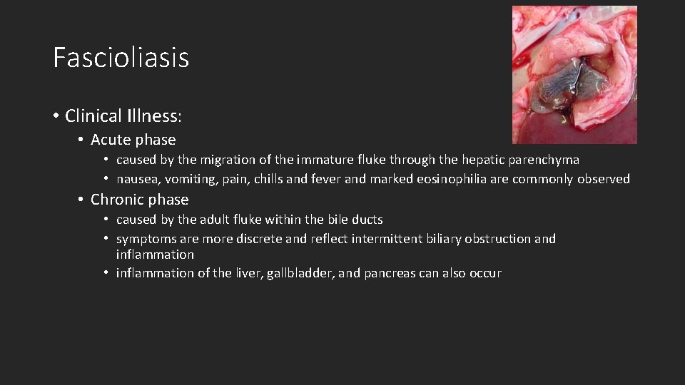
Fascioliasis • Clinical Illness: • Acute phase • caused by the migration of the immature fluke through the hepatic parenchyma • nausea, vomiting, pain, chills and fever and marked eosinophilia are commonly observed • Chronic phase • caused by the adult fluke within the bile ducts • symptoms are more discrete and reflect intermittent biliary obstruction and inflammation • inflammation of the liver, gallbladder, and pancreas can also occur
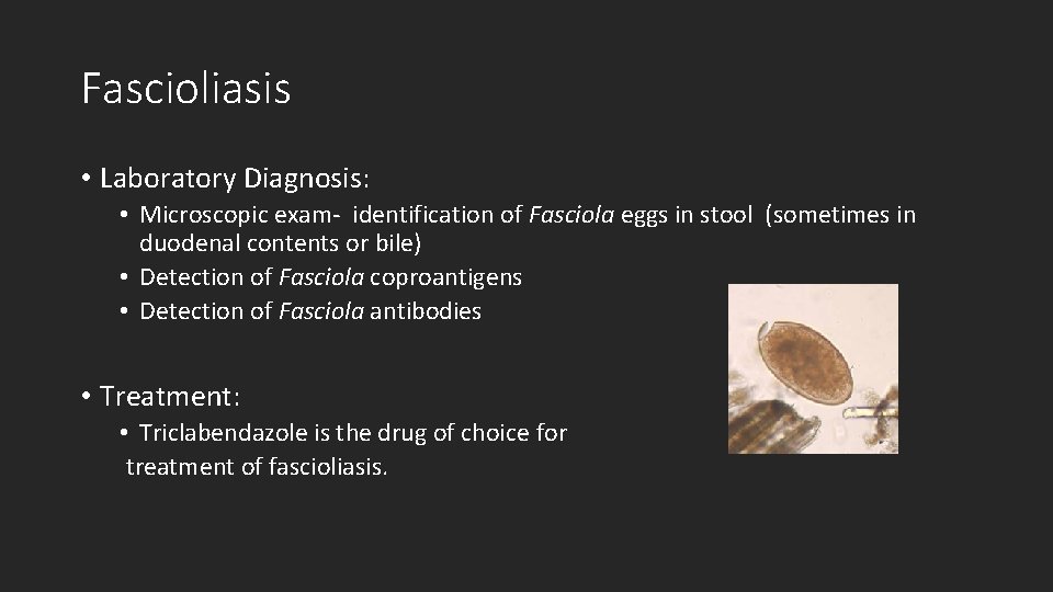
Fascioliasis • Laboratory Diagnosis: • Microscopic exam- identification of Fasciola eggs in stool (sometimes in duodenal contents or bile) • Detection of Fasciola coproantigens • Detection of Fasciola antibodies • Treatment: • Triclabendazole is the drug of choice for treatment of fascioliasis.
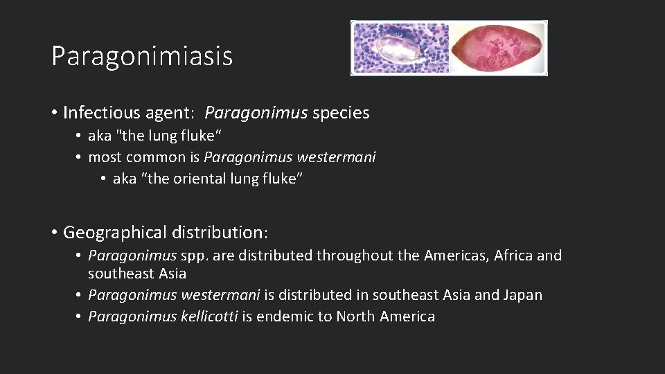
Paragonimiasis • Infectious agent: Paragonimus species • aka "the lung fluke“ • most common is Paragonimus westermani • aka “the oriental lung fluke” • Geographical distribution: • Paragonimus spp. are distributed throughout the Americas, Africa and southeast Asia • Paragonimus westermani is distributed in southeast Asia and Japan • Paragonimus kellicotti is endemic to North America
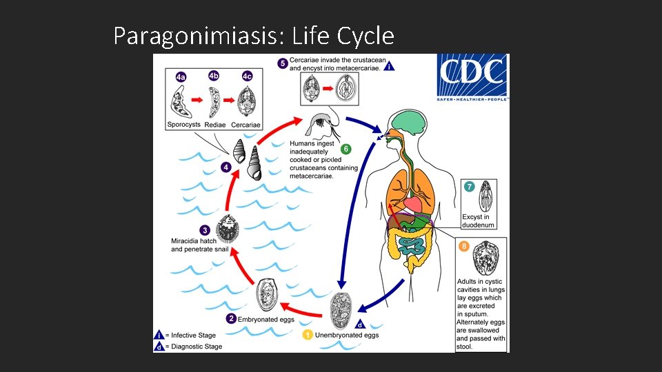
Paragonimiasis: Life Cycle
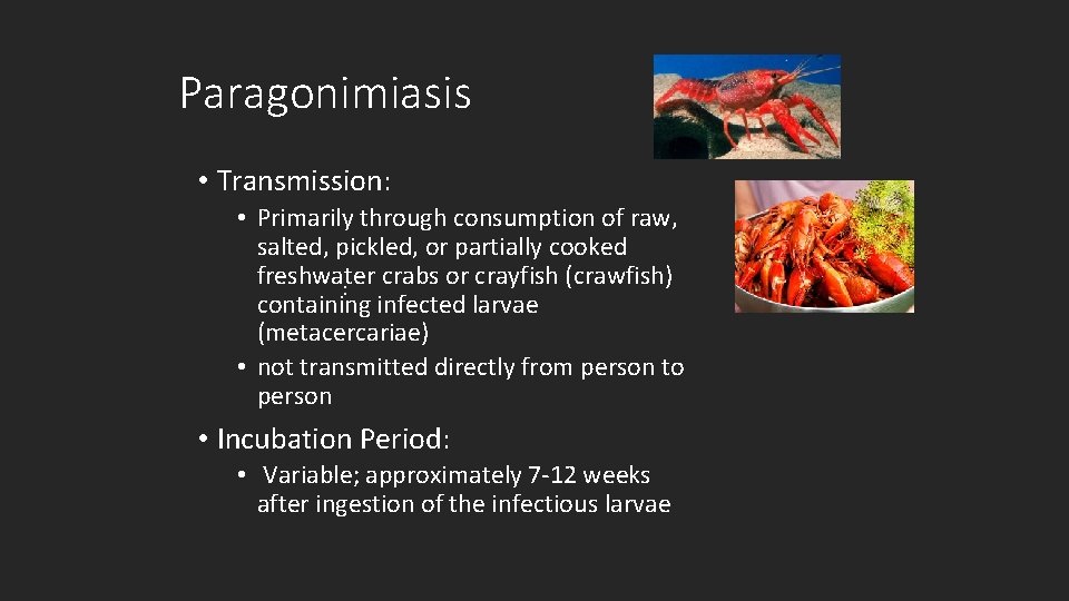
Paragonimiasis • Transmission: • Primarily through consumption of raw, salted, pickled, or partially cooked freshwater crabs or crayfish (crawfish) : containing infected larvae (metacercariae) • not transmitted directly from person to person • Incubation Period: • Variable; approximately 7 -12 weeks after ingestion of the infectious larvae
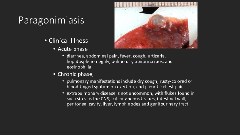
Paragonimiasis • Clinical Illness • Acute phase • diarrhea, abdominal pain, fever, cough, urticaria, hepatosplenomegaly, pulmonary abnormalities, and eosinophilia • Chronic phase, • pulmonary manifestations include dry cough, rusty-colored or blood-tinged sputum on exertion, and pleuritic chest pain • extrapulmonary disease is not uncommon, with flukes found in such sites as the CNS, subcutaneous tissues, intestinal wall, peritoneal cavity, liver, lymph nodes and genitourinary tract
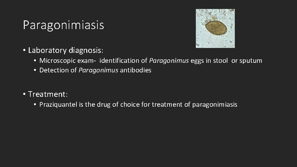
Paragonimiasis • Laboratory diagnosis: • Microscopic exam- identification of Paragonimus eggs in stool or sputum • Detection of Paragonimus antibodies • Treatment: • Praziquantel is the drug of choice for treatment of paragonimiasis
Zoonosis Control Branch NTDs Inger Vilcins MIPH, PHD
Echinococcosis
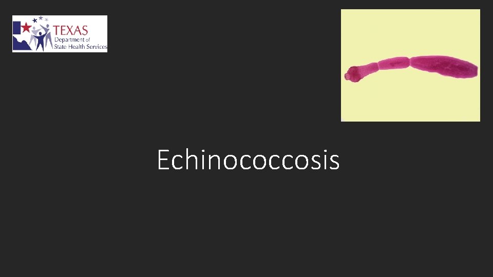
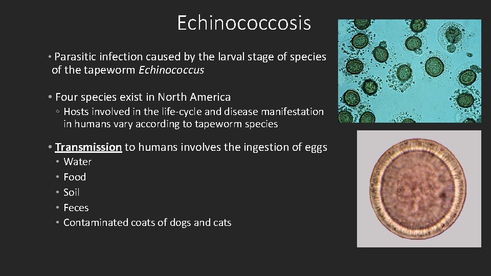
Echinococcosis • Parasitic infection caused by the larval stage of species of the tapeworm Echinococcus • Four species exist in North America ◦ Hosts involved in the life-cycle and disease manifestation in humans vary according to tapeworm species • Transmission to humans involves the ingestion of eggs • Water • Food • Soil • Feces • Contaminated coats of dogs and cats
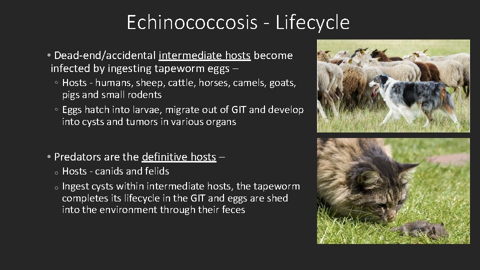
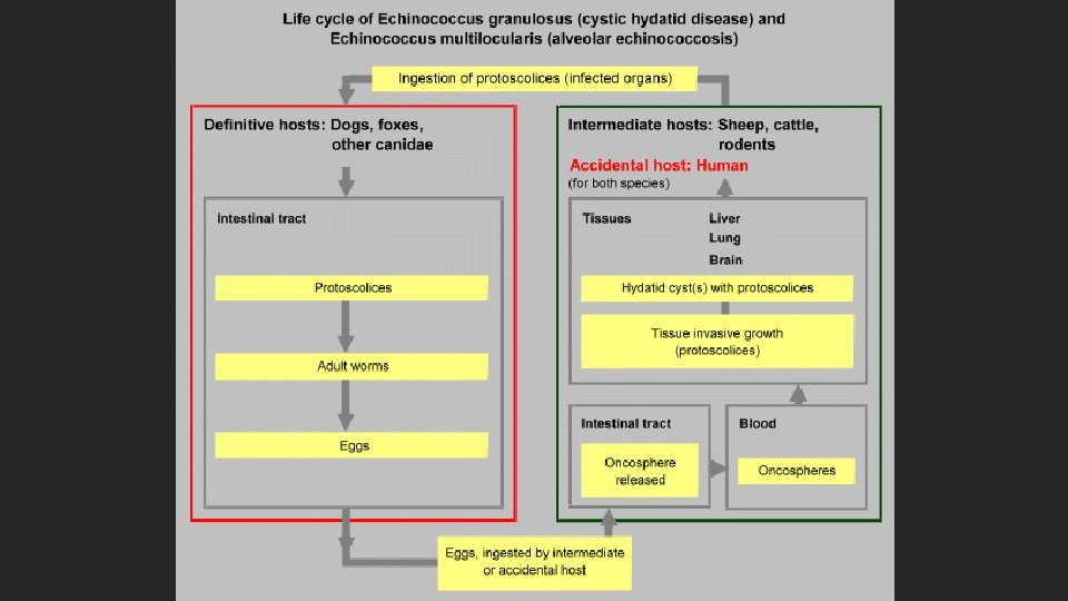
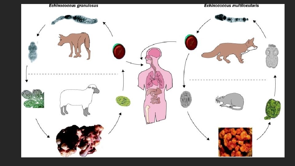
Echinococcosis - Lifecycle • Dead-end/accidental intermediate hosts become infected by ingesting tapeworm eggs – ◦ Hosts - humans, sheep, cattle, horses, camels, goats, pigs and small rodents ◦ Eggs hatch into larvae, migrate out of GIT and develop into cysts and tumors in various organs • Predators are the definitive hosts – Hosts - canids and felids o Ingest cysts within intermediate hosts, the tapeworm completes its lifecycle in the GIT and eggs are shed into the environment through their feces o
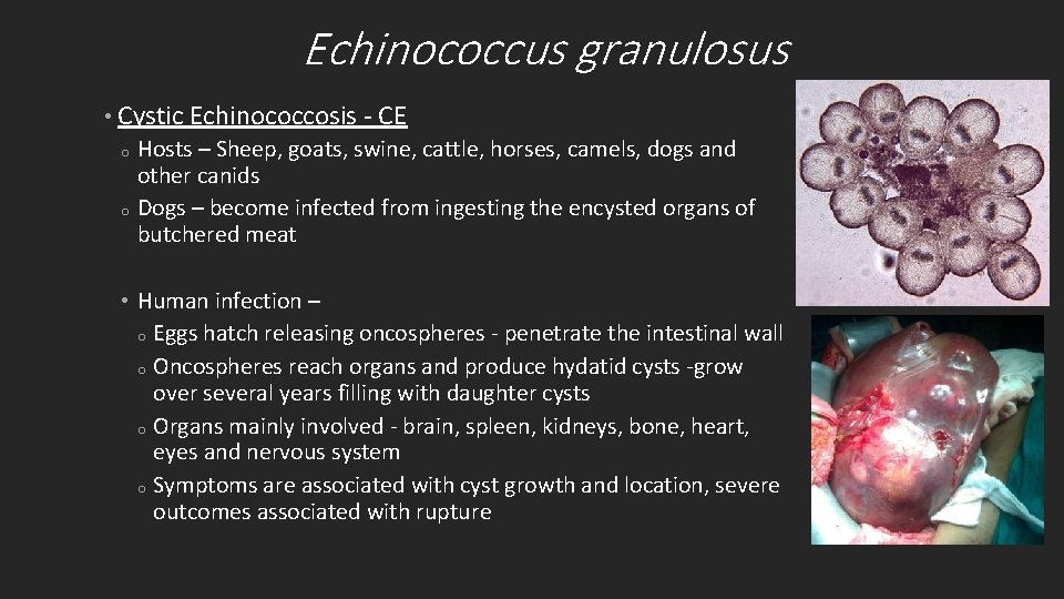
Echinococcus granulosus • Cystic Echinococcosis - CE o Hosts – Sheep, goats, swine, cattle, horses, camels, dogs and other canids o Dogs – become infected from ingesting the encysted organs of butchered meat • Human infection – o Eggs hatch releasing oncospheres - penetrate the intestinal wall o Oncospheres reach organs and produce hydatid cysts -grow over several years filling with daughter cysts o Organs mainly involved - brain, spleen, kidneys, bone, heart, eyes and nervous system o Symptoms are associated with cyst growth and location, severe outcomes associated with rupture
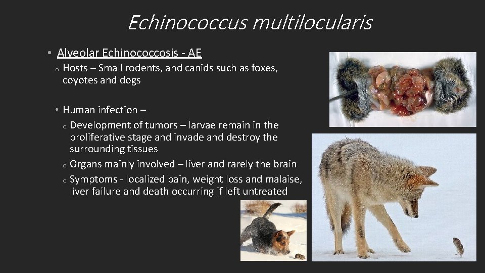
Echinococcus multilocularis • Alveolar Echinococcosis - AE o Hosts – Small rodents, and canids such as foxes, coyotes and dogs • Human infection – o Development of tumors – larvae remain in the proliferative stage and invade and destroy the surrounding tissues o Organs mainly involved – liver and rarely the brain o Symptoms - localized pain, weight loss and malaise, liver failure and death occurring if left untreated
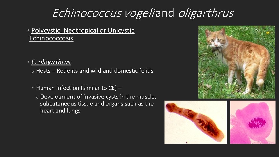
Echinococcus vogeli and oligarthrus • Polycystic, Neotropical or Unicystic Echinococcosis • E. oligarthrus o Hosts – Rodents and wild and domestic felids • Human infection (similar to CE) – o Development of invasive cysts in the muscle, subcutaneous tissue and organs such as the heart and lungs
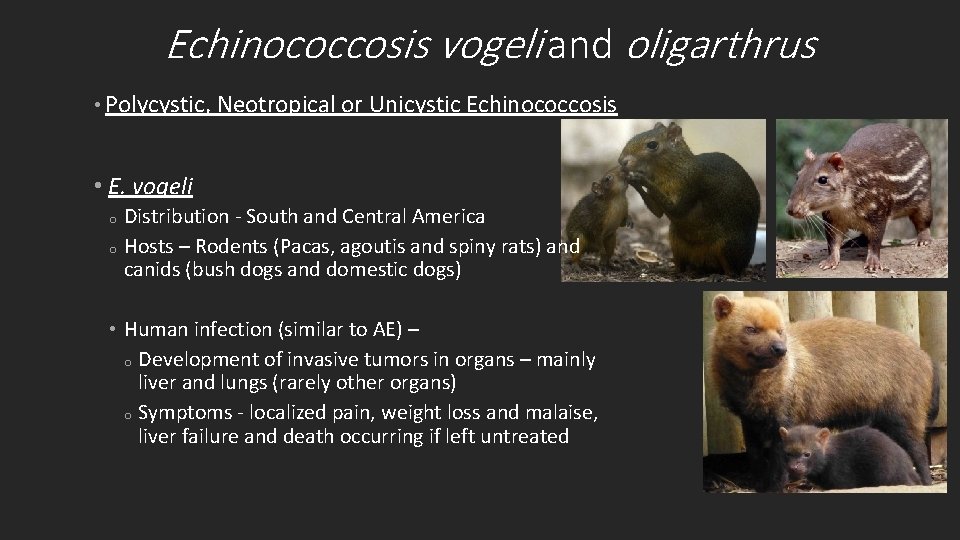
Echinococcosis vogeli and oligarthrus • Polycystic, Neotropical or Unicystic Echinococcosis • E. vogeli Distribution - South and Central America o Hosts – Rodents (Pacas, agoutis and spiny rats) and canids (bush dogs and domestic dogs) o • Human infection (similar to AE) – o Development of invasive tumors in organs – mainly liver and lungs (rarely other organs) o Symptoms - localized pain, weight loss and malaise, liver failure and death occurring if left untreated
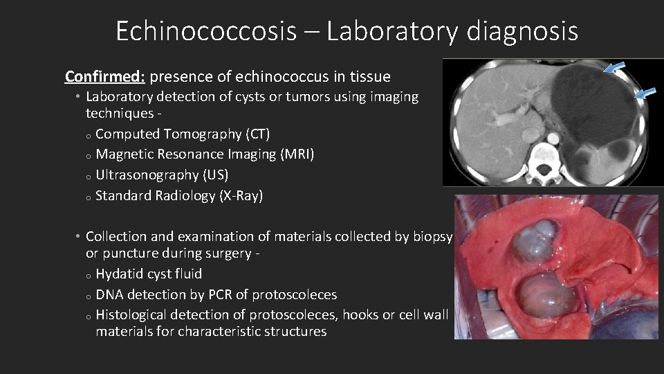
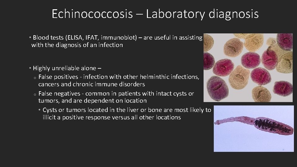
Echinococcosis – Laboratory diagnosis Confirmed: presence of echinococcus in tissue • Laboratory detection of cysts or tumors using imaging techniques o Computed Tomography (CT) o Magnetic Resonance Imaging (MRI) o Ultrasonography (US) o Standard Radiology (X-Ray) • Collection and examination of materials collected by biopsy or puncture during surgery o Hydatid cyst fluid o DNA detection by PCR of protoscoleces o Histological detection of protoscoleces, hooks or cell wall materials for characteristic structures
Echinococcosis – Laboratory diagnosis • Blood tests (ELISA, IFAT, immunoblot) – are useful in assisting with the diagnosis of an infection • Highly unreliable alone – o False positives - infection with other helminthic infections, cancers and chronic immune disorders o False negatives - common in patients with intact cysts or tumors, and are dependent on location • Cysts or tumors located in the liver or bone are most likely to illicit a positive response versus all other locations
Questions?