Fetal Pig Dissection Virtual Pig Dissection http www


 lies in the middle of the thoracic cavity, surrounded by a")
 up toward the head, you will be")
 is a soft sack embedded in the liver. In this")

 appears pinky in most fetal pigs. Remember that the fetus is")


 cavity, you will have to")


, lies between the two umbilical arteries (2). In this")

 is clearly visible as a smooth oval body inside the scrotum.")


- Slides: 38
Fetal Pig Dissection
Virtual Pig Dissection • http: //www. whitman. edu/offices_departme nts/biology/vpd/main. html • Go through each of the study guides and take the quizzes.
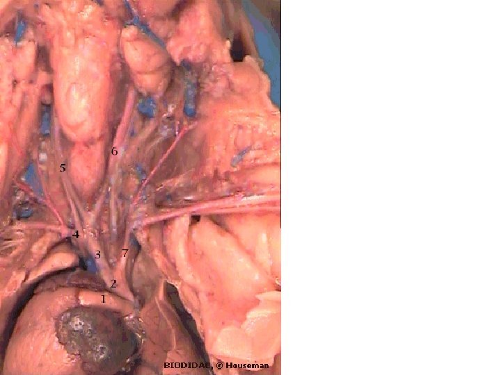
The heart (1) lies in the middle of the thoracic cavity, surrounded by a membrane called the pericardium. If you have damaged the pericardium beyond recognition, look at someone else's pig. By the time you are done the dissection, all the pericardia will have been removed. The top of the heart will be covered with the thymus gland, which extends up into the throat. Much of the thymus has been removed from the pig pictured here. On either side of the heart are the lungs (2). Note that the left lung is smaller. You will have cut the diaphragm (3), but it can still be seen, as can its attachment to the body wall. The large liver (4) may vary from reddish brown to blue. The former is its natural color, but blue latex often fills its numerous blood spaces. Just behind the liver on the left side, the stomach (5) can be seen, and the spleen (6) raps around the left side of the liver. The spleen is very delicate and may break when you try to separate it. It is not surprising that ruptured spleens are one of the most common types of internal injury. The large umbilical vein (7) can be seen entering the liver. You had to cut this vein during the initial incisions. Its other end can be seen on the flap carrying the umbilical cord. The intestines of a pig are arranged somewhat differently from those of a human. The pinkish colored jejunum (8) is seen on the top right, the greener ileum (9) at the lower centre, and the coiled up colon (10) at the top left. The urinary bladder (11) folds back with the umbilical cord, and is surrounded by the two large umbilical arteries (12). These arteries take blood from the fetus to the placenta, where it picks up nutrients and oxygen from the mother's blood, and loses waste products of the fetus's metabolism.
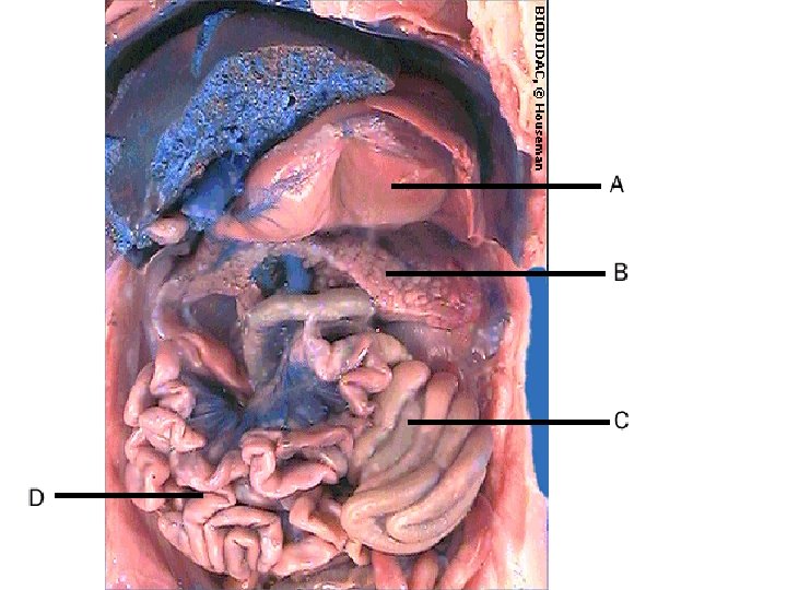
• By pushing the liver (1) up toward the head, you will be able to view many of the organs of the digestive system. • The gall bladder (2) is usually quite evident as a sort of blister on the third lobe of the liver. The next page looks at it more closely. • The stomach (3) is easily identified as a large floppy pouch under the left lobe of the liver. The duodenum (4) arches away from the stomach to the pig's right. A deep crease indicates the location of the pyloric sphincter (5). If you probe this area, you will be able to detect the strong muscular nature of this sphincter. The spleen (6) lies just below the stomach, and is attached to it by a thin membrane. You may have to break this membrane to see deeper structures. • The three major parts of the intestine, the jejunum (7), the ileum (8) and the colon (9) are still visible.
The gall bladder (1) is a soft sack embedded in the liver. In this view, with the liver turned back, you can just make out the attached ducts, the cystic duct (2) which drains the gall bladder, the hepatic duct (3) which carries bile from the liver to the gall bladder, and the common bile duct (4) which carries the bile from both organs to the duodenum (5). A person can live without the gall bladder, because the liver can drain bile directly into the duodenum, but a special restricted diet is required, since bile cannot be stored up for a large fatty meal.
• The pancreas can be viewed by pulling the bottom of the stomach up toward the head. It will be necessary to break some of the membranes holding the stomach and spleen in place. Use a blunt probe for this. • The pancreas (P) is a long, loosely organized gland, that has often been described as looking like a "bunch of grapes". If you look carefully, you may be able to find the pancreatic duct, which goes behind the duodenum (D) and joins the common bile duct before it enters the duodenum.
The jejunum (1) appears pinky in most fetal pigs. Remember that the fetus is not swallowing anything except a bit of amniotic fluid, so there is little in the intestine. The liver is secreting some bile. By the time the intestinal juices get to the ileum (2) some of the water has been absorbed and the bile is more concentrated. This makes the ileum look greenish. The ileum ends where the small intestine meets the colon (3) a a "T" junction. The other end of the "T" (4) is the caecum. Note that the caecum is large compared to the human appendix, but small compared to the pig's horselike relatives. The pig is descended from herbivores whose caecum was undoubtedly vital for digesting cellulose, but, like ourselves, it has become an omnivore and gets little if any nutrition from cellulose.
• The intestines are held in place by a pair of membranes called the mesenteries (1). The mesenteric arteries run between these parallel membranes, and give rise to a vast number of tiny arteries (2) that take blood to the jejunum and ileum. These in turn break up into the thousands of arterioles that supply the villi and help to absorb food. • Near the base of the mesenteries is a row of white, fatty-looking bumps. These are lymph nodes on the lymph ducts that drain the lacteals. Since you take so much foreign material into your gut, it makes sense that you should have a strong line of defense against any invading microorganisms that might escape the digestive processes.
• Close up…
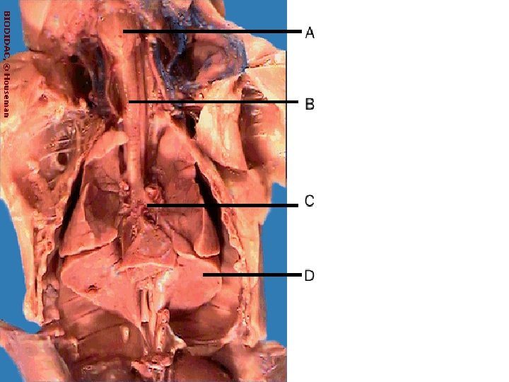
• • • To open the thoracic (chest) cavity, you will have to cut through the cartilage of the breast bone, and the collar bones. Try not to damage the underlying blood vessels as you do. When you first open the chest cavity, your view will be dominated by the heart (1) and lungs (2, 3). Usually, the right lung (2) will be larger than the left (3), though that is not obvious in the view shown here. Each lung consists of several lobes. The heart may be covered (or partly covered) by a transparent membrane, the pericardium, seen here almost intact. The chances are, you will have destroyed much of it while opening the chest. Feel the inside of the rib cage and the surface of the lungs with a gloved finger. They are very smooth and slippery, due to the presence of thin, moist membranes, the pleura. The pleura allow the lungs to slide across the walls of the thoracic cavity with almost no friction during inhalation and exhalation. Note the diaphragm (5) between the lungs and the liver. Remember that you cut the attachments of the diaphragm to the body wall during the initial cuts. If you have a small pig, the diaphragm may look like a translucent membrane with muscle fibers just beginning to grow into it from the edges. If your pig is older, it will be a definite layer of muscle, as shown here. Covering the top of the heart is part of the thymus (6), an important component of the fetal immune system. thymus tissue extends well up into the throat, and must be carefully picked away to see other features of the cervical region. The prominent thyroid, an endocrine gland, is visible as a brick red or slightly purplish round mass.
• • • The view shown here is one that you will not have, since it required removal of the heart. You will need the heart in place for the next part of the dissection. By moving it around and looking behind it, however, you should be able to see many of the features shown here, and perhaps to understand them in context a bit better. The cavity in which the heart sat is seen between the two lungs (1). The muscles, glands and membranes have been peeled away from the front of the trachea (2) which clearly shows the rings of cartilage that keep it open. The trachea branches into several main bronchi (3), of which three are visible here. The stumps of pulmonary arteries and veins are also visible (4). Note that the color of these vessels is not a reliable guide to their identity; you would be better to note whether they attach to the left atrium (veins) or the right ventricle (arteries).
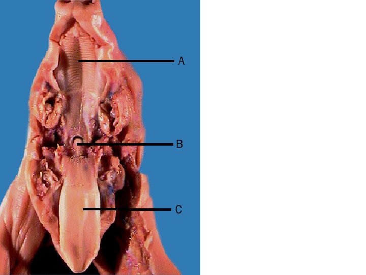
• • By cutting through the sides of the jaw, you can open the mouth wide enough to view the structures at the back. The ridged hard palate (1) is one of the most obvious features, grading into the soft palate (3) of the pharynx. Small "milk teeth" (2) are already erupted in the mouth. The large and muscular tongue (4) has been pulled forward and down in this picture. Papillae, which contain the taste buds, are visible, especially along the edge. At the back of the tongue is the epiglottis (5) which fits into an opening from the nasopharynx (6), allowing direct passage of air from the nose to the trachea. During swallowing, the epiglottis is pulled down to cover the entrance to the trachea, allowing food to pass over it to the esophagus. Depending on the position of your cut, you may be able to see the maxillary salivary glands (7).
• In this view, the tongue has been pulled far forward and down, causing the epiglottis (1) to pull out of the nasophrynx (4). The opening to the larynx and trachea is clearly visible, as is one of the cartilaginous vocal cords (2) that stretch across it. Vibrating vocal cords and the resonance of the larynx beneath them create sounds. Just behind the opening of the larynx is the opening of the esophagus (3)
• Externally, the differences between male and female fetal pigs are small. There is no external penis in the male, and the scrotum is not well developed until sexual maturity. In females, the entrance to the vagina is hidden beneath the tail. • The two specimens in this view show the small abdominal differences. The male (bottom) clearly shows the genital pore just behind the umbilical cord. This is the opening through which the erect penis will be everted for mating. • Running back from the genital pore along the midline of the abdomen, a faint white line is visible. This is the internal penis. • Slight development of the scrotum is visible between the legs. This is much clearer in the rear view, accessible by clicking at the end of this text. • In the female (top) there is a lack of the above features. The main external feature is the genital papilla, a triangular flap of tissue that covers the opening of the urogenital tract. It is found just ventral to the anus, beneath the tail.
• Back views
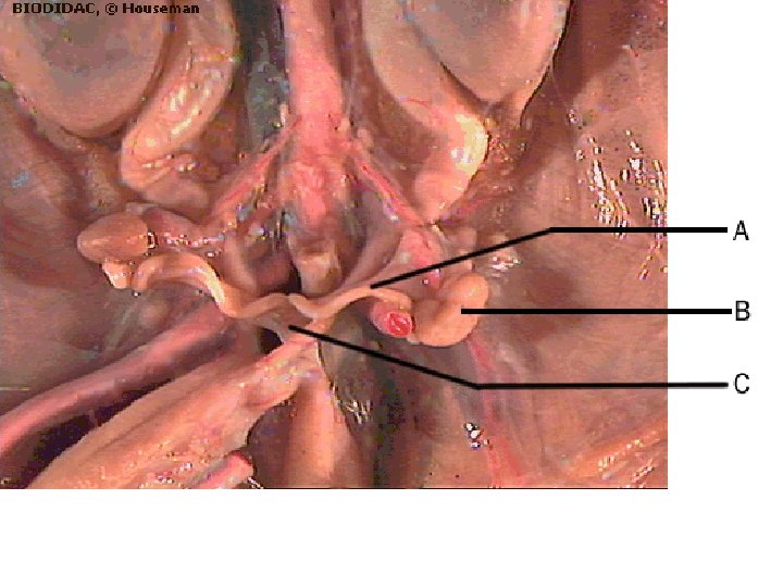
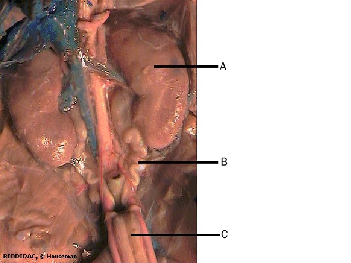
• • This close up view shows the primary reproductive organs of the female. The ovaries (1) are small kidney-shaped organs. They usually cover the fallopian tubes (2) which just show below the right ovary (left side!) in this view. In this view, showing the top end of the ovary (1), one can see the fallopian tube (2) terminating in the funnel-shaped funiculum (3). Note that the funiculum lies right next to the ovary, but is not attached. It must capture eggs released directly into the body cavity. Both the ovary and the fallopian tube are attached to the back wall of the abdomen by a strong ligament (4). The fallopian tubes bring ova to the uterine horns (3) which join in the midline to form the uterine body (4). The uterus is thus "Y-shaped". This allows for the attachment of placentas for a whole litter of young pigs along the uterine horns. The ovary and uterus are held in place by the broad ligament (5), which later in life will have to bear most of the weight during pregnancy
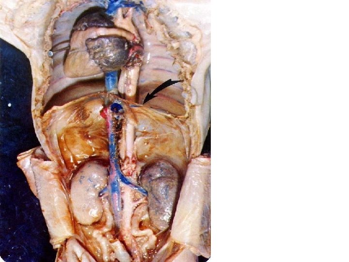
• The bladder (1), lies between the two umbilical arteries (2). In this view it is pulled over to the pig's right side. The left ureter (3), which drains urine from the kidney, can be seen entering the bladder • The ovary (4) lies just posterior to the kidney (not visible in this view). The uterine horns (5) pass over the umbilical arteries and the ureters, and join to form the uterine body (or common uterus)(6) • In this view, the pelvic canal has been dissected. The birth canal consists of the uterine body and the vagina (7). Urine leaves the body via the urethra (8). Note that, unlike in humans, the vagina and urethra join to form a short urogenital sinus (9) before reaching the outside.
• Close up…
The testis (1) is clearly visible as a smooth oval body inside the scrotum. The top end of the epididymis (2) is visible. The epididymis consists of thousands of tiny tubes that collect mature sperm from the tiny cavities in the testis and store them until ejaculation. The left vas deferens (3) can be seen ascending through the inguinal canal and passing over the umbilical artery. it will carry sperm to the urethra. The gonadal artery and vein run parallel to it, as do very sensitive nerves (not visible). Any blockage of the inguinal can cause sterility, pain, and varicose veins in the scrotum. The most common blockage is due to an inguinal hernia, in which a small bulge of the intestine is forced into the canal. Also visible in this view is the penis (4)
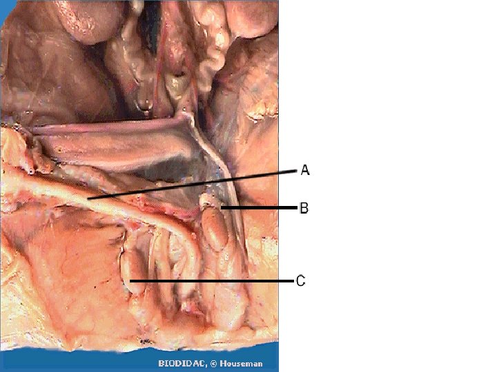
• This view of the male abdomen, with the intestines held out of the way, shows some features common to both sexes. A tough membrane, still intact on the pig's left side, separates the urinary organs from the digestive ones. This membrane has been removed on the pig's right side (your left) to show the kidney (1), which extracts nitrogen and other waste from the blood. The kidneys are drained by the ureters (2) which carry urine to the urinary bladder (3) for storage. • In this view, the bladder has been pulled back. In the intact animal, it would lie in front of the rectum. Note the umbilical arteries on either side of the bladder. After birth, these will atrophy and become ligaments connecting the aorta and the front wall of the abdomen. • Little of the reproductive system can be seen in the abdomen. The testes, which originated in the abdomen, have been pulled down into the scrotum through the inguinal canal (5). All that can be seen are the vasa deferentia (sing. vas deferns), which emerge from the inguinal canal, pass over the ureters and umbilical arteries, and enter the urethra (not clearly visible) just after it leaves the bladder.
• By dissecting through the pelvic bones, one can see the deeper structures of the male tract. The urethra (2) drains the urinary bladder (1) and enters the penis (3), allowing urine to be moved out of the body. The urethra appears very thick at "2" because it is surrounded by strong muscles which control urinary flow and also contract during ejaculation. • The left testis and epididymis are dissected out of the scrotum (6). The gubernaculum (7) has contracted to pull the testis into the scrotum. • Sperm travel from the testis to the epididymis, which loops beneath the testis an meets the vas deferens (8). The vas runs through the inguinal canal, over the umbilical artery and ureter, and joins the urethra just below the bladder. • Two sets of seminal glands can be seen attached to the urethra, the seminal vesicles (9) and the bulbourethral gland (10). These add nutritive and protective fluids to the semen as it passes on its way to the penis
Quiz…. • Identify the labeled parts and also identify the system each view represents.
Answers • • • • Set 1 A - stomach B - pancreas C - large intestine D - small intestine E - digestive Set 2 A - Uterine Horn (Fallopian Tube) B - Ovary C - Vagina D - Urogenital or Reproductive Set 3 A - penis B - epididymus C - teste Set 4 A - larynx B - trachea C - bronchial tube D - lung E - respiratory Set 5 A - kidney B - ureter C - bladder D - Urogenital (Excretory) Set 6 A - hard palate B - epiglottis C - tongue Set 7 A - carotid arteries B - Aorta C - circulatory
• Identify the artery labled #1 in the picture Your answer: a. conus arteriosus b. umbilical artery c. abdominal aorta d. femoral artery • Identify the artery labeled #2 in the picture. Your answer: a. pulmonary artery b. abdominal aorta c. renal artery d. external iliac • Identify the artery labeled #3 in the picture. Your answer: a. renal artery b. subclavian artery c. femoral artery d. external iliac
• Identify the arteries labeled #4 in the picture. Your answer: a. coronary arteries b. pulmonary arteries c. carotid arteries d. subclavian arteries • Identify the artery labeled #5 in the picture. Your answer: a. common carotid b. left subclavian artery c. right subclavian artery d. ductus arteriosis • Identify the artery labeled #6 in the picture. Your answer: a. left carotid artery b. left subclavian artery c. right subclavian artery d. aorta
• Identify the organ labeled #7 in the picture. Your answer: liver spleen pancreas stomach • Identify the organ labeled #8 in the picture. Your answer: liver large intestine small intestine pancreas • Identify the organ labeled #9 in the picture. Your answer: small intestine rectum stomach large intestine • Identify the organ labeled #10 in the picture. Your answer: large intestine stomach pancreas heart
• Identify the artery labeled #11 in the picture. Your answer: abdominal aorta renal artery carotid artery femoral artery • Identify the organ labeled #12 in the picture. Your answer: rectum pancreas kidney liver • Identify the vein labeled #13 in the picture. Your answer: renal vein femoral artery internal iliac artery abdominal aorta
• Identify the organs labeled #14 in the picture. Your answer: liver kidneys lungs intestines • Identify the organ labeled #15 in the picture. Your answer: heart spleen stomach lung