THE DIGESTIVE SYSTEM FUNCTIONS OF THE DIGESTIVE SYSTEM


- Slides: 37
THE DIGESTIVE SYSTEM
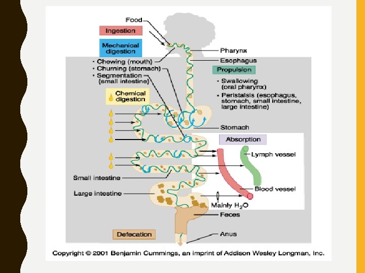
FUNCTIONS OF THE DIGESTIVE SYSTEM • Ingest food • Break down food into nutrient molecules • Absorb molecules into the bloodstream • Rid the body of indigestible remains
MAIN DIVISIONS OF THE DIGESTIVE SYSTEM • Alimentary Canal – Continuous, muscular digestive tube winding throughout the body – Digests and absorbs food particles – Contains the following organs: • Mouth, Pharynx, Esophagus, Stomach, Small and Large Intestines • Accessory Digestive Organs – Contains the following organs: • Teeth, Tongue, Gallbladder, Salivary Glands, Liver, and Pancreas
DIGESTIVE SYSTEM DIVISIONS
DIGESTIVE PROCESSES • Ingestion • Propulsion • Mechanical digestion • Chemical digestion • Absorption • Defecation
HISTOLOGY OF THE ALIMENTARY CANAL/GI TRACT • From esophagus to anus, the walls of the alimentary canal have the same 4 layers: – Mucosa (secretes hormones and mucus, absorbs end products of digestion, and protects against infection – Submucosa (contains lymphoid follicles and elastic tissue) – Muscularis externa (segmentation and peristalsis, contains inner circular layer and outer longitudinal layer, area where valves are found) – Serosa (same as visceral peritoneum, made of areolar connective tissue)
HISTOLOGY OF THE GI TRACT
FEATURES AND FUNCTIONS OF THE MOUTH • Buccal/oral cavity • Contains stratified squamous epithelium • Vestibule: area bounded by lips and cheeks externally and teeth and gums internally • Lips: posses no sweat or oil glands • Palate: forms roof of the mouth, soft and hard palate, uvula
ANATOMY OF THE MOUTH
FEATURES AND FUNCTIONS OF THE TONGUE • Helps grind food into a bolus which contains partially digested food and saliva • Helps form words and is a sensory organ for taste • Three surface features: – Filiform papillae (roughness and grip) – Fungiform papillae (contains taste buds) – Circumvallate papillae (contains taste buds)
ANATOMY OF THE TONGUE
FEATURES AND FUNCTIONS OF THE SALIVARY GLANDS • Main functions: – Produces and secretes saliva – Cleanses the mouth – Dissolves food chemicals so they can be tasted – Moistens food, compacting it into a bolus – Begins the chemical breakdown of food • Salivary amylase: starch
• Composition of Saliva: 97 -99. 5% water • p. H 6. 75 -7. 0 • Sodium, potassium, chloride, phosphate, and bicarbonate • Mucin • Salivary amylase
FEATURES AND FUNCTIONS OF THE TEETH • Break food into smaller parts, increasing surface area for digestion • Types of Teeth – Deciduous Teeth (“baby” teeth) – Permanent Teeth • • Incisors- cutting and shredding Canines- piercing and tearing Molars- grinding Premolars- grinding and crushing
FEATURES AND FUNCTIONS OF THE ESOPHAGUS • Muscular tube that propels food to stomach • Skeletal muscle (upper third for swallowing) and smooth muscle (lower third) for peristalsis • Esophageal sphincter – prevents backflow into oral cavity • Cardiac sphincter- prevents backflow into esophagus
ANATOMY OF THE ESOPHAGUS
DIGESTIVE PROCESSES IN THE MOUTH, PHARYNX, AND ESOPHAGUS • Mouth processes: – Ingestion – Mechanical digestion (e. g. salivary amylase) – Initiation of Propulsion – Mastication: chewing • Pharyngeal processes: – Deglutition = swallowing • Voluntary Buccal phase • Involuntary Pharyngeal-Esophageal Phase • Esophageal processes: – Peristalsis (rhythmic contractions, involuntary)
PERISTALSIS
FEATURES AND FUNCTIONS OF THE STOMACH • Temporary storage area for food and allows it to mix with gastric juice to produce chyme • Regions: cardiac, fundus, body, and pyloric • Greater and Lesser Curvatures: connected to greater and lesser omentums • Rugae folds: longitudinal folds in stomach wall - mucous b/w folds • Muscle layers arranged circularly, longitudinally, AND obliquely (aids in digestion)
ANATOMY OF THE STOMACH
GASTRIC MOTILITY AND EMPTYING • Peristaltic waves approach stomach and become stronger near pyloric region • Pyloric sphincter allows ~ 3 m. L of chyme to pass to duodenum and the rest to return to stomach for further mixing
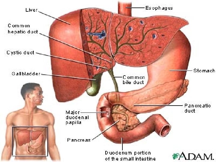
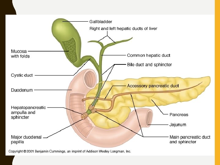
FEATURES AND FUNCTIONS OF THE SMALL INTESTINE • Receives chyme from stomach; performs majority of digestion and absorption of nutrients • Regions: – Duodenum (upper region receiving chyme from stomach and digestive enzymes from pancreas and bile from liver and gallbladder) – Jejunum/Ileum (lower regions where absorption occurs)
ANATOMY OF THE SMALL INTESTINE
FUNCTIONS OF THE LIVER • Largest internal organ • Functions: – Filters and processes nutrient-rich blood of carbohydrates, proteins, and lipids from intestine – Production and regulation of cholesterol – Production of bile which emulsifies fats – Removes drugs and hormones from circulation – Storage of vitamins and minerals
ANATOMY OF THE LIVER • Right and Left Lobes: separated by falciform ligament • Caudate and Quadrate Lobes: found on posterior side • Gallbladder: found underneath left lobe, stores bile
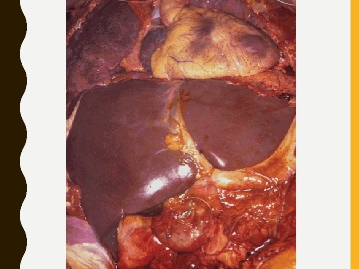
GROSS ANATOMY OF THE LIVER
FUNCTION AND REGULATION OF BILE • Bile emulsifies fats, separating them into smaller parts • Bilirubin: the chief bile pigment, a waste product of the heme of hemoglobin formed during the breakdown of worn-out erythrocytes
FEATURES AND FUNCTIONS OF THE PANCREAS • Pancreatic Juice secreted by acinar cells • Islets of Langerhans release insulin and glucagon (important in glucose metabolism) • Pancreatic Juice contains: – Sodium Bicarbonate (buffers HCl in stomach) – Proteases (break down polypeptides) – Pancreatic amylase (digests oligosaccarides and disaccharides into monosaccharides) – Pancreatic lipases (break down lipids into fatty acids and glycerol) – Pancreatic nucleases (break down nucleic acids)
FEATURES AND FUNCTIONS OF THE LARGE INTESTINE • Functions: – Reabsorption of remaining water and electrolytes – Production and absorption of Vitamins B and K – Elimination of feces • Diameter is only 7 cm but is larger than that of the small intestine
GROSS ANATOMY OF THE LARGE INTESTINE • Cecum: sac-like connection between the small and large intestines • Appendix: small structure containing lymphoid tissue; small immune function • Ascending, Descending, Transverse, and Sigmoid Colon • Rectum: storage area • Anus: regulates defecation with two sphincter muscles; internal and external
ANATOMY OF THE COLON
CLINICAL CORNER • Peptic ulcers - gastric and duodenal • Cirrohsis - scarred liver due to chronic inflammation • Hepatitis - A, B, C, D, and E (inflammation of the liver. The condition can be self-limiting or can progress to fibrosis (scarring), cirrhosis or liver cancer • Gall stones - crystals of cholesterol in bile • Borborygmus - rumbling noise caused by gas through intestines
• Cholecystitis - inflammation of gall bladder • Colitis - inflammation of colon • Dysphagia - difficulty in swallowing • Enteritis - inflammation of the intestines Flatuation/erucation