King Saud University College of medicine US of


- Slides: 44
King Saud University College of medicine
US of liver and gall stone Dr. sultan Alharbi MARAM MOBARA MD
outline: ► Introduction to US. ► Indications of liver and gall bladder US. ► Normal anatomy and radiological appearance. ► Pathology of liver and gall bladder. ► Common pathological cases.
Introduction to US
History OF US ► Piezoelectricity discovered by the Curies in 1880 using natural quartz. ► Piezoelectric Effect is the ability of certain materials to generate an electric charge in response to applied mechanical stress. ► (US) SONAR was first used in 1940’s wartime ► Diagnostic Medical applications in use since late 1950’s
Definition: ►a diagnostic technique in which highfrequency sound waves penetrate the body, bounce around, and produce multiple echoes; these echo patterns can be viewed as an image on a computer screen. ► Frequency ranges used in medical Ultrasound imaging are 2 - 15 MHz
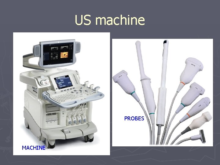
US machine PROBES MACHINE
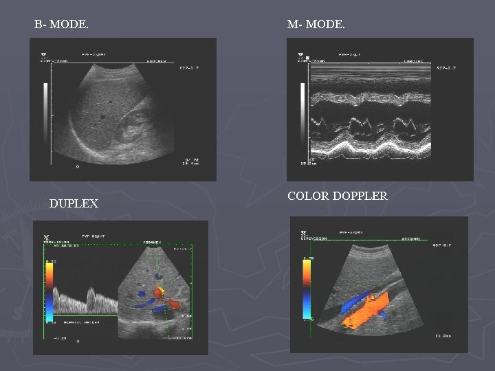
B- MODE. DUPLEX M- MODE. COLOR DOPPLER
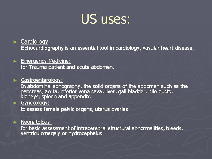
US uses: ► Cardiology ► Emergency Medicine: for Trauma patient and acute abdomen. Echocardiography is an essential tool in cardiology, vavular heart disease. Gastroenterology: In abdominal sonography, the solid organs of the abdomen such as the pancreas, aorta, inferior vena cava, liver, gall bladder, bile ducts, kidneys, spleen and appendix. ► Gynecology: to assess female pelvic organs, uterus ovaries ► ► Neonatology: for basic assessment of intracerebral structural abnormalities, bleeds, ventriculomegaly or hydrocephalus.
Cont. ► ► Neurology for assessing blood flow and stenoses in the carotid arteries (Carotid ultrasonography) ► ► Obstetrics: sonography is commonly used during pregnancy to check on the development of the fetus. ► ► Urology: to study a patient's bladder, prostate or testes. ► ► Musculoskeletal For assessing tendons, muscles, nerves, ligaments, soft tissue masses, and bone surfaces ► ► vascular system: To assess patency and possible obstruction of arteries Arterial doppler, diagnose DVT venous doppler and determine extent and severity of venous insufficiency
Advantages of US ► noninvasive ► inexpensive. ► Easy and available. ► Safe and non ionizing.
Disadvantages of US ► Inability to penetrate gas or bone. ► Operator dependant. ► Less sensitive in some situations.
Indications of liver and gall bladder US ► Right upper quadrant pain. ► Jaundice. ► High liver function test. ► Fever work up. ► Screening for metastasis.
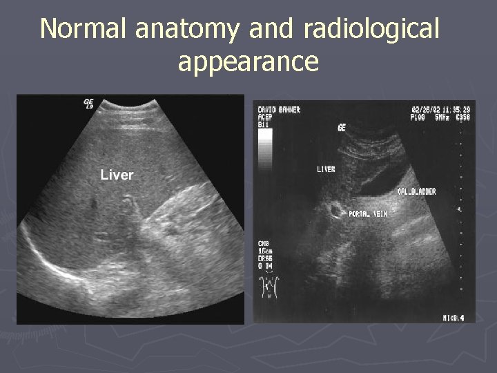
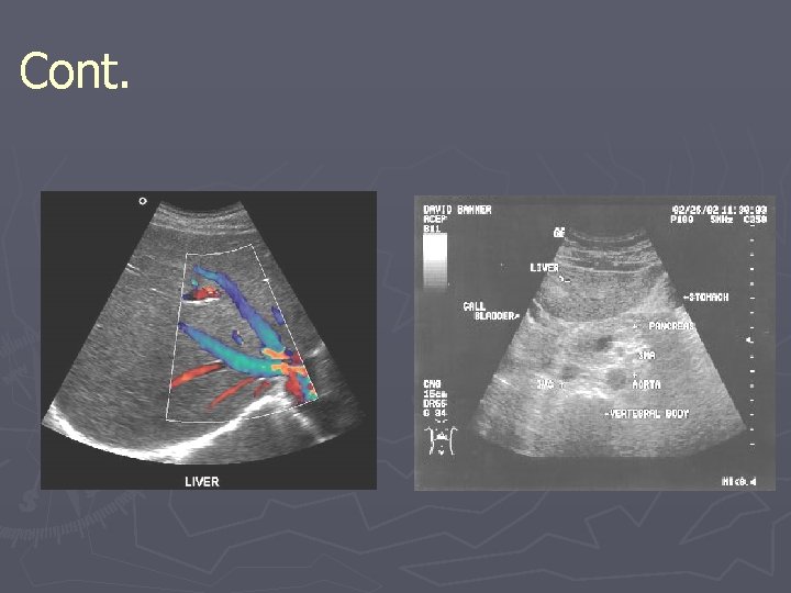
Normal anatomy and radiological appearance
Cont.
Pathology of the liver: ► Size. ► Diffuse liver disease. ► Focal liver disease. ► Hepatic vascularity. ► Biliary system obstruction/pathology.
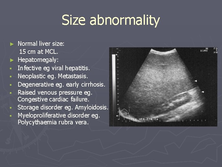
Size abnormality ► ► § § § Normal liver size: 15 cm at MCL. Hepatomegaly: Infective eg viral hepatitis. Neoplastic eg. Metastasis. Degenerative eg. early cirrhosis. Raised venous pressure eg. Congestive cardiac failure. Storage disorder eg. Amyloidosis. Myeloproliferative disorder eg. Polycythaemia rubra vera.
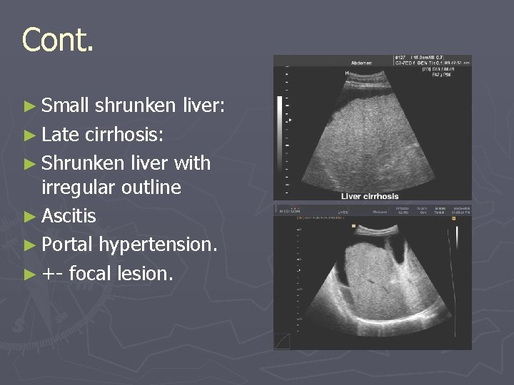
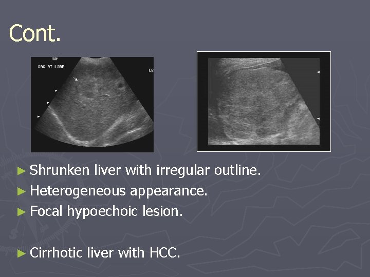
Cont. ► Small shrunken liver: ► Late cirrhosis: ► Shrunken liver with irregular outline ► Ascitis ► Portal hypertension. ► +- focal lesion.
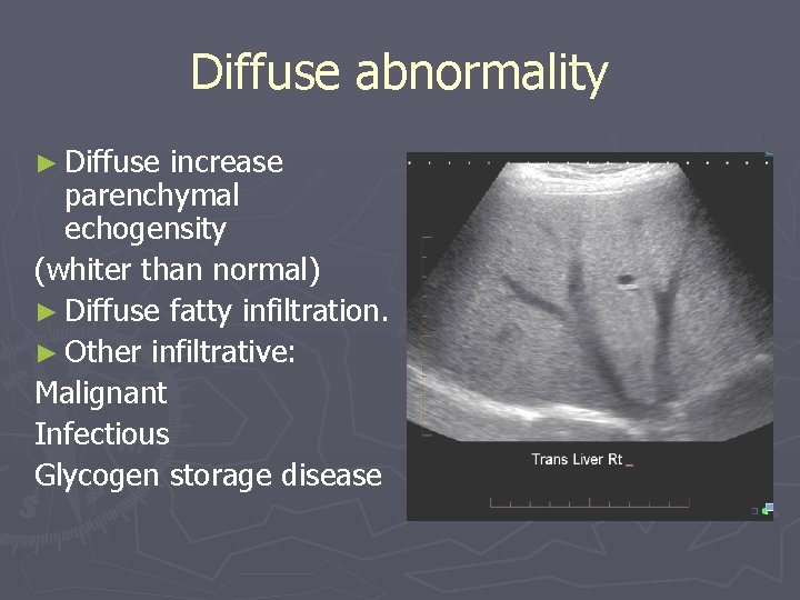
Diffuse abnormality ► Diffuse increase parenchymal echogensity (whiter than normal) ► Diffuse fatty infiltration. ► Other infiltrative: Malignant Infectious Glycogen storage disease
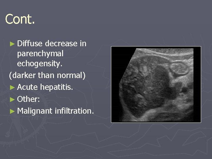
Cont. ► Diffuse decrease in parenchymal echogensity. (darker than normal) ► Acute hepatitis. ► Other: ► Malignant infiltration.
Focal liver lesions q Benign tumor: ► Hemangioma. q Malignant tumor: ► Primary eg. Hepatocellular carcinoma. ► Secondary metastasis eg. Colon breast. q Infective: ► Abscess ► hydated cyst. q Congenital: ► Hepatic cyst.
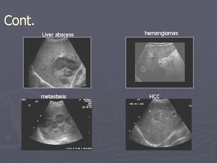
Cont. Liver abscess metastasis hemangiomas HCC
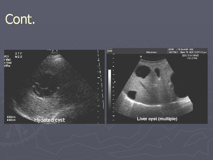
Cont. Hydated cyst
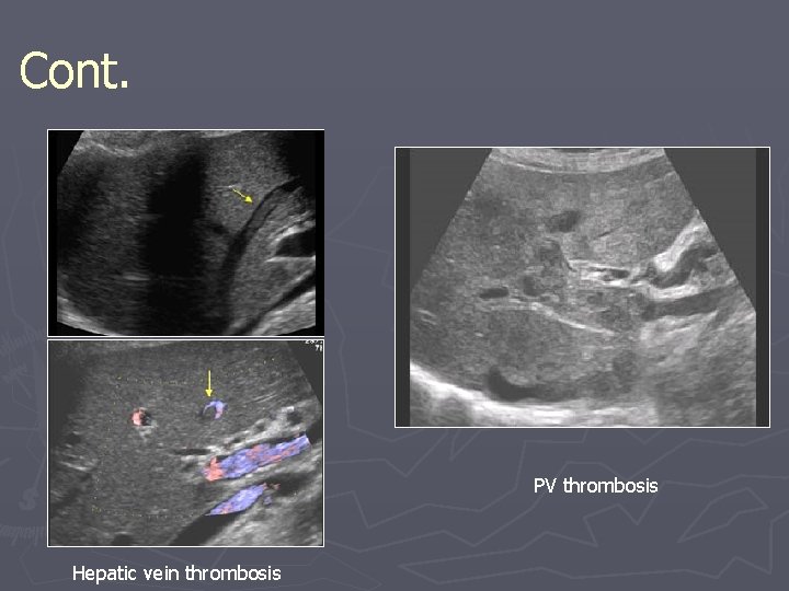
Vascular abnormality q Portal venous system: ► thrombosis. ► Portal hypertension. q Hepatic venous system: ► Thrombosis ► (budd chiari syndrome).
Cont. PV thrombosis Hepatic vein thrombosis
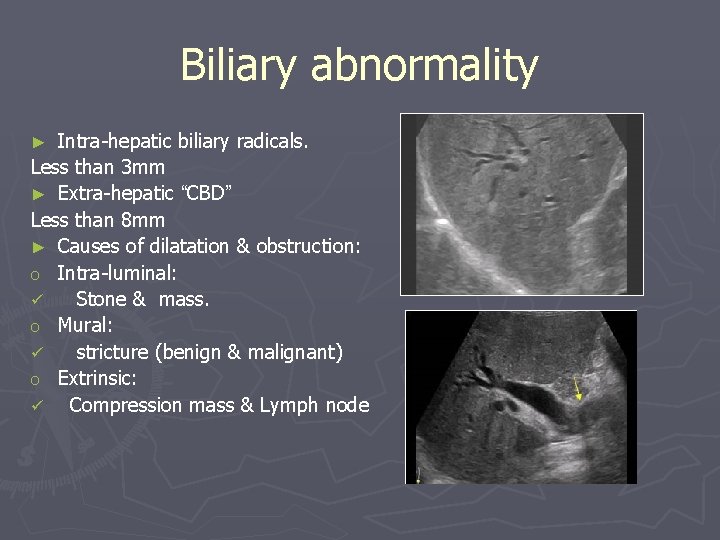
Biliary abnormality Intra-hepatic biliary radicals. Less than 3 mm ► Extra-hepatic “CBD” Less than 8 mm ► Causes of dilatation & obstruction: o Intra-luminal: ü Stone & mass. o Mural: ü stricture (benign & malignant) o Extrinsic: ü Compression mass & Lymph node ►
Pathology of gall bladder ► Intra-luminal pathology. ► Mural pathology.
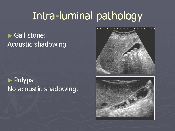
Intra-luminal pathology ► Gall stone: Acoustic shadowing ► Polyps No acoustic shadowing.
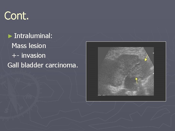
Cont. ► Intraluminal: Mass lesion +- invasion Gall bladder carcinoma.
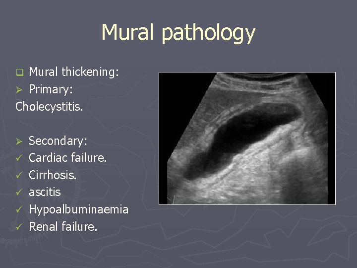
Mural pathology Mural thickening: Ø Primary: Cholecystitis. q Ø ü ü ü Secondary: Cardiac failure. Cirrhosis. ascitis Hypoalbuminaemia Renal failure.
Common pathological cases
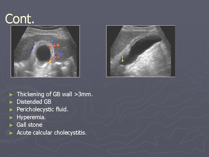
Case one ► Middle age women presented to ED with fever, RUQ pain ► On exam She looks ill, febrile and on pain Abdomen: RUQ tenderness ► Lab high LFTs & WBC.
Cont. ► ► ► Thickening of GB wall >3 mm. Distended GB Pericholecystic fluid. Hyperemia. Gall stone Acute calcular cholecystitis.
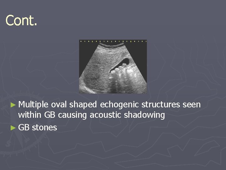
Case two ► Middle age women presented to surgical out patient clinic with 2 years history of recurrent RUQ pain mild to moderate in severity radiated to the right shoulder aggravated by fatty meal. ► On exam: obese lady well not distressed, febrile or jaundiced. ► Lab LFTs normal.
Cont. ► Multiple oval shaped echogenic structures seen within GB causing acoustic shadowing ► GB stones
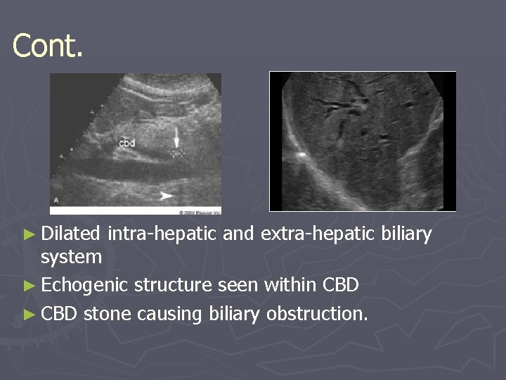
Case three ► Middle age man presented to ER with severe RUQ pain and yellowish discoloration of skin and sclera. ► On exam: he looks ill, jaundiced and on pain but not febrile ► Lab high LFTs.
Cont. ► Dilated intra-hepatic and extra-hepatic biliary system ► Echogenic structure seen within CBD ► CBD stone causing biliary obstruction.
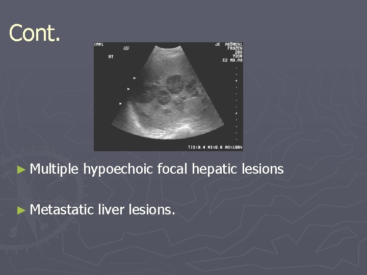
Case four ► Old man recently discovered to have colonic cancer presented to primary health care clinic with vague upper abdominal pain ► On exam: he was thin, ill not febrile or jaundiced. Mild abdominal tenderness enlarged liver with irregular outline. ► Lab mildly elevated LFTs.
Cont. ► Multiple hypoechoic focal hepatic lesions ► Metastatic liver lesions.
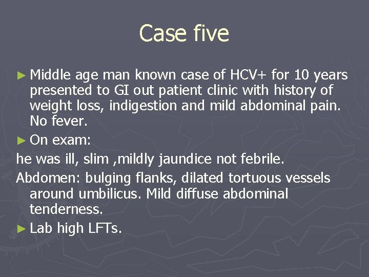
Case five ► Middle age man known case of HCV+ for 10 years presented to GI out patient clinic with history of weight loss, indigestion and mild abdominal pain. No fever. ► On exam: he was ill, slim , mildly jaundice not febrile. Abdomen: bulging flanks, dilated tortuous vessels around umbilicus. Mild diffuse abdominal tenderness. ► Lab high LFTs.
Cont. ► Shrunken liver with irregular outline. ► Heterogeneous appearance. ► Focal hypoechoic lesion. ► Cirrhotic liver with HCC.
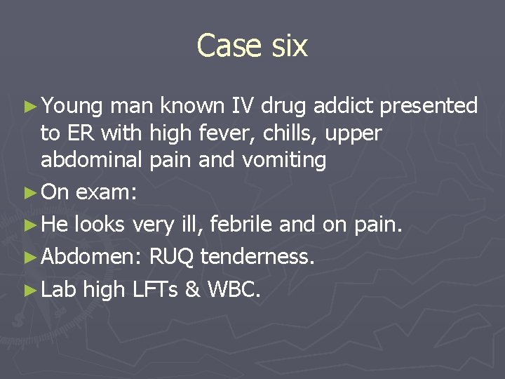
Case six ► Young man known IV drug addict presented to ER with high fever, chills, upper abdominal pain and vomiting ► On exam: ► He looks very ill, febrile and on pain. ► Abdomen: RUQ tenderness. ► Lab high LFTs & WBC.
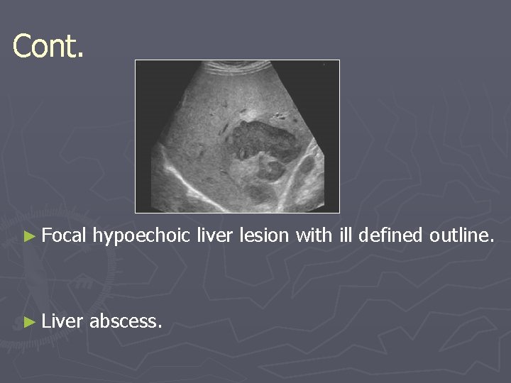
Cont. ► Focal hypoechoic liver lesion with ill defined outline. ► Liver abscess.