
Lackawanna College Diagnostic Medical Sonography Program Abdominal Sonography

Lackawanna College Diagnostic Medical Sonography Program Abdominal Sonography Liver Part I

Liver n n n Largest organ in the body Intraperitoneal structure Its parenchyma is used to evaluate other organs Occupies most of the right hypochondrium; part of the epigastrium; and extends into the left hypochondrium Its size and shape varies with body habitus, imprint of surrounding structures, and disease

Liver n Approximate diameters of the liver: ¨ Transverse- 20. 0 cm-22. 5 cm ¨ AP- 10. 0 cm-12. 5 cm ¨ Right lobe length- 15. 0 cm-17. 0 cm
Liver Surface The liver is covered by the Capsule of Glisson n Covered by the peritoneum except for a small portion called the ”bare area” n ¨ Bare Area- located to the right of the IVC and superior to the right kidney and adrenal gland ¨ Morrison’s Pouch- subhepatic space ¨ Subphrenic Space- subdiaphragmatic

SUPERIOR VIEW ANTERIOR VIEW Diaphragm Coronary ligament Falciform ligament Left triangular ligament Right triangular ligament Fundus of the gallbladder Left lobe Right lobe Left lobe Diaphragmatic surface Right lobe Falciform ligament Central surface Coronary ligament Ligamentum teres Gallbladder Inferior margin Left triangular ligament Caudete lobe IVC BARE AREA

RLL, in this diagram, is the right lower lobe of lung The bare area (BA) sign is demonstrated on this transverse sonogram, which shows pleural fluid posterior to the bare area of the liver. Ascites would not be present in this area because it is excluded from the peritoneum.

Perihepatic Relationships n n Diaphragm is seen as a dense echogenic line adjacent to the right lateral and cephalic aspect of the liver Liver is attached to the anterior abdominal wall, the diaphragm, retroperitoneum, and the lesser curvature of the stomach by seven ligaments Coronary, Falciform, Round(Teres Ligamentum), Right Triangular (lateral), Left Triangular (lateral), Gastrohepatic, and Hepatoduodenal
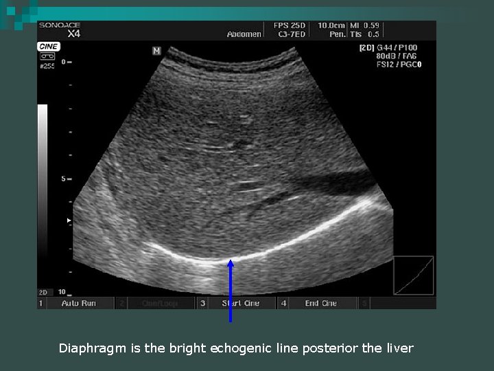
Diaphragm is the bright echogenic line posterior the liver
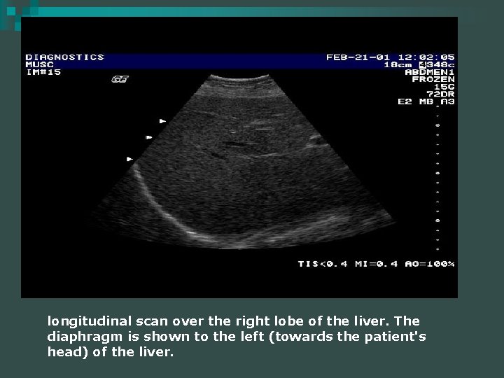
longitudinal scan over the right lobe of the liver. The diaphragm is shown to the left (towards the patient's head) of the liver.
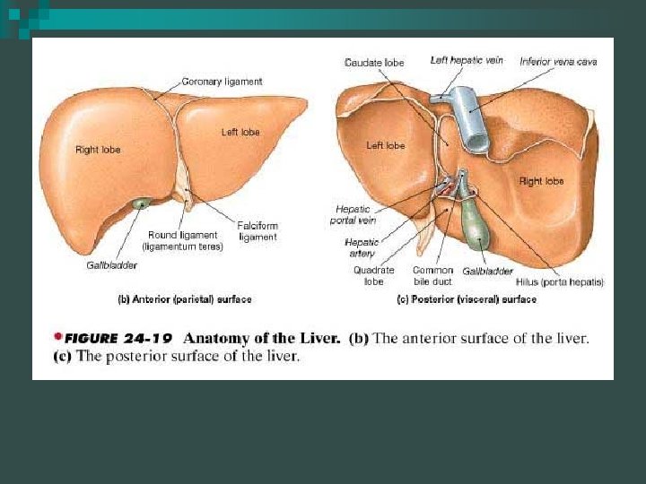
Liver Lobar Anatomy n Traditional anatomic system subdivides the liver into 4 lobes based on external landmarks ¨ Right ¨ Left ¨ Caudate ¨ Quadrate (actually the medial LLL)
Liver n Right Lobe (RLL) ¨ 6 times larger than the left ¨ Separated from the left lobe by the Middle Hepatic Vein and the Main Lobar Fissure ¨ Three fossae along the posterior and visceral surfaces Porta n Gallbladder n IVC n
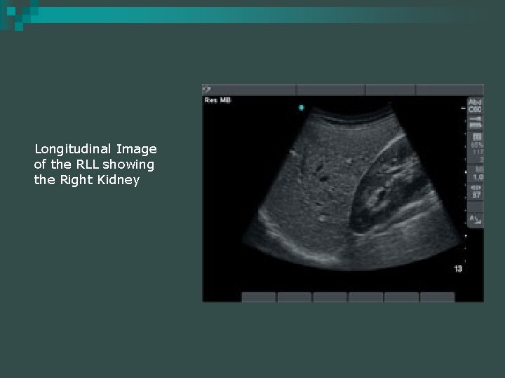
Longitudinal Image of the RLL showing the Right Kidney
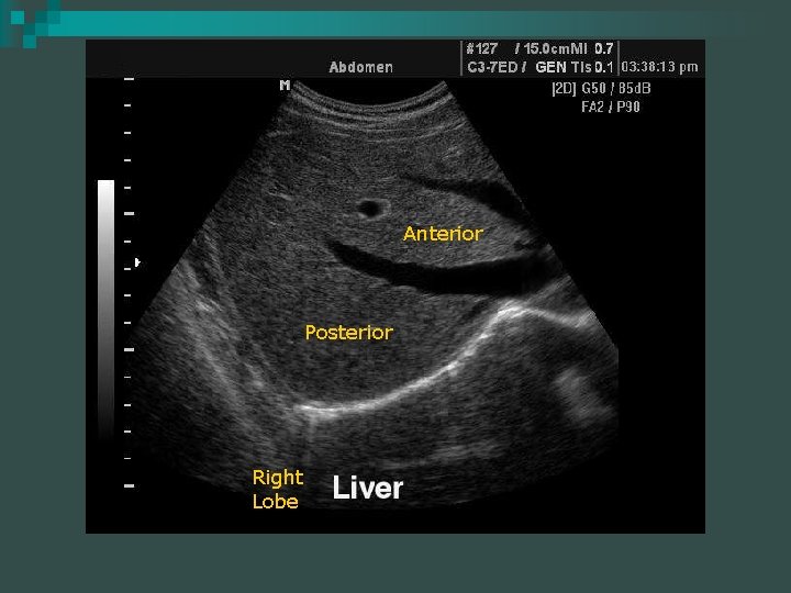
Anterior Posterior Right Lobe
Liver Lobar Anatomy n Left Lobe (LLL) ¨ Lies left of the falciform ligament ¨ Lies left of the ascending portion of the proximal left portal vein n Quadrate Lobe (medial aspect of the LLL) ¨ Lies along the visceral surface of the liver ¨ Anterior to the porta hepatis ¨ Lies between the GB and the falciform ligament fissure for the ligamentum teres
Liver Lobar Anatomy n Caudate Lobe ¨ Lies on the Posterior-Superior surface of the liver between the IVC and Medial LLL ¨ Lies anterior and medial to the IVC ¨ Posterior to the ligamentum venosum ¨ Superior to the Main Portal Vein
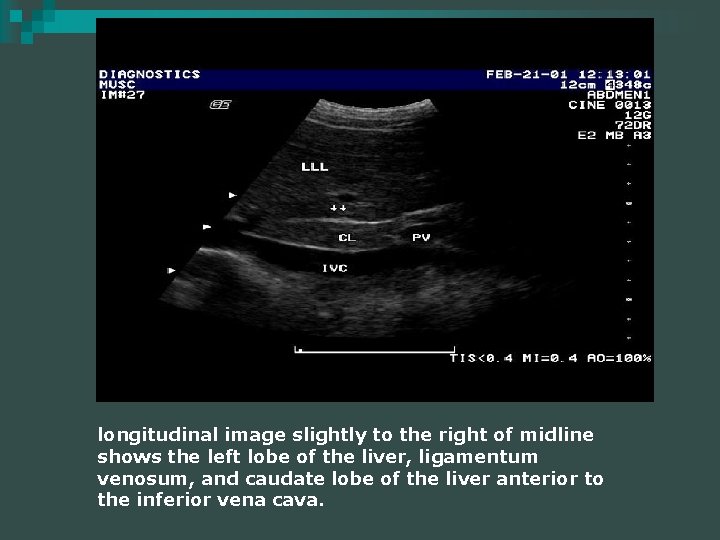
longitudinal image slightly to the right of midline shows the left lobe of the liver, ligamentum venosum, and caudate lobe of the liver anterior to the inferior vena cava.
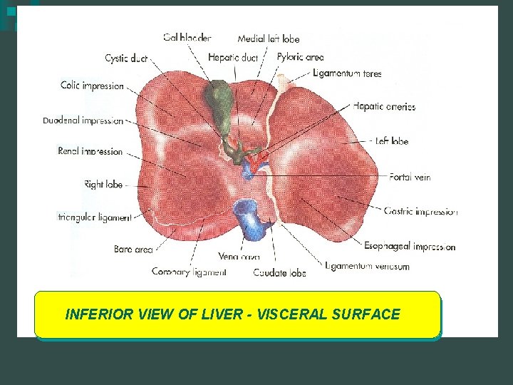
INFERIOR VIEW OF LIVER - VISCERAL SURFACE
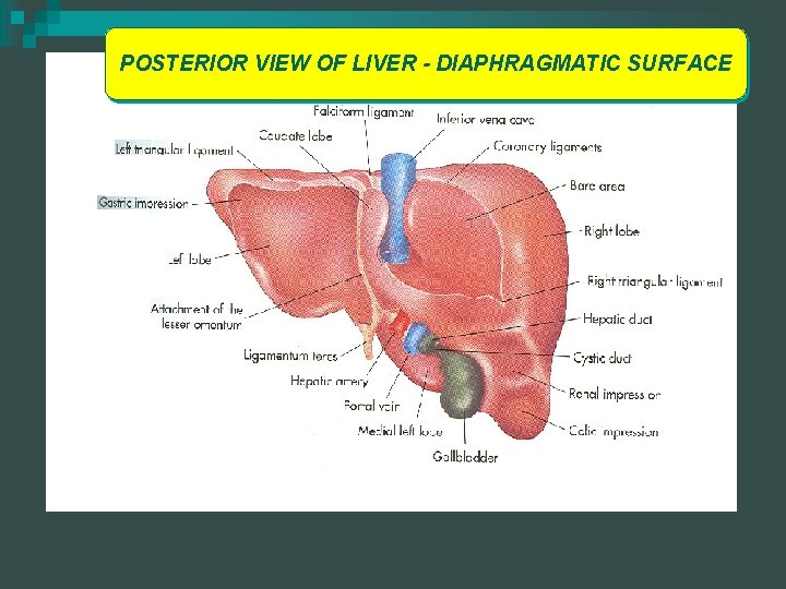
POSTERIOR VIEW OF LIVER - DIAPHRAGMATIC SURFACE
Functional Lobar Anatomy – Segmental Anatomy n n Functional System divides the liver into the right, left and caudate lobes based on vascular supply Divisional landmarks can be seen on ultrasound ¨ Hepatic Veins course between lobes (interlobar) and between segments (intersegmental) ¨ Portal Veins course within the segments except for the proximal left portal vein which is interlobar
Functional Lobar Anatomy – Segmental Anatomy n Right Lobe- bulk of hepatic tissue ¨ Divided from the left lobe by the main lobar fissure ¨ Separated superiorly from the medial segment of the LLL by the middle hepatic vein ¨ Inferiorly from the LLL by a plane running between the GB fossa and the IVC within the Main Lobar Fissure
Functional Lobar Anatomy – Segmental Anatomy ¨ Subdivided into anterior and posterior segments These segments are divided by the right intersegmental fissure in which the right hepatic veins runs through n Anterior branch of the Right Portal Vein within the anterior segment of the Right Lobe of the Liver n Posterior branch of the Right Portal Vein courses within the posterior segment of the RLL n
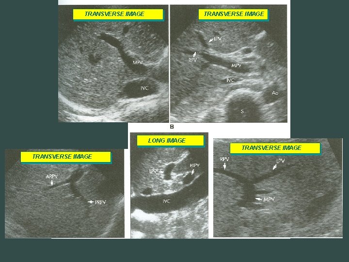
TRANSVERSE IMAGE LONG IMAGE TRANSVERSE IMAGE
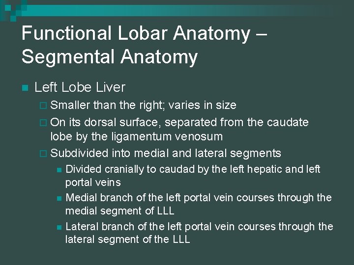
Functional Lobar Anatomy – Segmental Anatomy n Left Lobe Liver ¨ Smaller than the right; varies in size ¨ On its dorsal surface, separated from the caudate lobe by the ligamentum venosum ¨ Subdivided into medial and lateral segments n n n Divided cranially to caudad by the left hepatic and left portal veins Medial branch of the left portal vein courses through the medial segment of LLL Lateral branch of the left portal vein courses through the lateral segment of the LLL
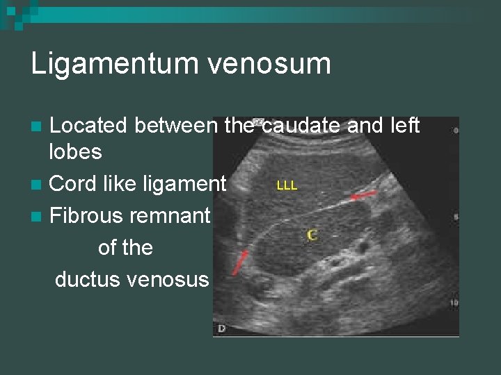
Ligamentum venosum Located between the caudate and left lobes LLL n Cord like ligament n Fibrous remnant of the ductus venosus n
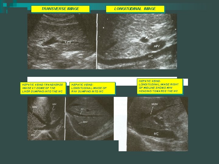
TRANSVERSE IMAGE HEPATIC VEINS-TRANSVERSE IMAGE AT DOME OF THE LIVER DUMPING INTO THE IVC HEPATIC VEINSLONGITUDINAL IMAGE OF RHV DUMPING INTO IVC LONGITUDINAL IMAGE HEPATIC VEINSLONGITUDINAL IMAGE RIGHT OF MIDLINE SHOWS MHV HEADING TOWARDS THE IVC
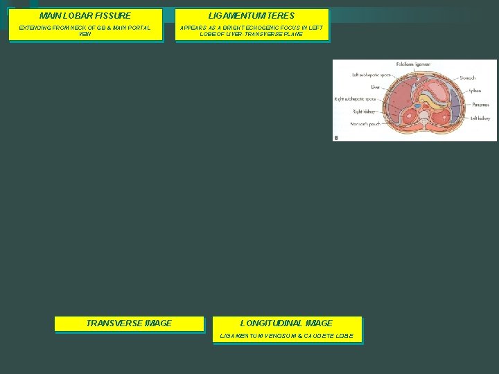
MAIN LOBAR FISSURE LIGAMENTUM TERES EXTENDING FROM NECK OF GB & MAIN PORTAL VEIN APPEARS AS A BRIGHT ECHOGENIC FOCUS IN LEFT LOBE OF LIVER-TRANSVERSE PLANE TRANSVERSE IMAGE LONGITUDINAL IMAGE LIGAMENTUM VENOSUM & CAUDETE LOBE
Functional Lobar Anatomy – Segmental Anatomy n Caudate Lobe ¨ Located posterior to the porta hepatis, between the ligamentum venosum and the IVC ¨ The left margin forms the hepatic boundary of the superior recess of the lesser sac ¨ Right margin extends in a tongue-like projection, the caudate process, between the IVC and Portal Vein
Liver: Ligaments and Fissures n Liver is attached to the anterior abd. wall, diaphragm, retroperitonieum, and the lesser curvature of the stomach by 7 ligaments ¨ Main Lobar ¨ Right and Left Segmental ¨ Falciform Ligament n Ligamentum Teres ¨ Ligamentum n Venosum Gastrohepatic ¨ Middle third or Left Intersegmental ¨ Coronary Ligament
Couinaud’s Anatomy Universal nomenclature for hepatic lesion localization n Divided into eight segments n Each segment has its own blood supply n
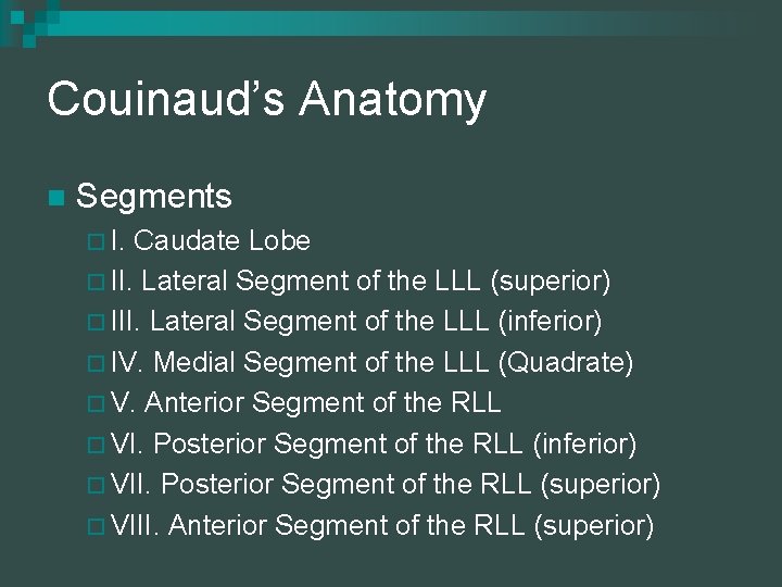
Couinaud’s Anatomy n Segments ¨ I. Caudate Lobe ¨ II. Lateral Segment of the LLL (superior) ¨ III. Lateral Segment of the LLL (inferior) ¨ IV. Medial Segment of the LLL (Quadrate) ¨ V. Anterior Segment of the RLL ¨ VI. Posterior Segment of the RLL (inferior) ¨ VII. Posterior Segment of the RLL (superior) ¨ VIII. Anterior Segment of the RLL (superior)
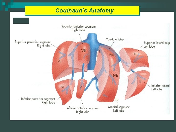
Couinaud’s Anatomy
Liver Vascular Anatomy n Dual blood supply ¨ Portal vein- supplies nutrients to the liver from the intestine; carries 70 -80% of oxygenated blood to the liver n n Main Portal Right and Left ¨ Hepatic Artery- supplies 20 -25% of blood to the liver (low resistant) The portal vein, common hepatic artery and common bile duct make up the portal triad
Portal Veins Interlobar; follow a transverse course n Collagenous connective sheaths n Hepatopedal with little or no pulsation n Normal caliper of the Main Portal Vein is at the level anterior to the IVC is 11. 0 mm (+/2 mm) at normal respiration n
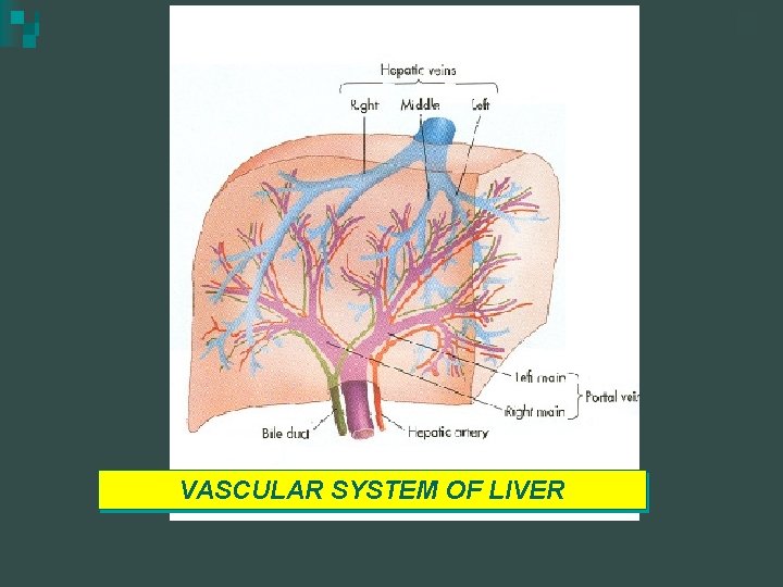
VASCULAR SYSTEM OF LIVER
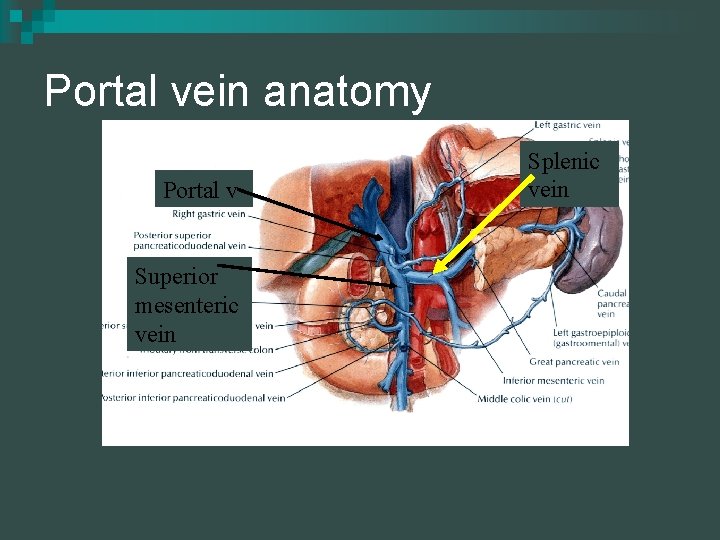
Portal vein anatomy Portal v Superior mesenteric v vein Splenic v vein
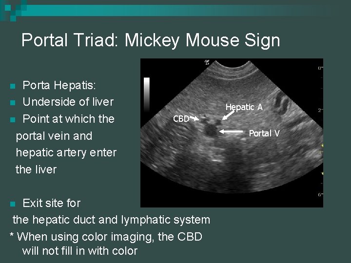
Portal Triad: Mickey Mouse Sign Porta Hepatis: n Underside of liver n Point at which the portal vein and hepatic artery enter the liver n Hepatic A CBD Exit site for the hepatic duct and lymphatic system * When using color imaging, the CBD will not fill in with color n Portal V
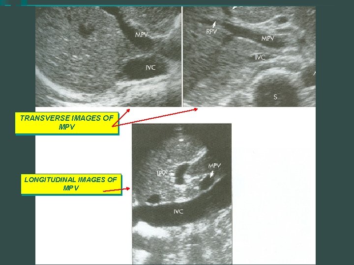
TRANSVERSE IMAGES OF MPV LONGITUDINAL IMAGES OF MPV
Portal Vein n Portal Vein further divides into Right and Left Branches ¨ Right Portal Vein n Anterior and Posterior Branches ¨ Left n Portal Vein Medial and Lateral Branches
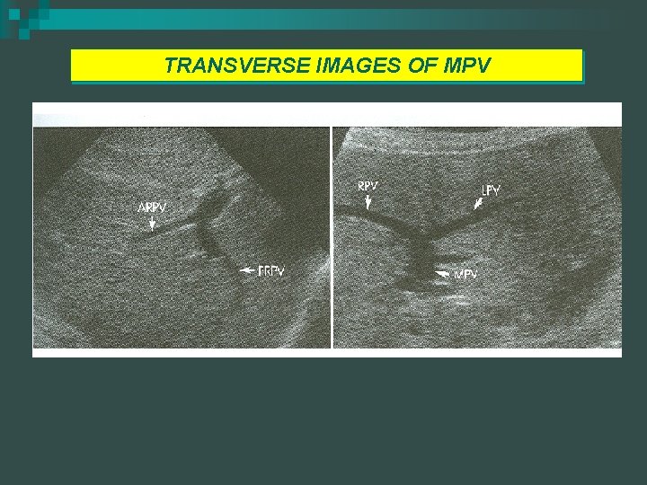
TRANSVERSE IMAGES OF MPV
Portal Vein - normal n n n Low velocity (20 -40 cm/s), continuous flow No filling defects with color Doppler. Hepatopetal direction (toward liver)
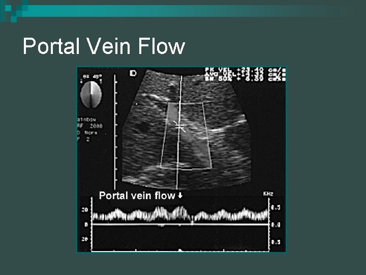
Portal Vein Flow
Hepatic Veins n Intersegmental and interlobar; follow a longitudinal course ¨ Right, n n Middle and Left Little or no collagen Caliper of the hepatic veins increase as they course towards the IVC Flow is dependent on the right atrium of the heart. Hepatofugal with pulsations
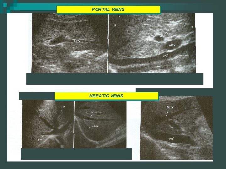
PORTAL VEINS HEPATIC VEINS
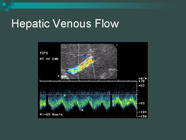
Hepatic Venous Flow

Normal HV Doppler

COMPARISON OF PV/HV
Anatomic Variants Reidel’s Lobe – right lobe extends below the lower pole of the right kidney towards the iliac crest (tongue-like projection) more common in women than men n Situs Inversus n Pseudofissures n
Microscopic Anatomy n n Consists of many functional and structural units called hepatic lobules Hepatic A and Portal Veins bring blood to the Hepatic Sinusoids ¨ n Within the sinusoids n They are lined with Kupffer cells which remove any debris, bacteria, and worn out red blood cells n Blood is then delivered into the Hep. vv. to the IVC Liver cells secrete bile into bile canaliculi which unite to form ductules, which in turn merge to form intrahepatic ducts
Liver Functions n Liver Functions ¨ Secretes bile aiding in digestion n Bile formation n Bile compostion n Bile function ¨ Removes nutrients from the blood ¨ Converts glucose to glycogen and stores it ¨ Stores iron and other important vitamins, A, D, E and K ¨ Cholesterol is a major constituent of bile and is manufactured in the liver ¨ Converts excess amnio acids to fatty acids and urea ¨ Metabolizes proteins, fats and carbohydrates ¨ Produces plasma proteins
Liver Functions n In addition: ¨ Detoxifies drugs and toxins ¨ Regulates blood clotting, including heparin production and regulation, fibrinogen, and prothrombin ¨ Activity of Reticuloendothelial Tissues n Blood production (hempoiesis) n Production of plasma proteins and antibodies n Destruction of red blood cells n Phagocytosis
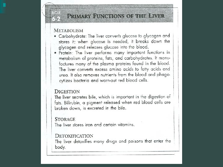
Liver Functions (continued) ¨ Metabolism of Carbohydrates, Lipids, and Proteinspreparatory use or storage n n n n Maintaining normal blood sugar Desaturation of fats and conversion to phosolipids Synthesis of amino acids Formation of serum albumin, serum globulin, antibodies, heparin, fibrogen, and prothrombin Conversion of protein to carbohydrate and fat Formation of urea and uric acid, destruction of uric acid Cholesterol from lipids
Liver Functions (continued) n Storage Depot ¨ Glycogen/Glucogon ¨ Amino Acids ¨ Fats ¨ Vitamins A, D and B ¨ Iron and Copper n n complex Blood Reservoir Detoxification ¨ Indol, Skatol, Phenol ¨ Morphine, Strychnine, Nicotine ¨ Steroids ¨ Bilirubin (uptake, conjugation, excretion)
Bile n n Excretory process of the Liver Formed continuously by the hepatocytes Bile is transferred to the GB and then to the intestine by the bile ducts Components of Bile ¨ Water, Bile Salts, Bile Pigments, Cholesterol, Lecithin, Protein n Bile emulsifies fat in the intestines
Lab Values for Liver Function Tests (LFT’s) 1. AST: Aspartate aminotransferase ¨ 2. ALT: Alanine aminotransferace ¨ 3. aka SGOT serum glutamic-oxaloacetic transaminase aka SGPT serum glutamic-pyruvic transaminase Bilirubin o o o Direct Indirect Elevated levels of bilirubin could lead to jaundice n Obstructive Jaundice n Non-Obstructive Jaundice
Lab Values for Liver Function Tests (LFT’s) 4. Serum Protein 5. ALP: Alkaline Phosphatase 6. LDH: Lactic Acid Dehydrogenase 7. PT: Prothrombin time 8. AFP: Alpha-fetal Protein 9. Albumin and Globulins 10. GGT: Gamma Glutamyl Transpeptidase
Labs n AST ¨ Formally known as SGOT ¨ Elevation of AST by itself is non-specific for liver disease ¨ Very Sensitive in almost all Hepatocellular Diseases. Will be Moderate to Severely Elevated in HCD ¨ Mild Increase in Biliary Disease n ALT ¨ Formally known as SGPT ¨ More specific for liver disease than AST ¨ Will be Moderate to Severely Elevated in HCD ¨ Mild Increase in Biliary Disease ¨ Elevated AST and LDH (lactic dehydrogenase) normal ALT rules out liver disease with
Labs n Bilirubin When bilirubin is high a condition called jaundice occurs ¨ Breakdown of hemoglobin in tired RBC ¨ 2 fraction of bilirubin is measured ¨ n n n Direct ¨ n Direct Indirect Reacts chemically in an aqueous medium and consisted of conjugated bile Indirect ¨ Inconjugated bile that is released from the reticularendothelial system and reacts in a non-aqueous medium
Labs n n Direct Bilirubin predominates in HCD and Biliary Obstruction. Usually related to obstructive jaundice from a stone in the biliary system or a neoplasm The liver may receive more bilirubin than it can detoxify. The byproducts are converted into bile pigments and if the cycle is disturbed ¨ Destruct RBC’s ¨ Malfunction of liver cells ¨ Blockage of the ducts n This disruption causes a rise in Bilirubin which leak into the tissues and causes Jaundice
LABS n Serum Albumin ¨ Decreased with HCD ¨ Normal with Biliary Obstruction n Alk Phos ¨ Minimum to Moderate with HCD ¨ Severe with Billiary Obstruction n AFP ¨ Marked Elevations occur with Hepatocelluar Carcinoma
LABS n GGT ¨ Elevation can indicate both HCD and Biliary Obstruction ¨ Rise in GGT and ALP is an indication of Biliary Disease
- Slides: 62