Chapter 16 Lecture Slides Copyright The Mc GrawHill


 formed in mouth and pushed into")


- Slides: 68
Chapter 16 Lecture Slides Copyright © The Mc. Graw-Hill Companies, Inc. Permission required for reproduction or display.
Functions 1. 2. 3. 4. 5. Take in food Break down food Absorb digested materials Provide nutrients Eliminate waste
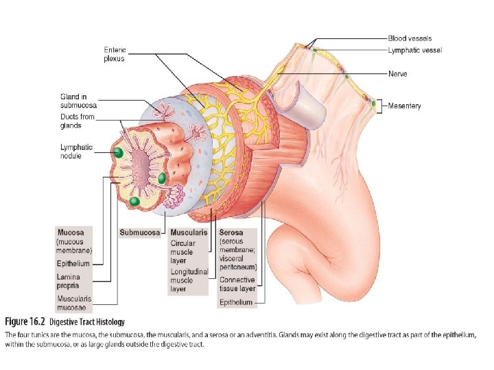
Layers of Digestive System • Digestive system is one large tube from mouth to anus plus the accessory organs 1. Mucosa: - innermost layer - secretes mucus 2. Submucosa: - above mucosa - contains blood vessels, nerves, small glands
3. Muscularis: - above submucosa - longitudinal, circular, and oblique muscles 4. Serosa/adventitia: - outermost layer - peritoneum is present called serosa - no peritoneum then called adventitia (Ex. Esophagus)
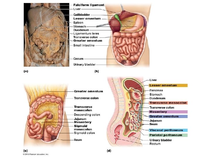
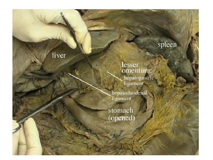
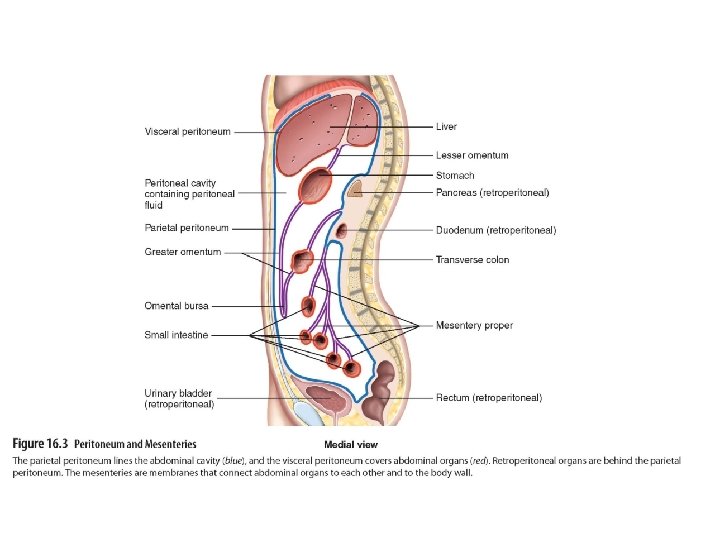
Peritoneum • Layer of smooth epithelial tissue • Mesenteries: connective tissue of organs in abdominal cavity • Lesser omentum: mesentery connecting lesser curvature of stomach to liver and diaphragm • Greater omentum: mesentery connecting greater curvature of stomach to transverse colon and posterior body wall
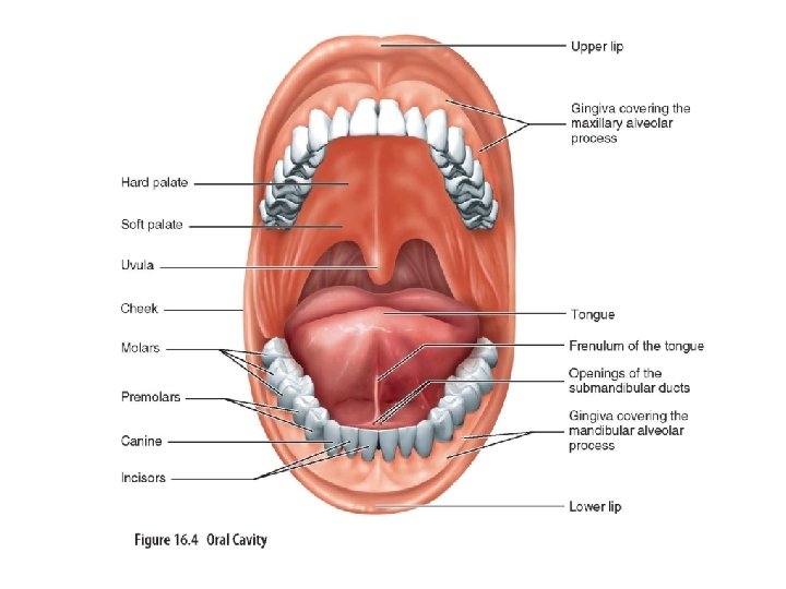
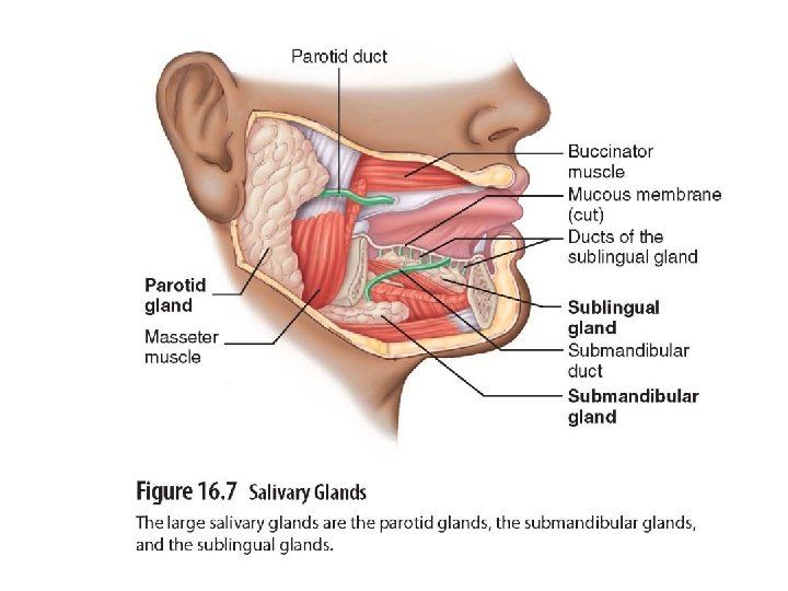
Oral Cavity • First part of digestive system • Contains stratified squamous epithelia • Salivary glands: - produce saliva which contains enzymes to breakdown carbohydrates into glucose - cleanse mouth - dissolve and moisten food
• Amylase: salivary enzyme that breaks down carbohydrates • Lysozyme: salivary enzymes that are active against bacteria • Tongue: house taste buds and mucus
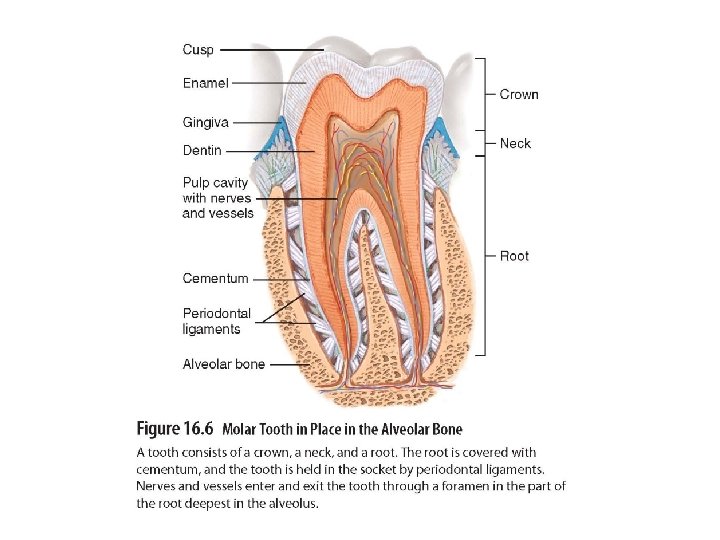
Teeth • • • 32 teeth in normal adult Incisors, canine, premolars, wisdom 20 primary teeth (baby teeth) Each tooth has crown, cusp, neck, root Center of tooth is pulp cavity Enamel is hard covering protects against abrasions • Cavities are breakdown of enamel by acids from bacteria
Figure 16. 5
Palate • Palate: roof of oral cavity • Hard palate: anterior part • Soft palate: posterior part
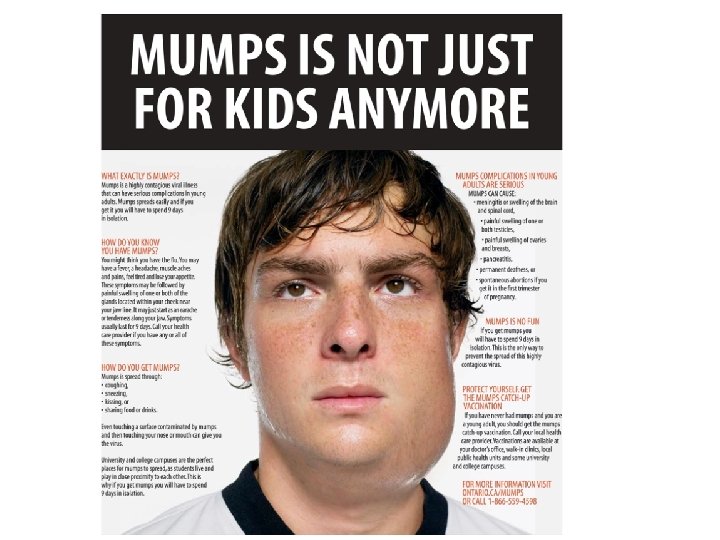
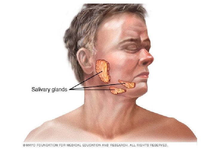
Salivary Glands • Salivary Glands: - includes submandibular, sublingual, parotid - produce saliva contains enzymes to breakdown food - mumps is inflammation of parotid gland
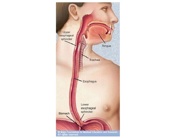
Pharynx • Throat • Connects mouth to esophagus
Esophagus • • Tube that connects pharynx to stomach Transport food to stomach Joins stomach at cardiac opening Heartburn: - occurs when gastric juices regurgitate into esophagus - caused by caffeine, smoking, or eating or drinking in excess
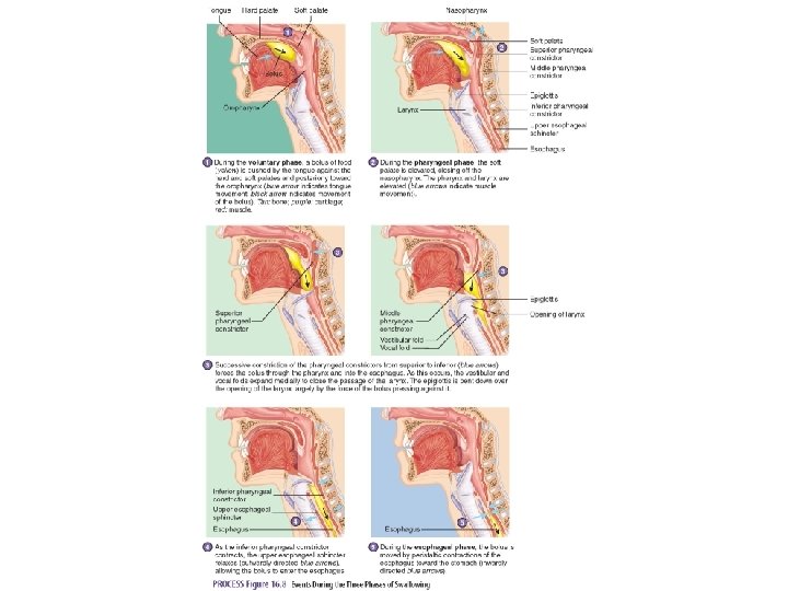
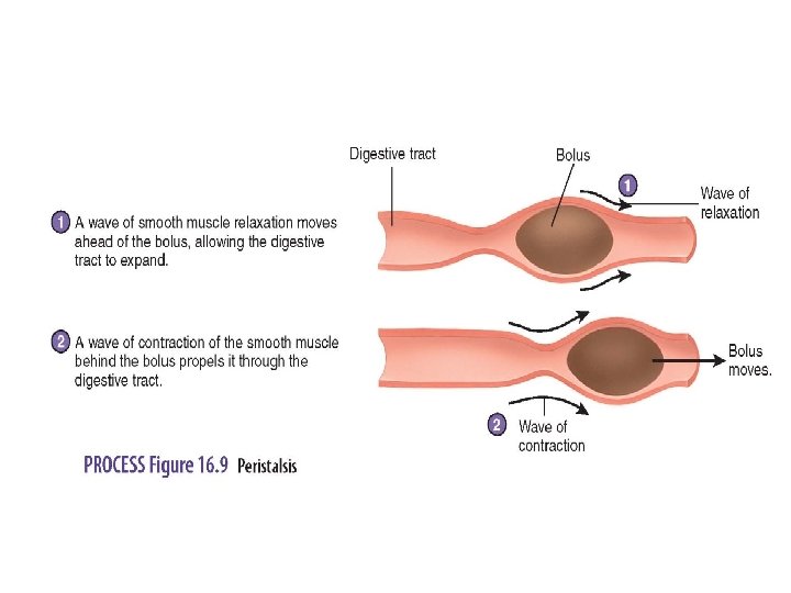
Swallowing • Voluntary phase: bolus (mass of food) formed in mouth and pushed into oropharynx • Pharyngeal phase: swallowing reflex initiated when bolus stimulates receptors in oropharynx • Esophageal phase: moves food from pharynx to stomach • Peristalsis: wave-like contractions moves food through digestive tract
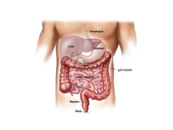
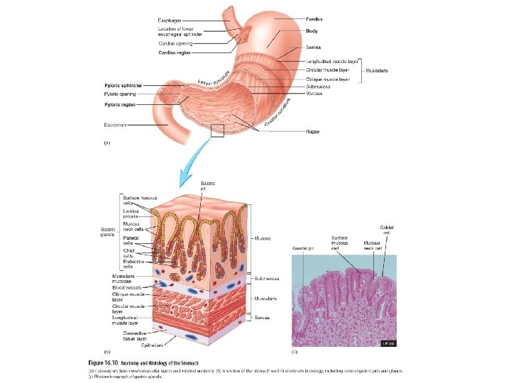
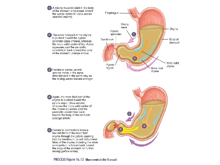
Stomach • • Located in abdomen Storage tank for food Can hold up to 2 liters of food Produces mucus, hydrochloric acid, protein digesting enzymes • Contains a thick mucus layer that lubricates and protects epithelial cells on stomach wall form acidic p. H (3)
• 3 muscular layers: outer longitudinal, middle circular, and inner oblique to produce churning action • Rugae: large folds that allow stomach to stretch • Chyme: paste-like substance that forms when food begins to be broken down
• Pyloric opening: opening between stomach and small intestine • Pyloric sphincter: thick, ring of smooth muscle around pyloric opening • Hunger pangs: stomach is stimulated to contract by low blood glucose levels usually 12 -24 hours after a meal
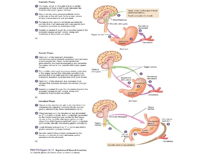
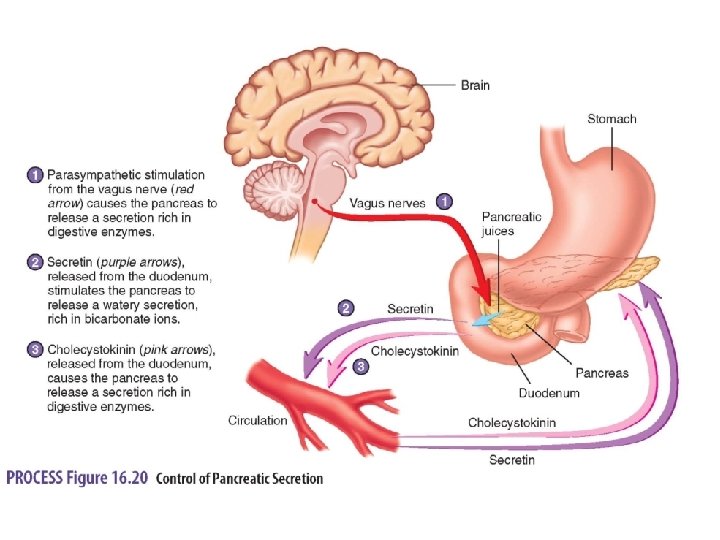
Regulation of Stomach Secretions • Parasympathetic stimulation, gastrin, histamine increase stomach secretions • Cephalic phase: - 1 st phase - stomach secretions are initiated by sight, smell, taste, or food thought
• Gastric phase: - 2 nd phase - partially digested proteins and distention of stomach promote secretion • Intestinal phase: - 3 rd phase - acidic chyme stimulates neuronal reflexes and secretions of hormones that inhibit gastric secretions by negative feedback loops
Movement in Stomach • Mixing waves: - weak contraction - thoroughly mix food to form chyme • Peristaltic waves: - stronger contraction - force chyme toward and through pyloric sphincter • Hormonal and neural mechanisms stimulate stomach secretions • Stomach empties every 4 hours after regular meal, and 6 -8 hours after high fatty meal
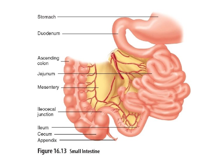
Small Intestine • • Measures 6 meters in length Major absorptive organ Chyme takes 3 -5 hours to pass through Contains enzymes to further breakdown food • Contains secretions for protection against chyme’s acidity
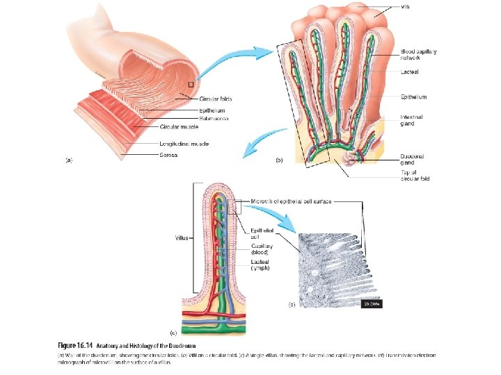
Parts of Small Intestine • Duodenum: - 25 cm long - contains absorptive cells, goblet cells, granular cells, endocrine cells - contains microvilli and many folds - contains bile and pancreatic ducts • Jejunum: 2. 5 meters long and absorbs nutrients • Ileum: 3. 5 meters long
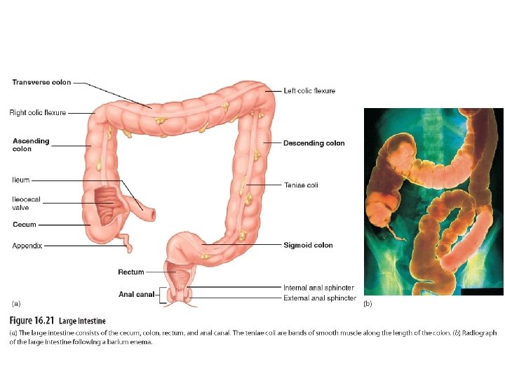
Large intestine • Function is to absorb water from indigestible food • Contains cecum, colon, rectum, anal canal • Cecum: - joins small intestine at ileocecal junction - has appendix attached • Appendix: 9 cm structure that is often removed
• Colon: - 1. 5 meters long - contains ascending, transverse, descending, sigmoid regions • Rectum: straight tube that begins at sigmoid and ends at anal canal
• Anal canal: last 2 -3 cm of dig. tract • Food takes 18 -24 hours to pass through • Feces is product of water, indigestible food, and microbes • Microbes synthesize vitamin K
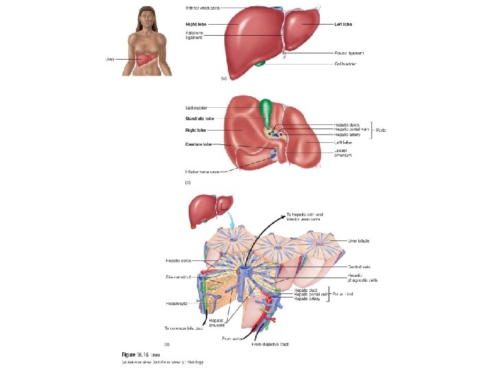
Liver Anatomy • Weighs about 3 lbs. • In right upper quadrant of abdomen under diaphragm • Right, left, caudate, quadrate lobes • Porta: gate where blood vessels, ducts, nerves enter and exit • Receives blood from hepatic artery
• Lobules: divisions of liver with portal triads at corners • Portal triad: contain hepatic artery, hepatic portal vein, hepatic duct • Hepatic cords: - between center margins of each lobule - separated by hepatic sinusoids
• Hepatic sinusoids: contain phagocytic cells that remove foreign particles from blood • Central vein: - center of each lobule - where mixed blood flows towards - forms hepatic veins
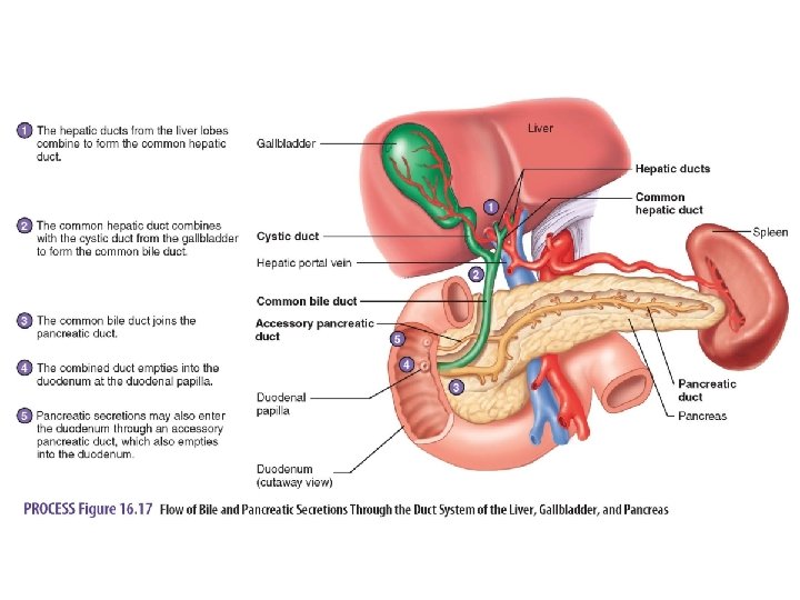
Liver Ducts • Hepatic duct: transport bile out of liver • Common hepatic duct: formed from left and right hepatic duct • Cystic duct: - joins common hepatic duct - from gallbladder • Common bile duct: formed from common hepatic duct and cystic duct
Gallbladder • Small sac on inferior surface of liver • Stores and concentrates bile
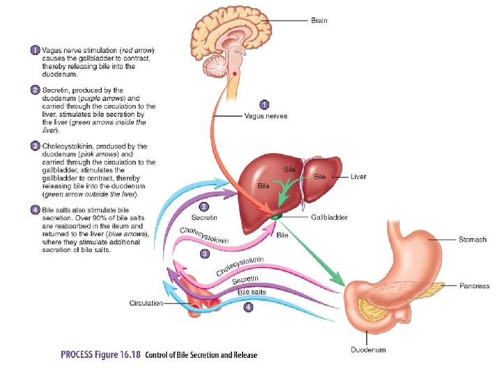
Functions of Liver • • • Digestive and excretory functions Stores and processes nutrients Detoxifies harmful chemicals Synthesizes new molecules Secretes 700 ml of bile each day Bile: dilutes and neutralizes stomach acid and breaks down fats
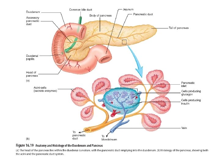
Pancreas • Located posterior to stomach in inferior part of left upper quadrant • Head near midline of body • Tail extends to left and touches spleen • Endocrine tissues have pancreatic islet that produce insulin and glucagon • Exocrine tissues produce digestive enzymes
Digestive Process 1. Digestion: breakdown of food occurs in stomach and mouth 2. Propulsion: moves food through digestive tract includes swallowing and peristalsis 3. Absorption: primarily in duodenum and jejunum of small intestine 4. Defecation: elimination of waste in the form of feces
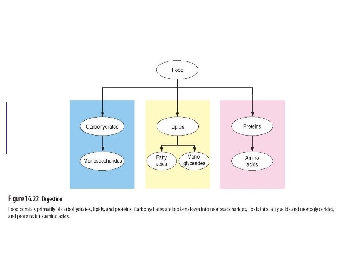
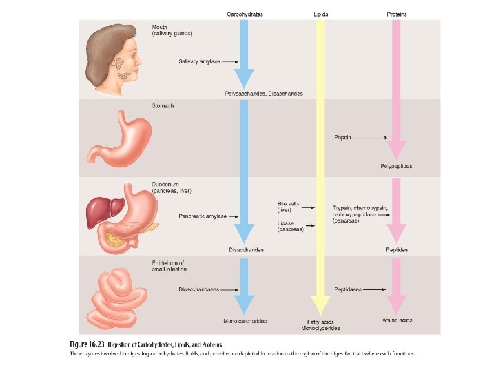
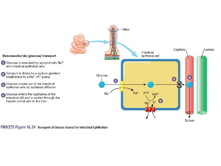
Carbohydrate Digestion • Polysaccharides split into disaccharides by salivary and pancreatic amylases • Disacchardies broken down into monosaccharides by disaccharidases on surface of intestinal epithelium • Glucose is absorbed by cotransport with Na+ into intestinal epithelium • Glucose is carried by hepatic portal vein to liver and enters most cells by facilitated diffusion
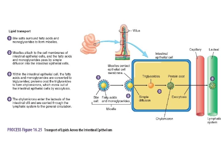
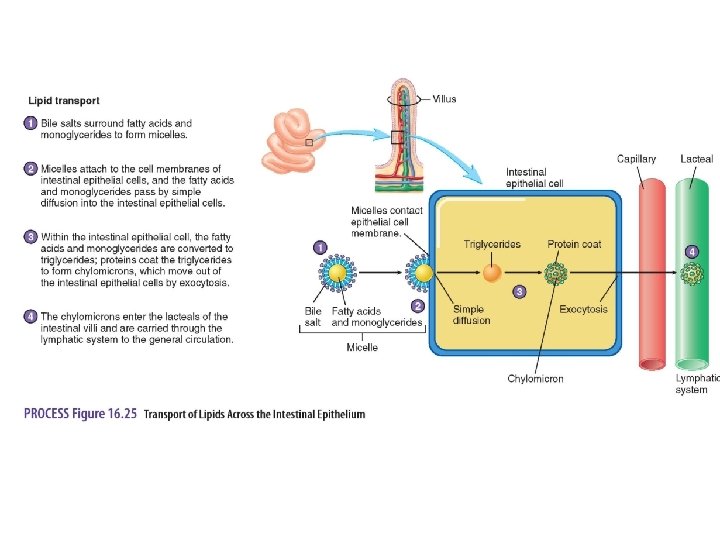
Lipid Digestion • Bile salts emulsify lipids • Lipase breaks down lipids which form micelles • Micelles are in contact with intestinal epi. and diffuse with cells where they are packaged and released into lacteals • Lipids are stored in adipose tissue and liver
Proteins Digestion • Proteins are split into polypeptides by enzymes secreted by stomach and pancreas • Peptides and amino acids are absorbed into intestinal epi. cells • Amino acids are actively transported into cells (help from GH and insulin) • Amino acids used to build new proteins
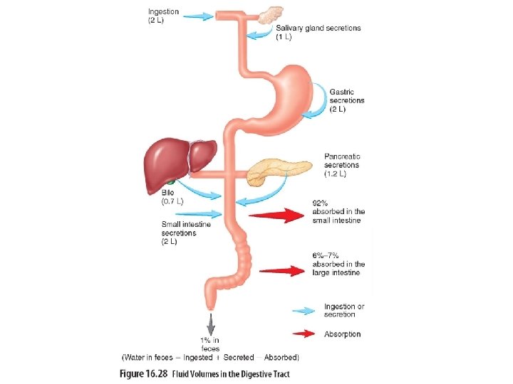
Water and Minerals • Water can move across intestinal wall in either direction • Depends on osmotic conditions • 99% of water entering intestine is absorbed • Minerals are actively transported across wall of small intestine