Myringoplasty Tympanoplasty Department of Otorhinolaryngoglogy the 2 nd

- Slides: 40
Myringoplasty Tympanoplasty Department of Otorhinolaryngoglogy the 2 nd Hospital affliatted to Medical college Zhejiang University Xu Yaping
overview Define terms Ø History Ø Anatomy Ø Preoperative evaluation Ø Techniques Ø Complications Ø Results Ø
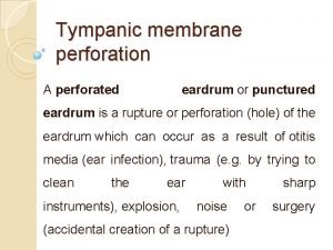
Definition Ø Myringoplasty and tympanoplasty are descriptive terms defining surgical procedures that address pathology of the tympanic membrane (TM) and middle ear.
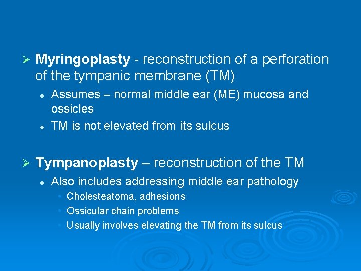
Ø Myringoplasty - reconstruction of a perforation of the tympanic membrane (TM) l l Ø Assumes – normal middle ear (ME) mucosa and ossicles TM is not elevated from its sulcus Tympanoplasty – reconstruction of the TM l Also includes addressing middle ear pathology • • • Cholesteatoma, adhesions Ossicular chain problems Usually involves elevating the TM from its sulcus
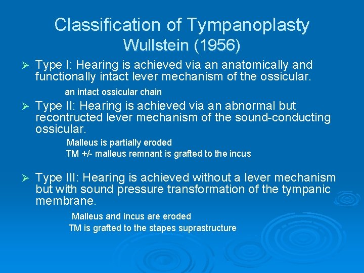
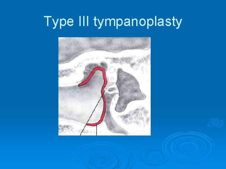
Classification of Tympanoplasty Wullstein (1956) Ø Type I: Hearing is achieved via an anatomically and functionally intact lever mechanism of the ossicular. an intact ossicular chain Ø Type II: Hearing is achieved via an abnormal but recontructed lever mechanism of the sound-conducting ossicular. Malleus is partially eroded TM +/- malleus remnant is grafted to the incus Ø Type III: Hearing is achieved without a lever mechanism but with sound pressure transformation of the tympanic membrane. Malleus and incus are eroded TM is grafted to the stapes suprastructure
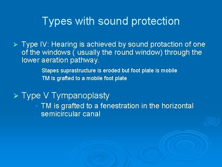
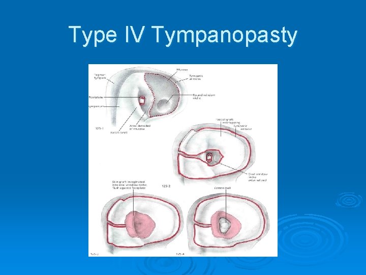
Types with sound protection Ø Type IV: Hearing is achieved by sound protaction of one of the windows ( usually the round window) through the lower aeration pathway. Stapes suprastructure is eroded but foot plate is mobile TM is grafted to a mobile foot plate Ø Type V Tympanoplasty • TM is grafted to a fenestration in the horizontal semicircular canal
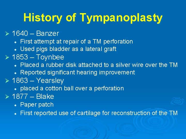
History of Tympanoplasty Ø 1640 – Banzer l l Ø 1853 – Toynbee l l Ø Placed a rubber disk attached to a silver wire over the TM Reported significant hearing improvement 1863 – Yearsley l Ø First attempt at repair of a TM perforation Used pigs bladder as a lateral graft placed a cotton ball over a perforation 1877 – Blake l l Paper patch First reported use of cartilage for reconstruction of the TM
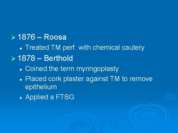
Ø 1876 – Roosa l Treated TM perf. with chemical cautery Ø 1878 – Berthold l l l Coined the term myringoplasty Placed cork plaster against TM to remove epithelium Applied a FTSG
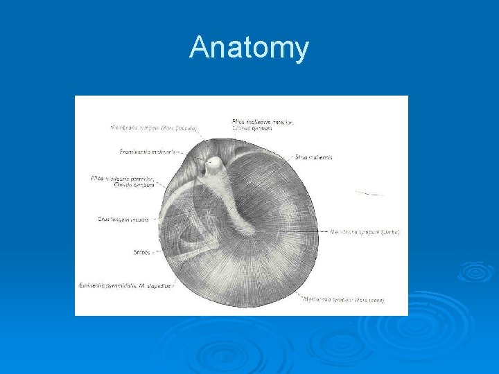
Anatomy
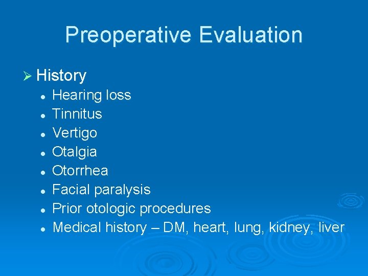
Preoperative Evaluation Ø History l l l l Hearing loss Tinnitus Vertigo Otalgia Otorrhea Facial paralysis Prior otologic procedures Medical history – DM, heart, lung, kidney, liver
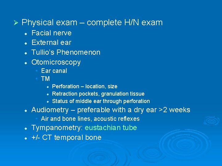
Ø Physical exam – complete H/N exam l l Facial nerve External ear Tullio’s Phenomenon Otomicroscopy • Ear canal • TM l l Perforation – location, size Retraction pockets, granulation tissue Status of middle ear through perforation Audiometry – preferable with a dry ear >2 weeks • Air and bone lines, acoustic reflexes l l Tympanometry: eustachian tube +/- CT temporal bone
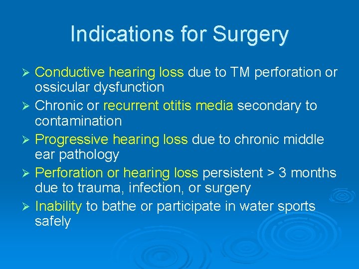
Indications for Surgery Conductive hearing loss due to TM perforation or ossicular dysfunction Ø Chronic or recurrent otitis media secondary to contamination Ø Progressive hearing loss due to chronic middle ear pathology Ø Perforation or hearing loss persistent > 3 months due to trauma, infection, or surgery Ø Inability to bathe or participate in water sports safely Ø
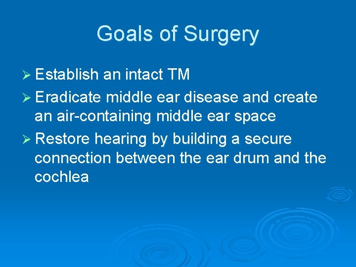
Goals of Surgery Ø Establish an intact TM Ø Eradicate middle ear disease and create an air-containing middle ear space Ø Restore hearing by building a secure connection between the ear drum and the cochlea
Ø Myringoplasty
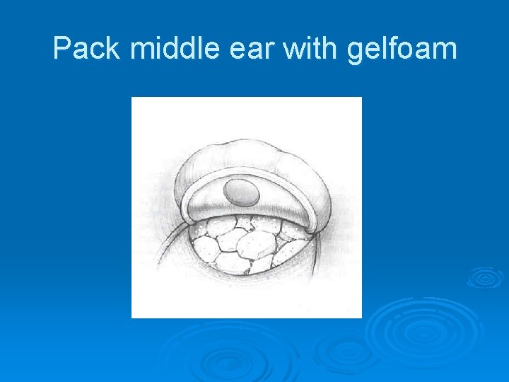
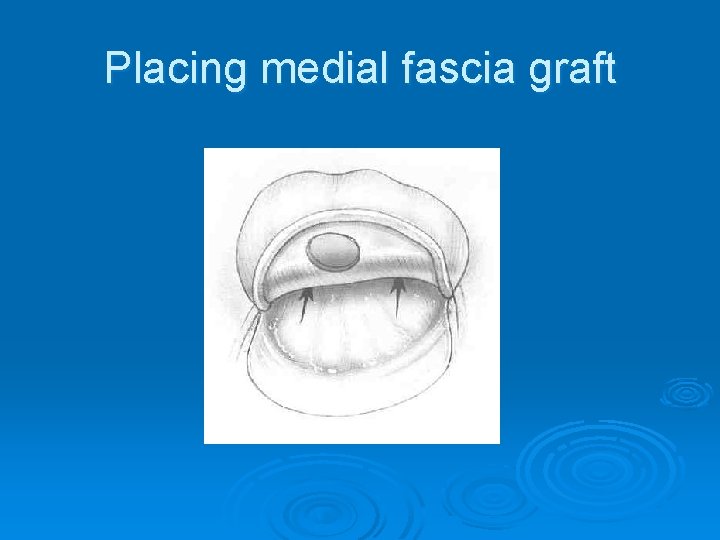
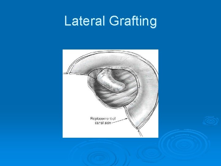
Techniques Ø Overlay technique (lateral grafting) Ø Underlay technique (medial grafting)
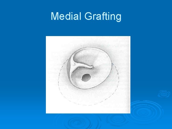
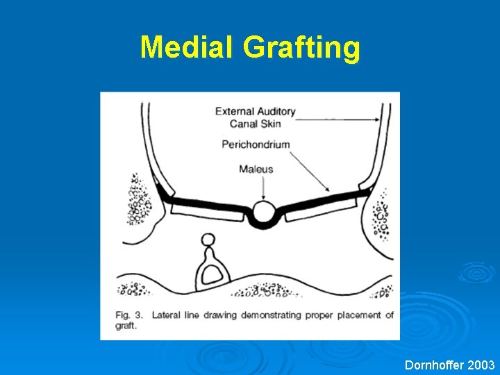
Medial Grafting
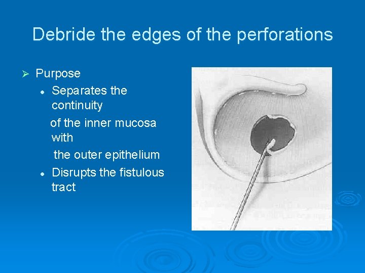
Debride the edges of the perforations Ø Purpose l Separates the continuity of the inner mucosa with the outer epithelium l Disrupts the fistulous tract
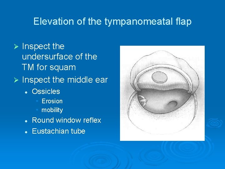
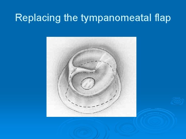
Elevation of the tympanomeatal flap Inspect the undersurface of the TM for squam Ø Inspect the middle ear Ø l Ossicles • Erosion • mobility l l Round window reflex Eustachian tube
Pack middle ear with gelfoam
Placing medial fascia graft
Replacing the tympanomeatal flap
Lateral Grafting
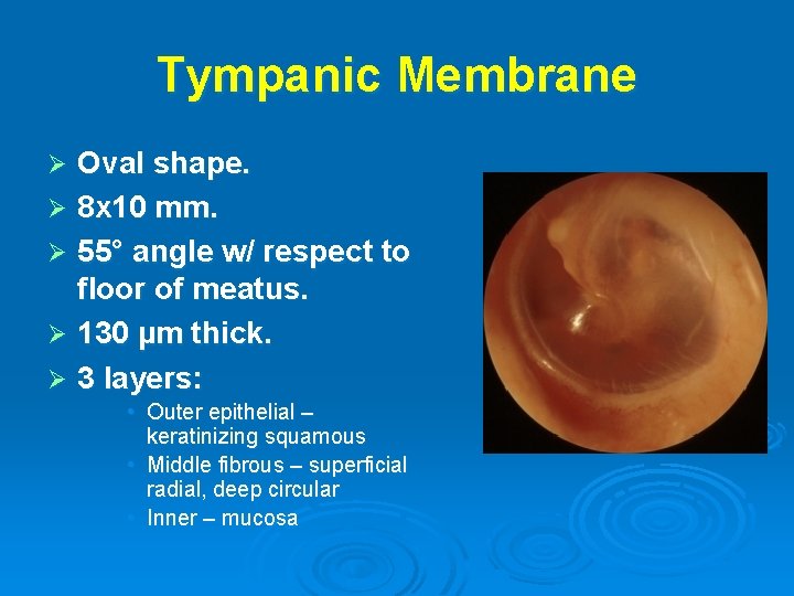
Tympanic Membrane Oval shape. Ø 8 x 10 mm. Ø 55° angle w/ respect to floor of meatus. Ø 130 µm thick. Ø 3 layers: Ø • Outer epithelial – keratinizing squamous • Middle fibrous – superficial radial, deep circular • Inner – mucosa
Graft Materials Ø Fascia Ø Perichondrium: tragal cartilage Ø Vein Ø Dura Ø Skin Ø Cartilage: tragal cartilage
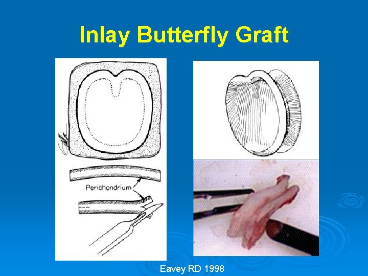
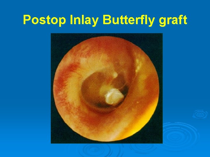
Inlay Butterfly Graft Eavey RD 1998
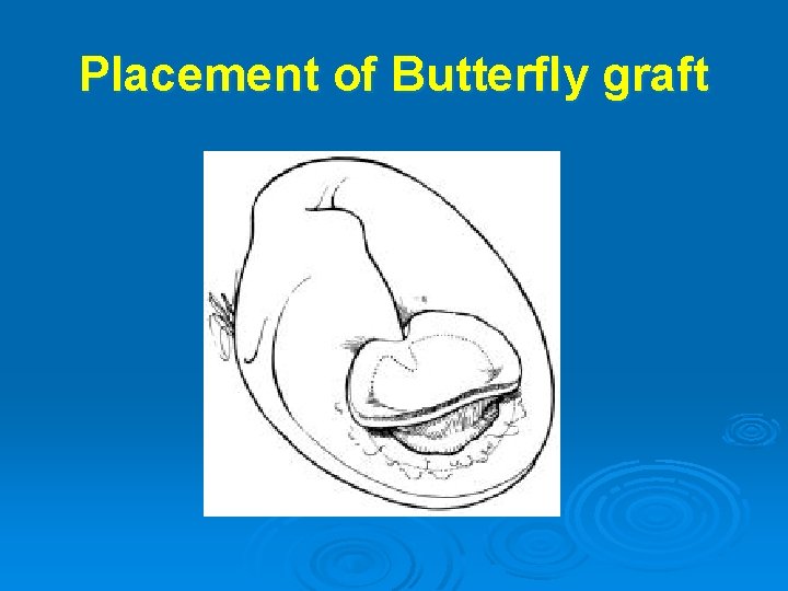
Placement of Butterfly graft
Postop Inlay Butterfly graft
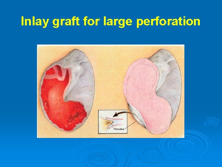
Inlay graft for large perforation
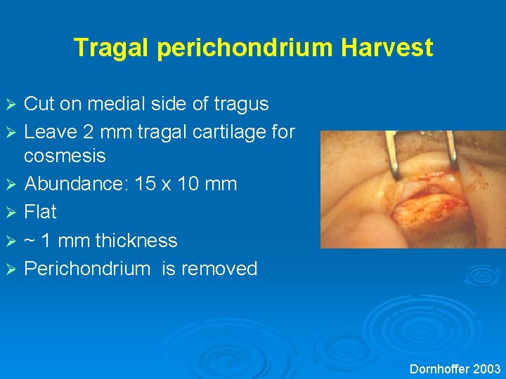
Tragal perichondrium Harvest Cut on medial side of tragus Ø Leave 2 mm tragal cartilage for cosmesis Ø Abundance: 15 x 10 mm Ø Flat Ø ~ 1 mm thickness Ø Perichondrium is removed Ø Dornhoffer 2003
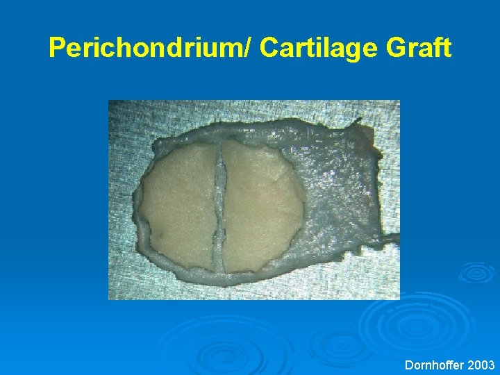
Perichondrium/ Cartilage Graft Dornhoffer 2003
Medial Grafting Dornhoffer 2003
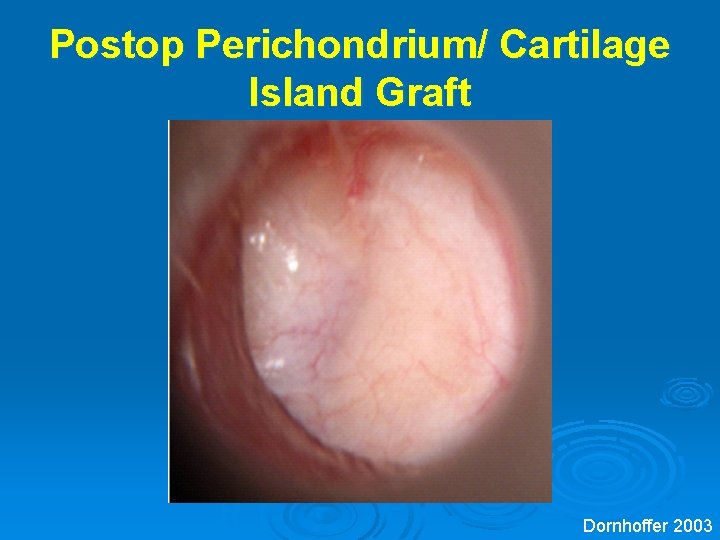
Postop Perichondrium/ Cartilage Island Graft Dornhoffer 2003
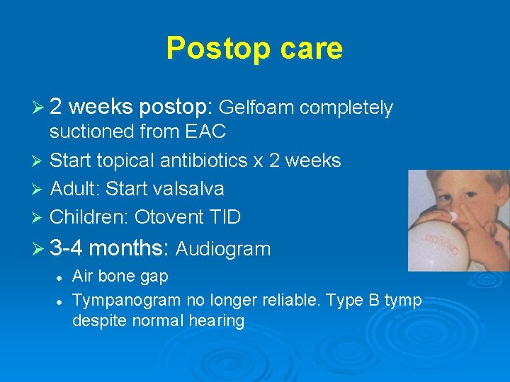
Postop care Ø 2 weeks postop: Gelfoam completely suctioned from EAC Ø Start topical antibiotics x 2 weeks Ø Adult: Start valsalva Ø Children: Otovent TID Ø 3 -4 months: Audiogram l l Air bone gap Tympanogram no longer reliable. Type B tymp despite normal hearing
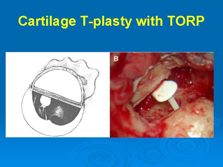
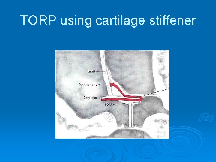
Cartilage T-plasty with TORP
Type III tympanoplasty
TORP using cartilage stiffener
Type IV Tympanopasty
Complications Ø Infection l l l Ø Poor aseptic technique Prior contamination Graft failure is associated with postop infection Graft failure l l l Infection Inadequate packing (anterior mesotympanum) Inadequate overlay of graft with TM remnant (underlay)
Chondritis Ø Injury to the chorda tympani nerve Ø SNHL and vertigo Ø l Ø Excessive manipulation of the ossicles Increased conductive hearing loss l l Unrecognized eroded ISJ Blunting • Thick graft extending onto the anterior canal wall in lateral grafting l Ø Lateralization of the TM from the malleus handle External auditory canal stenosis l Lateral grafting
Conclusion Ø A high rate of success in closing tympanic membrane perforations and improving hearing Ø Patients should be chosen carefully based on the indications for a dry ear prior to surgery Ø Patients should be thoroughly counseled preoperatively about the expectations and goals of the surgery Ø Tympanoplasty in the pediatric age group is controversial (less successful than adults, higher incidence of ETD -eustachian tube dysfunction and otitis media) Ø Both underlay and overlay techniques for grafting are effective, however, the surgeon should do what he/she is most experienced and successful