Muscle tissue 1 Striated skeletal muscle tissue 2
 Striated skeletal muscle tissue. 2) Striated cardiac muscle tissue. 3) Smooth")

 myocyte (muscle cell) sarx/sarcós (meat): cell membrane = sarcolemma cytoplasm =")
 Perimysium – around")


 – elongated, cylindrical")


 < rhabdomyocyte > Used terms: Muscle fiber = myofiber =")

![Myofibril [Ø 0. 5 – 1. 5 ]: elongated structure in sarcoplasm, oriented paralelly](https://slidetodoc.com/presentation_image_h2/8af44b7828f988e878b736a6e24adf74/image-16.jpg "Myofibril [Ø 0. 5 – 1. 5 ]: elongated structure in sarcoplasm, oriented paralelly")
 – light, Z-line (telophragma) anizotropic parts (A-bands) – dark,")


 Light")
 via T-tubule (= invagination of sarcolemma) Change of")


 Nexus (quick")


 with myofilaments not arranged into myofibrils (no")


 Heart muscle (64, 65 –")


 organs Cross section")


 2.")


- Slides: 109
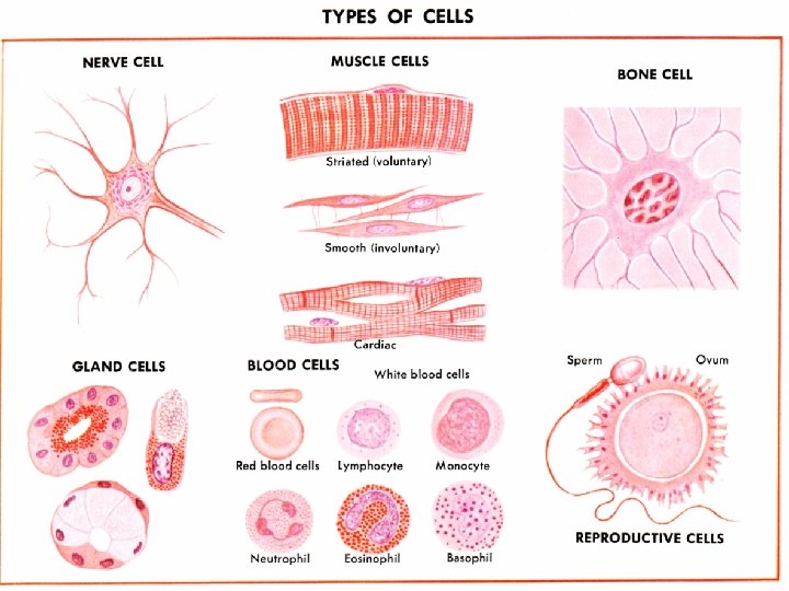
Muscle tissue 1) Striated skeletal muscle tissue. 2) Striated cardiac muscle tissue. 3) Smooth muscle tissue.
General characteristic of muscle tissue Origin: mesoderm and mesenchyme Excitability Contraction + relaxation cause movement Composition: muscle cells + connective tissue (+blood vessels + nerves) contractile proteins in sarcoplasm – actin and myosin Long axis of cells is usually oriented paralelly with direction of contraction
Nomenclature mys/myos (muscle) myocyte (muscle cell) sarx/sarcós (meat): cell membrane = sarcolemma cytoplasm = sarcoplasm smooth ER = sarcoplasmic reticulum
Connective tissue of muscle Endomysium – around each muscle cell (fiber) Perimysium – around among the primary bundles of muscle cells Epimysium – connective tissue „capsule“ covering the surface of muscle
Connective tissue in skeletal muscle contains vessels and nerves
rhabdomyocyte MUSCLE CELLS cardiomyocyte Ø 3 -10 µm Ø 15 µm Ø 25 -100 µm leiomyocyte
Occurrence: Skeletal muscles Heart Inner organs (their wall
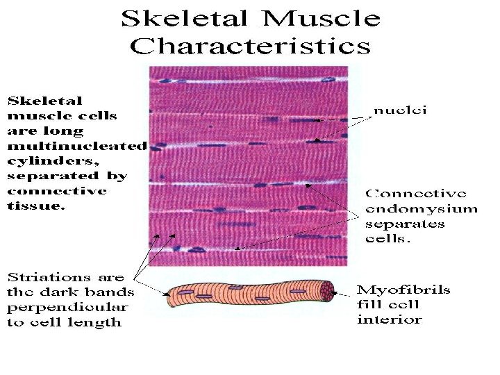
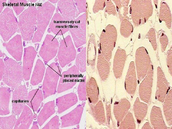
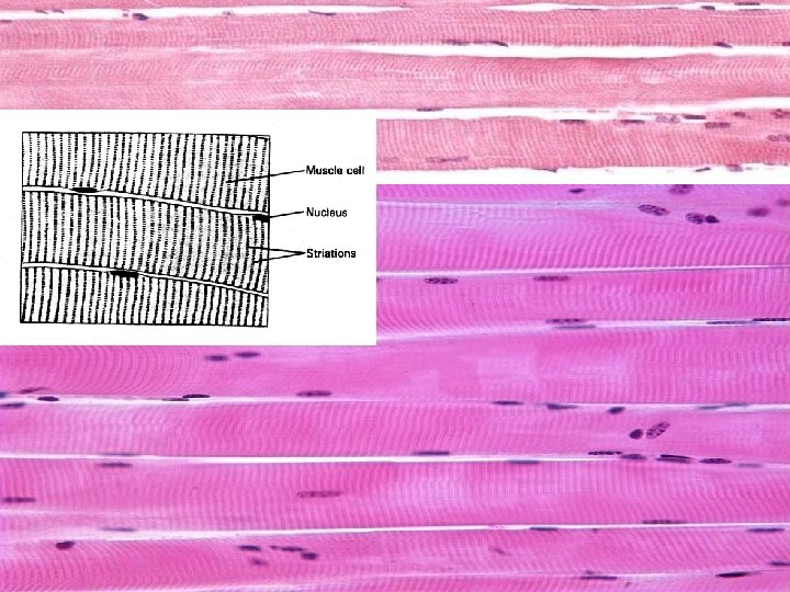
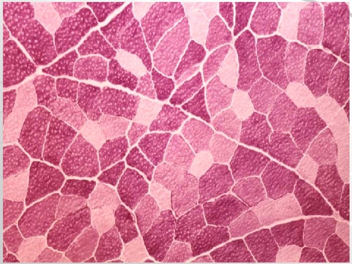
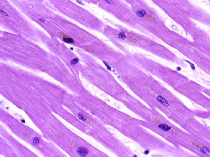
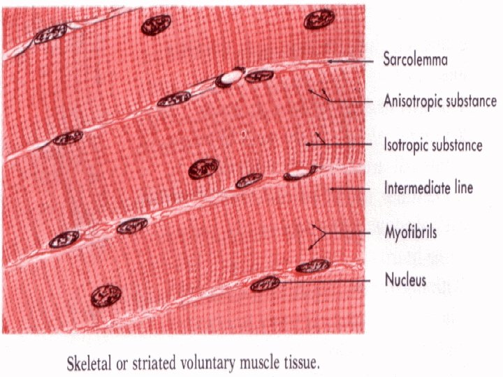
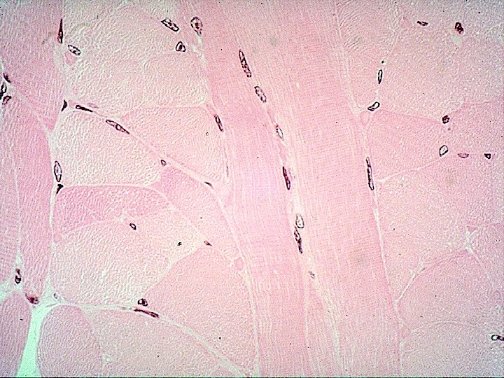
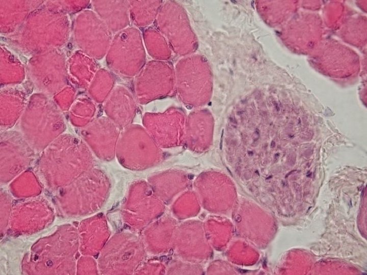
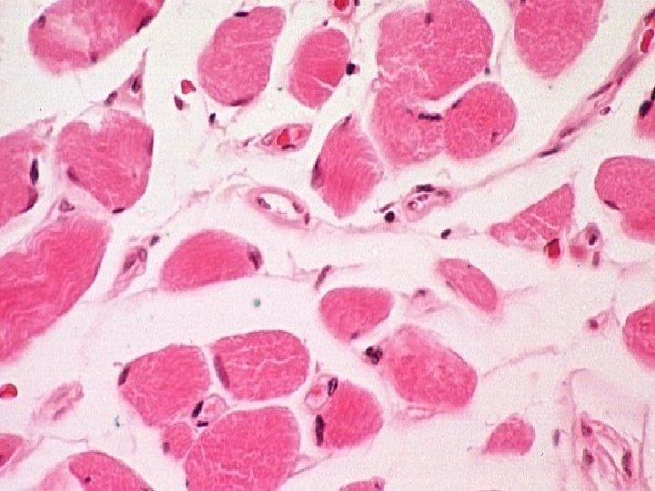
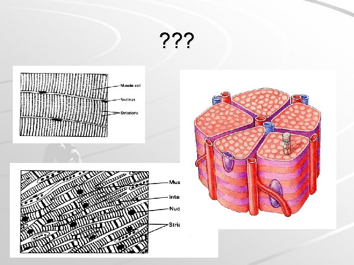
Cross-striated skeletal muscle tissue morphological and functional unit: muscle fiber (rhabdomyocyte) – elongated, cylindrical shape, multinucleated cell (=syncytium) – nuclei are located at the periphery (beneath sarcolemma), myofibrils show cross striation diameter of muscle fiber: 25 -100 m length: milimeters - centimeters (up 15)
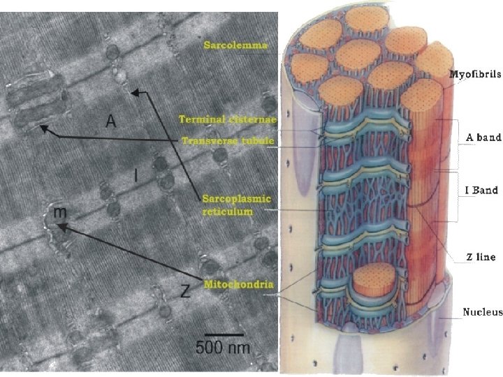
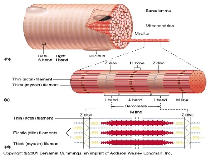
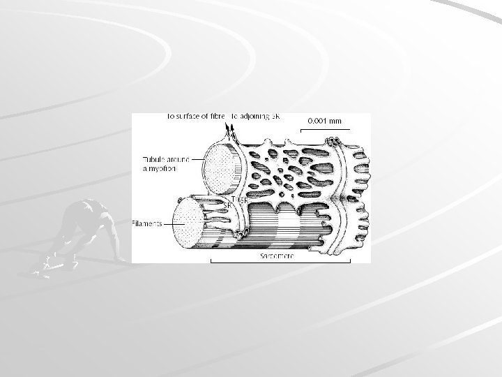
Structure of rhabdomyocyte sarcolemma + T-tubules nuclei (25 -40 per 1 mm of the length) sarcoplasm: Ø myoglobin (protein with heme (iron-containing porphyrin) prostetic group, and is the primary oxygen-carrying pigment Ø myofibrils (cross-striated 1– 2 µm thick fibrils) Ø organelles: mitochondria, Golgi apparatus, sarcoplasmic reticulum (Ca 2+ depot , sarcotubules, terminal cisternae) Ø inclusion (glycogen)
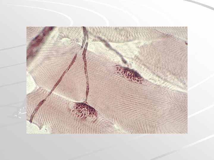
Rhabdomyocyte
Skeletal muscle cell (fiber) < rhabdomyocyte > Used terms: Muscle fiber = myofiber = syncitium = rhabdomyocyte Muscle fiber – morphologic and functional unit of skeletal muscle [Ø 25 – 100 ] Myofibrils – compartment of fiber sarcoplasm [Ø 0. 5 – 1. 5 ] Myofilaments – actin and myosin, are organized into sarcomeres (several in the length of myofibril) [Ø 8 and 15 nm] Sarcomere – the smallest contractile unit [2. 5 m in length]
Bundles of muscle fibrers Muscle fiber – morphologic and functional unit of skeletal muscle [Ø 25 – 100 ] Myofibrils – compartment of fiber sarcoplasm [Ø 0. 5 – 1. 5 ] Myofilaments – actin++myosin are organized into sarcomeres (several in the length of myofibril) [Ø 8 and 15 nm]
Myofibril [Ø 0. 5 – 1. 5 ]: elongated structure in sarcoplasm, oriented paralelly to the length of cell, composed of 2 types of myofilaments – actin and myosin – arranged into: Sarcomere: the smallest contractile unit between Z-lines on myofibril (organization of myofilaments causes cross striation of myofibrils) Z-line
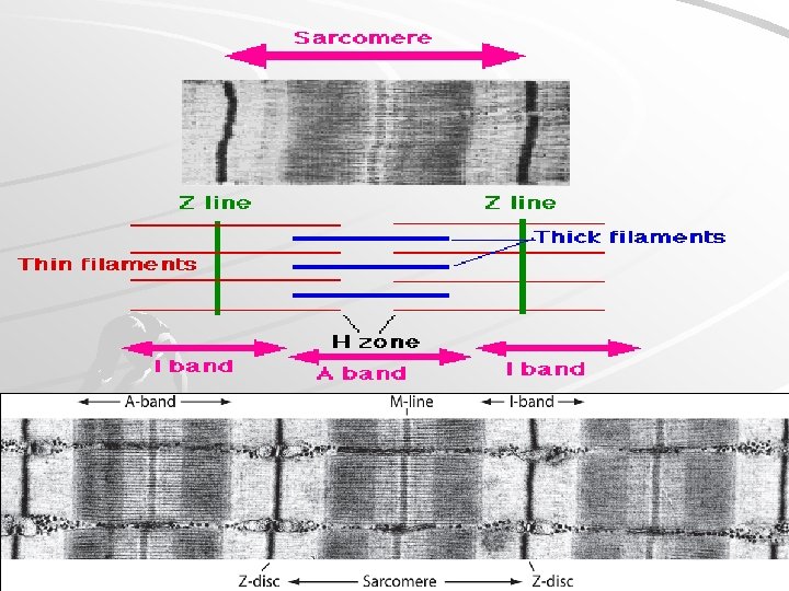
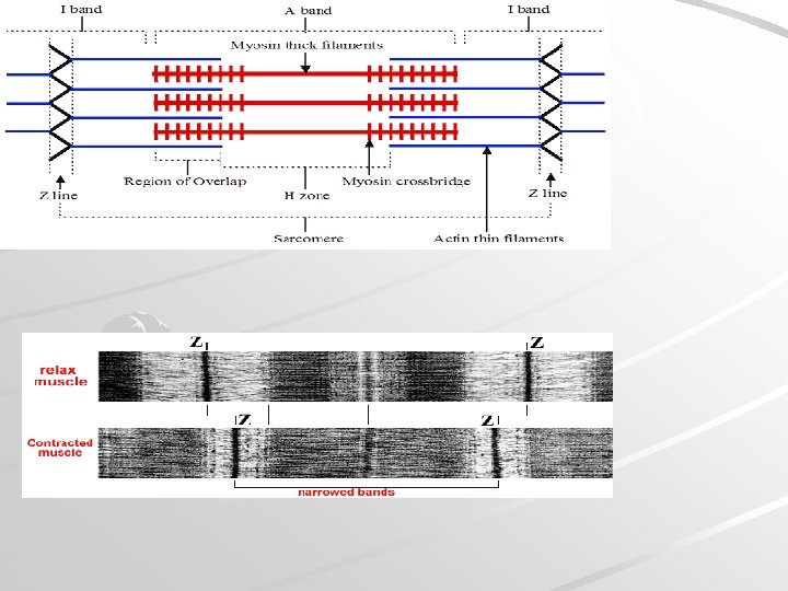
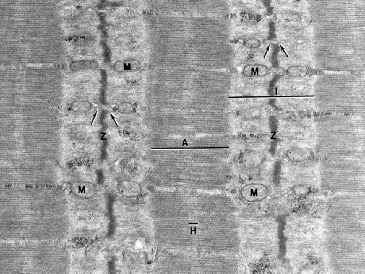
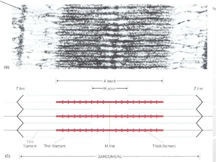
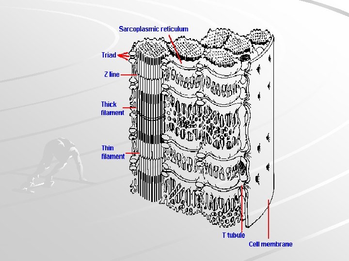
Myofibril structure izotropic parts (I-bands) – light, Z-line (telophragma) anizotropic parts (A-bands) – dark, M-line (mesophragma), H-band Myofilaments Thick: only in A-band, myosin, 15 nm thick, 1. 6– 1. 8 µm long Thin: in I-band at periphery of A-band), F-actin and regulatory proteins (troponin, tropomyosin), 6 nm thick, 1. 5 µm long Principle of myofibril shortening during muscle contraction: sliding mechanismu the length of myofilaments does not change during contraction
SARCOMERE MYOFIBRIL Z – line M – line Z – line H - zone Myofilaments: actin I - band actin – myosin – actin A – band actin I - band
Myofibrils - TEM sarcoplasm myofibrils sarcoplasm strips
Sarcomere H-zone I–band ½ I-band A–band
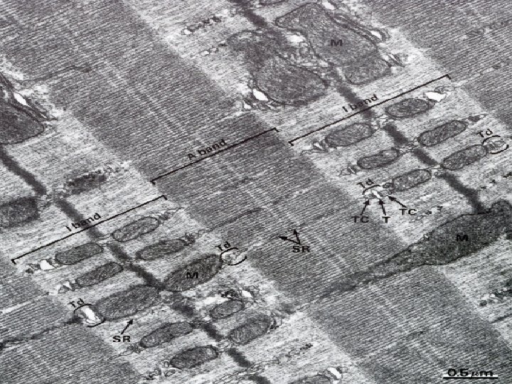
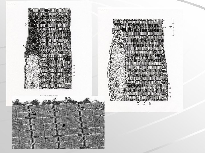
Skeletal muscle in EM
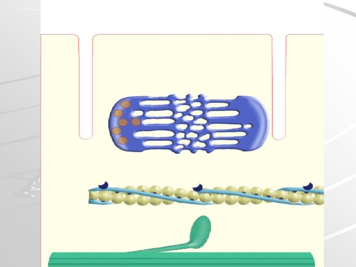
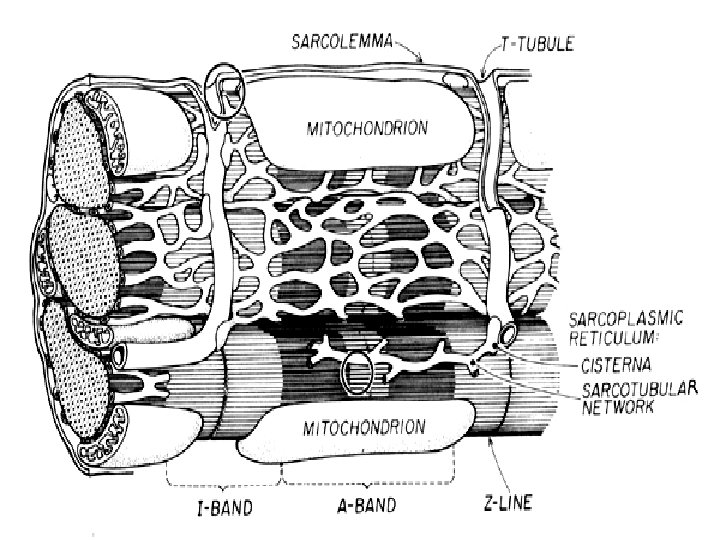
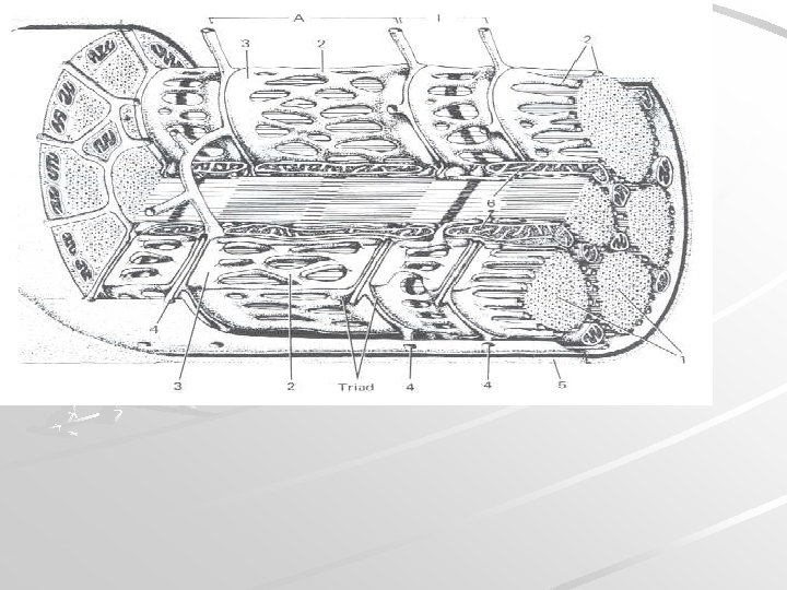
Sarcoplasmic reticulum, t-tubule Forms transversal terminal cisternae and longitudinal tubules. Function: reservoir of Ca ions Terminal cisterna T-tubule Terminal cisterna TRIAD T-tubules are invaginations of sarcoplasm and leads action potential to terminal cisternae (it changes permeability of membrane for Ca ions)
I-band A-band I-band
T-tubule terminal cisterna mitochondria sarcolemma myofibrils tubules + cisternae of s. ER
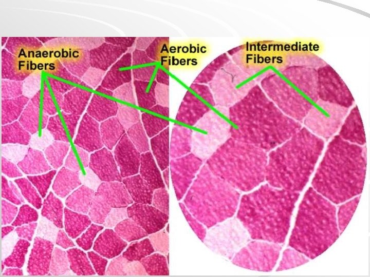
Characteristic of skeletal muscle fiber types Fibers: Red Intermediary White Colour Dark (red) Light (white) Content of myoglobin Number of mitochondria ATPase activity High Intermediate Low Many small Mi Many large Mi Little small Mi Low High Type of metabolism Contraction Oxidative Anaerobic Slow Aerobic and anaerobic Quick Defatigability Very small Small High Capilary density High Low Quick
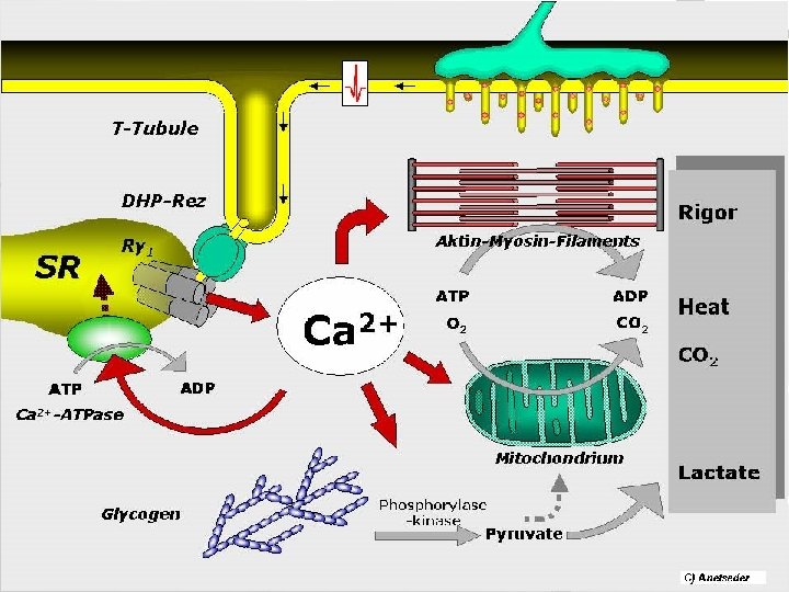
Contraction Propagation of action potential (depolarization) via T-tubule (= invagination of sarcolemma) Change of terminal cisternae permeability – releasing of Ca+ ions increases their concentration in sarcoplasm Myosin contacts actin and sarcomera shortens by sliding movement – contraction Relaxation: repolarization, decreasing of Ca 2+ ions concentration, inactivation of binding sites of actin for myosin
TRIAD Ca 2+ Calsequestrin is to Ca 2+ as haemoglobin is to O 2 The T-tubules have special Ca 2+ channels termed 'dihydropyridine receptors'. Ca 2+ Other Ca 2+ channels ('ryanodine receptors') in the membrane of the SR cisternae. Opening of these channels causes Ca 2+ to be dumped from the cisternae into the sarcoplasm.
Mechanism of muscle contraction During contraction the thick /M/ and thin /A/ slide past each other /sliding filament theory/ and the Z-discs are brought near the ends of the thick filaments.
Mechanism of muscle contraction Players:
Mechanism of contraction: sliding of myofilaments myosin actin
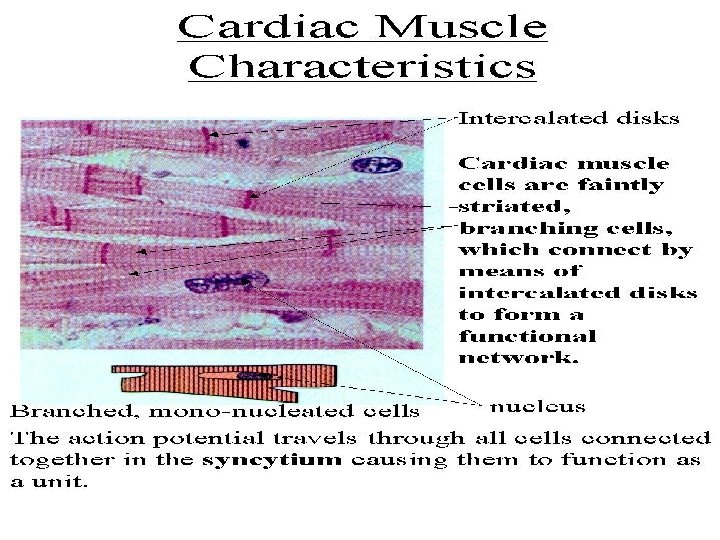
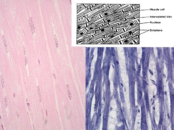
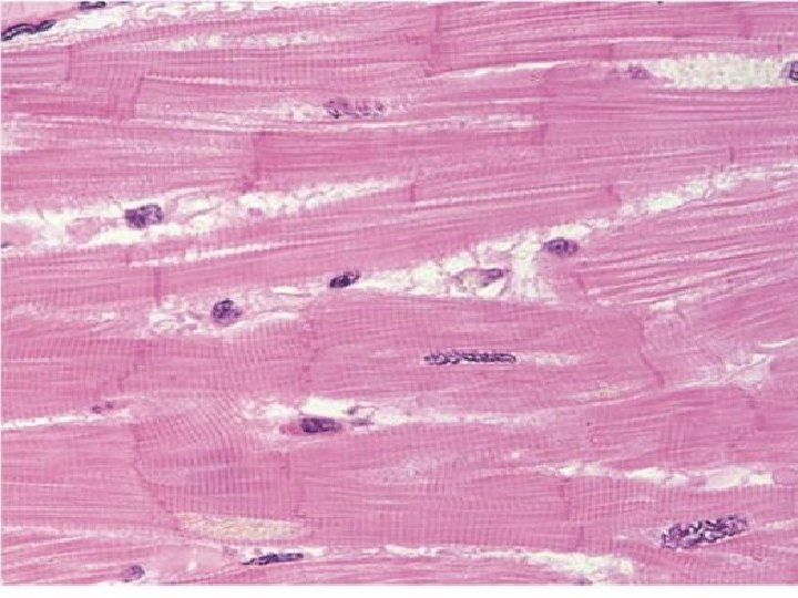
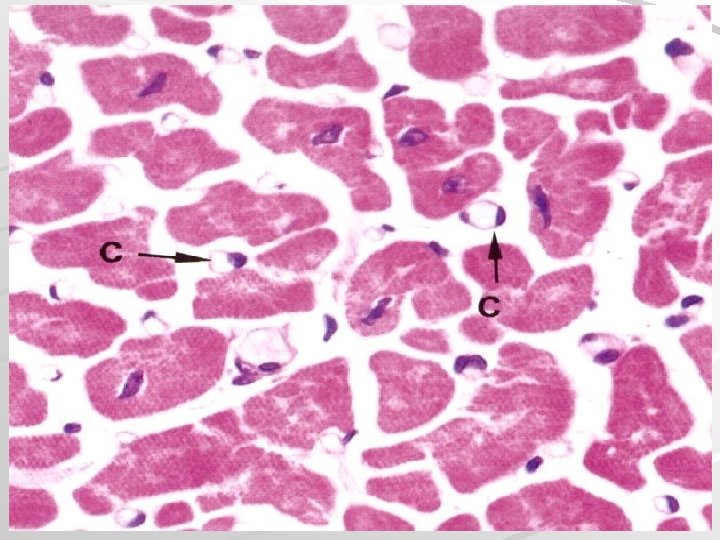
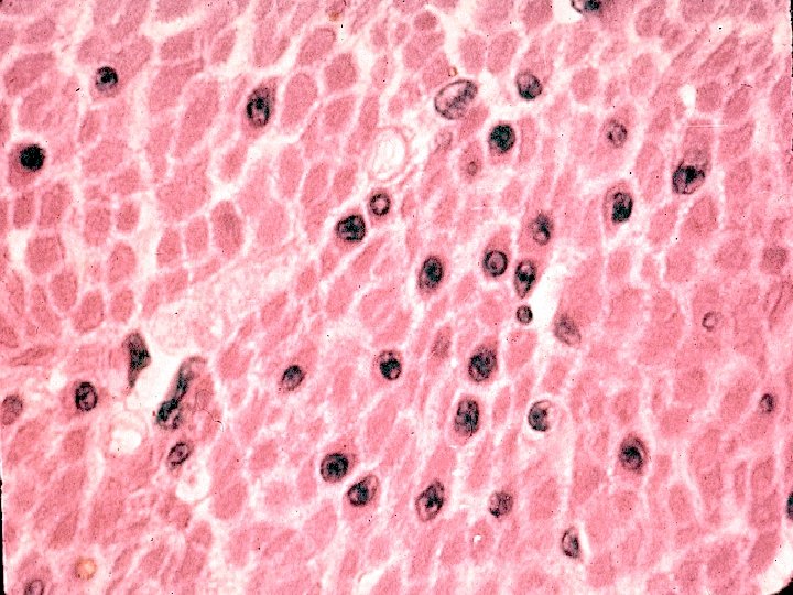
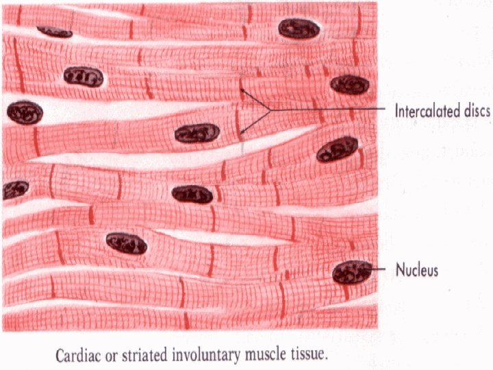
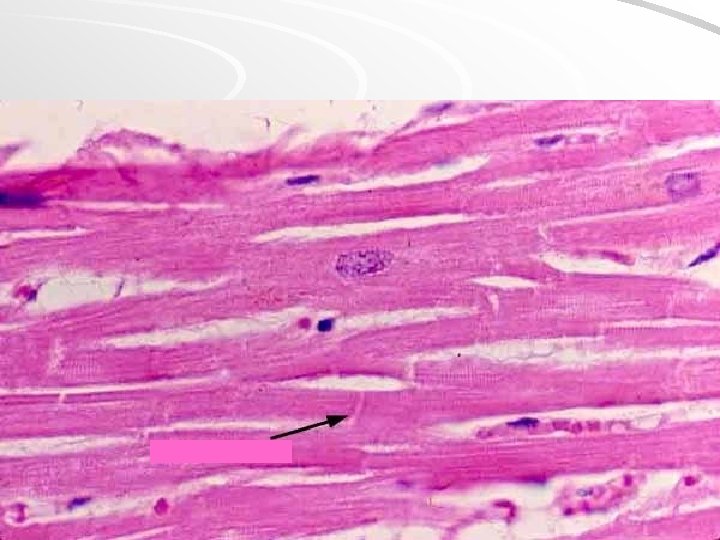
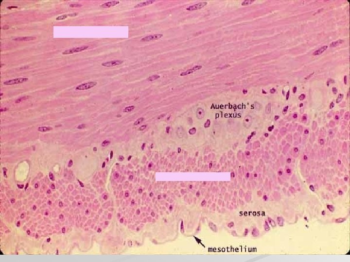
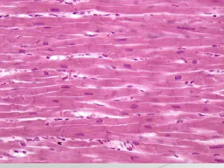
Cardiac muscle - myocardium is made up of long branched fibers, composed of cells – cardiomyocytes, cardiomyocytes are cylindrical cells, which can be branched on one or both ends (Y, X shaped cells), Sarkoplasm: 1 nucleus in the center of cell, striated myofibrils, numerous mitochondria, cells are attached to one another by end-toend junctions – intercalated discs.
chains of cardiomyocytes blood capillary with erythrocytes Intercalated disc
Diagram and electron micrograph of a part of cardiomyocyte
DIFFERENCES BETWEEN CARDIAC AND SKELETAL MUSCLE TISSUES there are no triads, but diads: 1 t-tubule + 1 cisterna t-tubules encircle the sarcomeres at the Z lines rather than at the zone of overlap. sarcoplasmic reticulum via its tubules contact sarcolemma as well as the t-tubules cardiac muscle cells are totally dependent on aerobic metabolism to obtain the energy needed to continue contracting. The sarcoplasm thus contains large numbers of mitochondria and abundant reserves of myoglobin (to store oxygen). Energy reserves are maintained in the form of glycogen and lipid inclusions.
Intercalated disc „scalariform“ shape of cell ends fasciae adherentes (adhesion of cells) Nexus (quick intercellular communication – transport of ions, electric impulses, informations)
striated cardiomyocytes intercalated discs – desmosomes, fasciae adherentes, nexuses
Actin myofilaments Cell 2 Cell 1 Intercalated disc: nexus fascia adherens
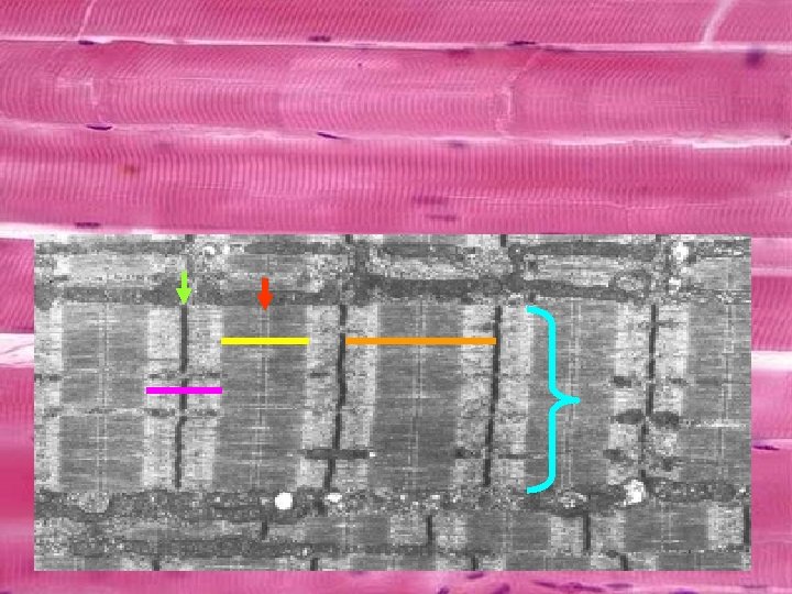
Myofibril of cardiomyocyte Actin + myosin myofilaments Sarcomere Z-line M-line and H-zone I-band, A-band T-tubule + 1 cisterna = diad (around Z-line)
Purkinje‘s fibers are located in the inner layer of heart ventricle wall are specialized cells fibers that conduct an electrical stimuli or impulses that enables the heart to contract in a coordinated fashion numerous sodium ion channels and mitochondria, fewer myofibrils
Natriuretic hormone in atrial granules in sarcoplasm of kardiomyocytes in ventricles 300 – 400 nm Ø this hormone is involved in regulation of volume of circulating blood
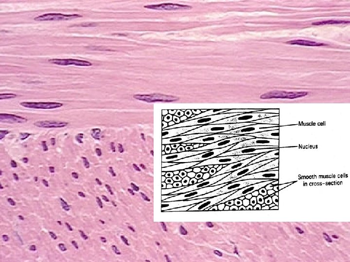
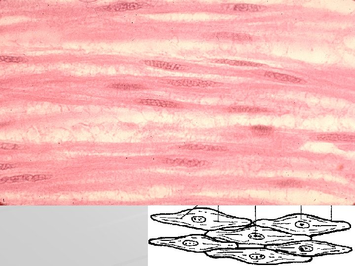
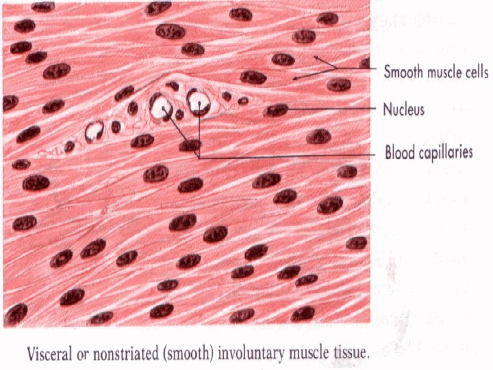
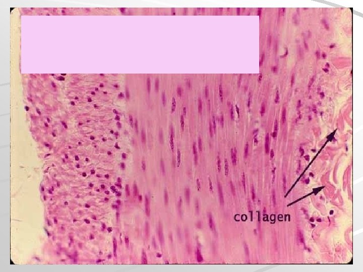
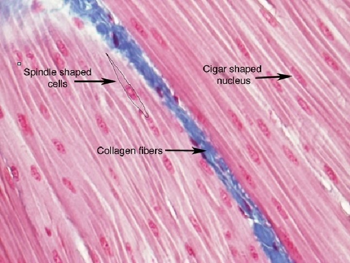
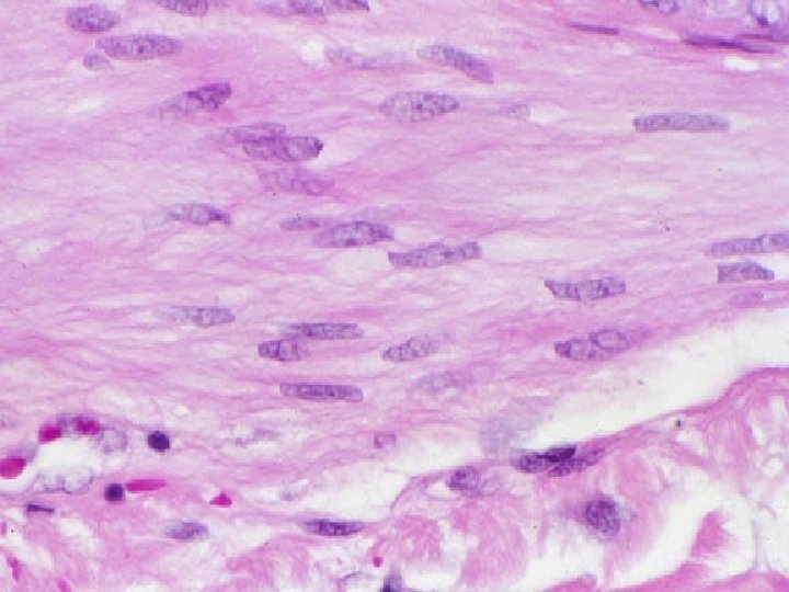
Smooth muscle tissue spindle shaped cells (leiomyocytes) with myofilaments not arranged into myofibrils (no striation), 1 nucleus in the centre of the cell myofilaments form bands throughout the cell actin filaments attach to the sarcolemma by focal adhesions or to the dense bodies substituing Z-lines in sarcoplasm calmodulin (has function as troponin) sarcoplasmic reticulum forms only tubules Caveolae (have function as T-tubules) zonulae occludentes and nexuses connect cells
Leiomyocyte Caveolae are equivalent to t-tubule and in their membrane ions channel are present to bring Ca needed fo Contraction. Caveolae are in contact with sarcoplasmic reticulum.
Leiomyocyte: contractile filaments Myosin flaments Actin filaments + nexuses
Muscle tissue Slides: Skeletal muscle (2 – Apex linguae) Heart muscle (64, 65 – Myocardium) Smooth muscle (16 – Intestinum tenue)
Smooth muscle
intestinum tenue – smooth muscle
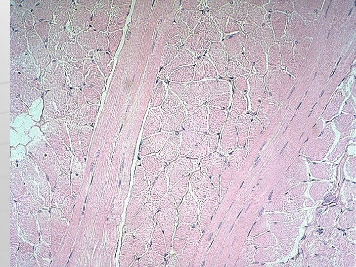
Leiomyocytes are arranged into laeyers of wall of hollow (usually tubular) organs Cross section Longitudinal section
Development of rhabdomyocyte Origin of skeletal muscle: paraaxial mesoderm – somites – myotomes Fusion of myoblasts (primitive muscle cells in myotomes) into multinucleated muscle fiber
Development of cardiomyocytes and leiomyocytes
Practice and Quiz
Rhabdomyocyte Endomysium Sarcomere T-tubule Troponin Myofilament Myofibril Sarcoplasmic reticulum Actin Sarcolemme Perimysium
Leiomyocyte Myosin Epimysium Triad Intercalated disc A-band Cardiomyocyte M-line Myosin Fascia adherens Motor-end-plate
Smooth muscle cell Basal lamina
Structure or rhabdomyocyte Sarcolemme + t-tubules, In sarcoplasm: Nuclei, Mitochondria, Golgi apparatus, Glycogen (beta granules) (sarcoplasm with organelles forms columns among myofibrils) Sarcoplasmic reticulum (smooth ER) – reservoir of Ca 2+ Myofibrils (parallel to the length of the muscle fiber)
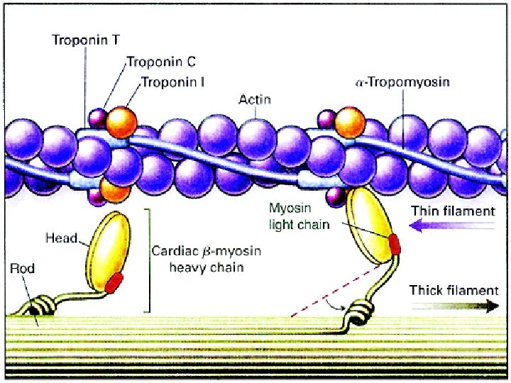
Mechanism of contraction: The chemical players in muscle contraction are: 1. myosin (protein) 2. actin (protein) 3. tropomyosin (protein) 4. troponin (protein) 5. ATP (nucleotide) 6. calcium ions
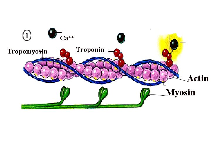
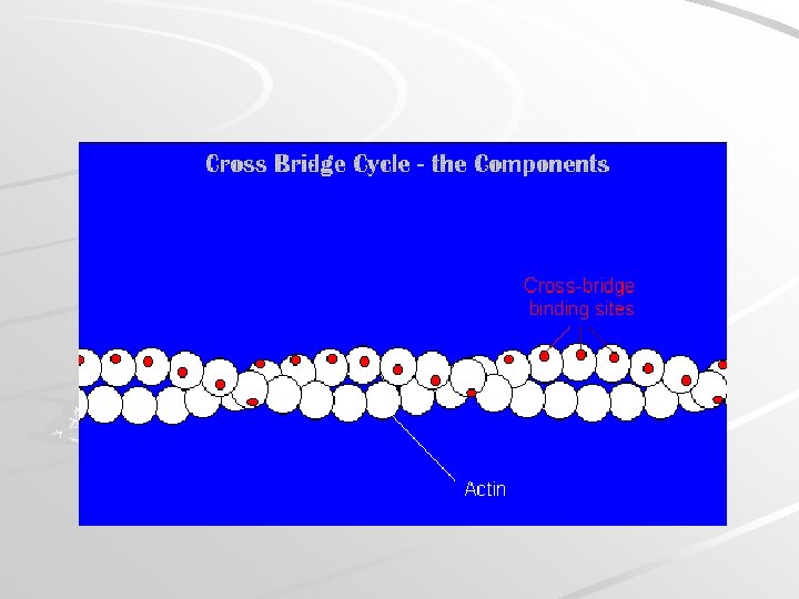
- There are two binding sites on each myosin head, one for ATP and one for actin. ATP is hydrolyzed into ADP + phosphate and energy is transferd into the head of myosin. - Thin filaments are made of these three protein molecules: 1. actin, 2. tropomyosin, 3. troponin. - The major component of the thin filament, actin is composed of a double strand of actin subunits each of which contain myosin binding sites. - The regulatory protein tropomyosin twists around the actin. When the sarcomere is not shortening, the position of the tropomyosin covers the binding sites on the actin subunits and prevents myosin cross bridge binding. - Troponin, which is found periodically along the tropomyosin strand, functions to move the tropomyosin aside, exposing the myosin binding sites. - Role of calcium in muscle contraction: action potential occurs - calcium ions are released from the terminal cisternae - calcium ions then bind to troponin – tropomyosin moves away from the myosin binding sites on actin.
Review of participants in the Cross Bridge Cycle: Participant 1. Myosin 2. Actin 3. Tropomyosin 4. Troponin 5. ATP 6. Calcium ions Will bind to: ATP, Actin Myosin, Troponin Calcium, Actin, Tropomyosin Myosin Troponin
Detail of steps required to expose the binding sites on actin: Presence of an action potential in the muscle cell membrane. Release of calcium ions from the terminal cisternae. Calcium ions rush into the cytosol and bind to the troponin. There is a change in the conformation of the troponin-tropomyosin complex. This tropomyosin slides over, exposing the binding sites on actin.
Summary of the role that ATP plays in the contraction of muscle: 1. ATP transfers its energy to the myosin cross bridge, which in turn energizes the power stroke. 2. ATP disconnects the myosin cross bridge from the binding site on actin. 3. ATP fuels the pump that actively transports calcium ions back into the sarcoplasmic reticulum.
ATP is required to cleave the avidly formed bond between actin and myosin. In the absence of ATP the cross linking becomes fixed, and the muscle becomes rigid and inextensible. This condition is seen shortly after death when the muscles' energy supply is used up, and is known as rigor mortis.
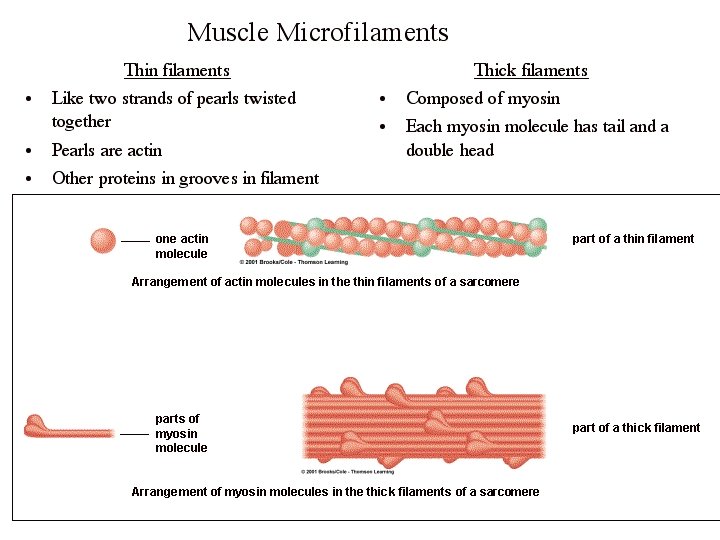
The thin actin filament is a dimeric polymer of G-actin sub-units arranged like two strings of beads twisted together. Attached to the actin chain of the thin filament, are the proteins troponin (Tn) and tropomyosin. A tropomyosin molecule runs along each actin chain, bound to the actin. Each tropomyosin sub-unit covers about 7 G-actin sub-units. The troponin molecule has three sub-units: Tn. T that binds to tropomyosin near the ends of the tropomyosin sub-units; Tn. I that binds to the actin; and Tn. C that binds to the Tn. I and Tn. T sub-units, and which also has a strong affinity for Ca 2+ at four binding sites.
The thick filaments, at either end, have sticking off at regular intervals, the fibrillar necks and globular, ATPase heads of the myosin subunits - the polymerized tails of which make the backbone of the filament. The heads at the opposite ends of the thick filament stick off in opposite directions leaving a bare middle region (pseudo H-zone) on the filament. Each successive myosin head is staggered so that six rows of heads stick off in six directions (60 o intervals) around the filament. Each myosin filament is surrounded by six actin filaments and each actin filament by three myosins.
Each myosin filament is surrounded by six actin filaments and each actin filament by three myosins.