The Muscular System Functions of Muscular Tissue Like


 Skeletal muscle (b) Cardiac muscle (c) Visceral smooth")
 G.")

 are arranged into bundles called fascicles Fascicles are bound by")


 – a type of ER")


")


 ADP Pi 1 Myosin head attaches")


")


- Slides: 71
The Muscular System
Functions of Muscular Tissue • Like nervous tissue, muscles are excitable or "irritable” they have the ability to respond to a stimulus • Unlike nerves, however, muscles are also: Contractible (they can shorten in length) Extensible (they can extend or stretch) Elastic (they can return to their original shape)
Three Types of Muscular Tissue (a) Skeletal muscle (b) Cardiac muscle (c) Visceral smooth muscle
Three Types of Muscular Tissue Location Skeletal skeleton Cardiac heart Visceral (smooth muscle) G. I. tract, uterus, eye, blood vessels Function movement, heat, posture Appearance Control striated, multinucleated (eccentric), voluntary fibers parallel pump blood striated, one central continuously nucleus involuntary Peristalsis, blood pressure, no striations, one pupil size, central nucleus erects hairs involuntary
Muscular System • Skeletal muscle is the only organ of the muscular system • Skeletal muscle is composed of skeletal muscle tissue and also contains nervous tissue, blood vessels and connective tissue • Half of the body’s weight is muscle tissue – Skeletal muscle = 40% in males, 32% in females – Cardiac muscle = 10%
Skeletal muscle fibers (cells) are arranged into bundles called fascicles Fascicles are bound by connective tissue Four different connective tissue coverings Deep fascia Surrounds entire skeletal muscle and extends beyond its length Perimysium Surrounds each fascicle Epimysium Closely surrounds skeletal muscle, binds fascicles together Endomysium Surrounds each muscle fiber (cell)
Organization of Muscle Tissue A muscle, a fasciculus, and a fiber all visualized
Organization of Muscle Tissue Organization of a fasciculus
Organization of Muscle Tissue Organization of a muscle fiber
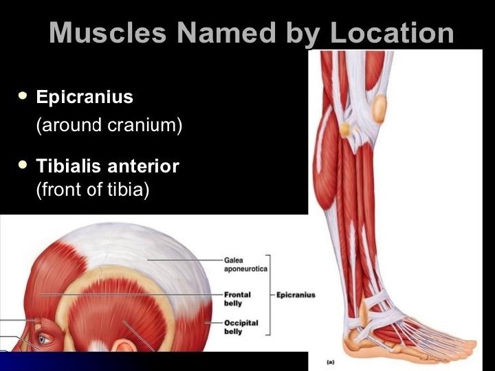
Connective Tissue Coverings • Fascia – Surrounds an individual skeletal muscle, separating it from other muscles – Fascia may extend beyond the ends of the muscle to become a tendon – Fascia may connect muscle to muscle and is called an aponeurosis
Organization of Muscle Tissue • Many large muscle groups are encased in both a superficial and a deep fascia Real Anatomy, John Wiley and Sons
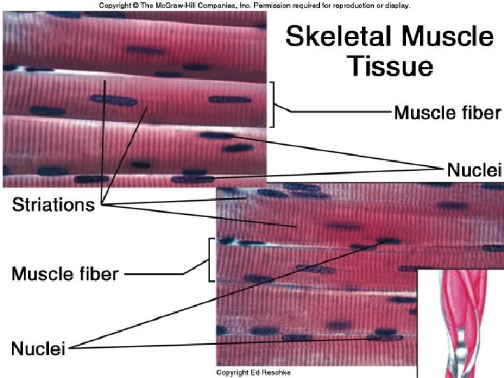
Characteristics of Skeletal Muscle Tissue • Long, thin contractile fibers that are Striated – Striations due to arrangement of thick and thin filaments • Under voluntary control • Attached to the bones of the skeleton by tendons • Allow for movement, facial expressions, breathing, swallowing, writing, talking and singing, posture, heat production, joint stability
Skeletal Muscle Arrangement • A single muscle cell is a muscle fiber – Fibers are made up of myofibrils – Myofibrils are made up of thick and thin filaments • Sarcolemma – muscle cell membrane • Sarcoplasm • muscle cell cytoplasm
Skeletal Muscle Cells • Myofibrils are striated – Striations due to arrangement of thick and thin filaments • Seen as alternating areas of light and dark bands • The length of each myofibril is divided into repeating units called sarcomeres – A sarcomere is the functional unit of skeletal muscle
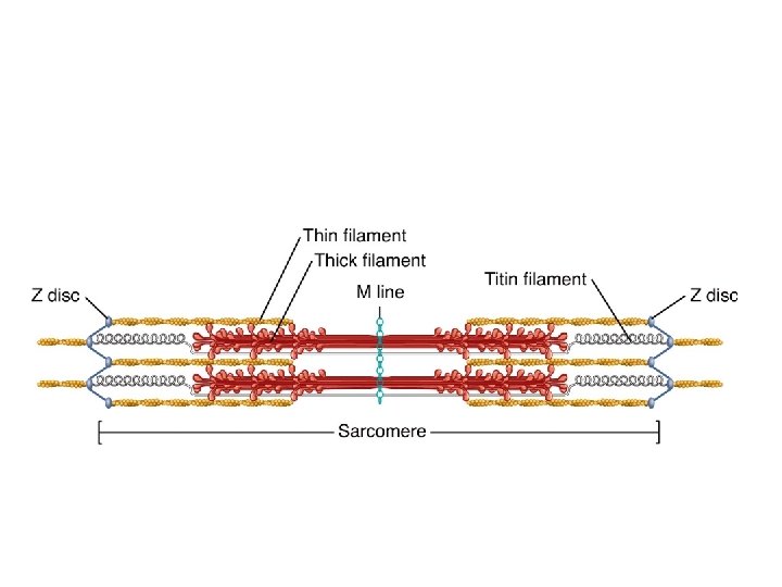
Sarcomere Arrangement
Sarcomere Structure • Sarcomere exists from Z-line to Z-line • A-Band is dark middle band – Overlapping think and thin filaments • I-Band – ends of A-Band, thin filaments only • Z-line is in the middle if the I-Band • Myosin filaments are held to the Z-line by titin proteins
Microscopic anatomy of a skeletal muscle fiber Z disc H zone Z disc Thin (actin) filament Thick (myosin) filament (c) I band Thin (actin) filament A band Sarcomere I band M line Z disc Elastic (titin) filaments Thick (myosin) filament (d) (e) I band thin filaments only H zone thick filaments only M line thick filaments linked by accessory proteins Outer edge of A band thick and thin filaments overlap
Thick Filament Structure • Composed of many myosin molecules – Each myosin molecule has a tail region and 2 globular heads (crossbridges)
Thin Filament Structure • Composed of actin protein – 2 strands of globular actin molecules twisted into a helix – Actin filaments have binding sites for myosin cross bridges Troponin complex Tropomyosin Actin – Tropomyosin protein spirals around actin helix – Troponin protein (3 subunits) is attached to actin and holds tropomyosin in place • Call this the troponin-tropomyosin complex
Specialized Organelles of Skeletal Muscle • Sarcoplasmic Reticulum (SR) – a type of ER – Surrounds each myofibril, running parallel to it – Stores calcium, when stimulated, calcium diffuses into sarcoplasm • Transverse Tubules (TT) – Extends into sarcoplasm as invaginations continuous with sarcolemma • T tubules run between cisternae of SR – Filled with extracellular fluid – Cisternae of SR and TT form a triad near where thick and thin filaments overlap
Relationship of the sarcoplasmic reticulum and T tubules to myofibrils of skeletal muscle I band A band I band Z disc H zone Z disc M line Part of a skeletal muscle fiber (cell) Sarcolemma Triad Mitochondrion Myofibril Sarcolemma Myofibrils Tubules of sarcoplasmic reticulum Terminal cisterna of the sarcoplasmic reticulum T tubule
Skeletal Muscle Contraction
Motor Neuron • Nerve cell that innervates skeletal muscle tissue • Dendrite – Receives information • Axon • – Transmits information – Has vesicles containing neurotransmitter that will stimulate or inhibit muscle contraction Neuromuscular Junction -Site where branch of motor neuron (motor nerve ending) comes in contact with sarcolemma of skeletal muscle fiber • A type of synapse
NMJ Motor end-plate Sarcolemma of muscle fiber directly beneath motor nerve ending Contains an abundance of mitochondria and nuclei
Neuromuscular Junction
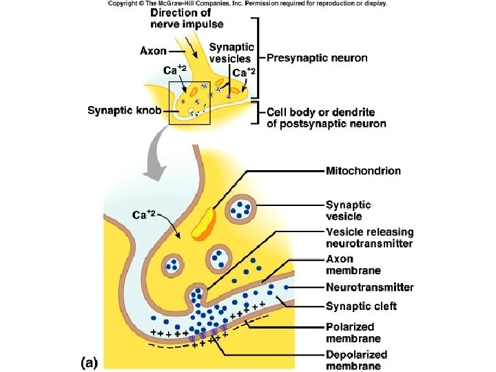
Synapse • Junction between the axonal end of one neuron and the dendrite of another neuron OR membrane of another cell type • Synaptic cleft – Separation that exists between the axonal ending of the motor nerve and the sarcolemma of the muscle cell fiber • The axon ending contains vesicles of neurotransmitter
Neurotransmitter • Chemical substance released from vesicles in the motor nerve ending (axonal ending) – Acetylcholine (Ach) is the neurotransmitter released by motor neurons • When stimulated by a nerve impulse, Ach is released, travels across the synaptic cleft and binds receptors on the motor end plate • Stimulates contraction
Sliding Filament Theory • A sarcomere is the functional unit of skeletal muscle • When a skeletal muscle contracts, sarcomeres shorten • This is described by the sliding filament theory
Sliding Filament Theory • Sarcomeres shorten because thick and thin filaments slide past one another • Thin filaments move towards the center of the sarcomere from both ends
The Sliding-Filament Mechanism • The “sliding” of actin on myosin (thick filaments on thin filaments) can be broken down into a 4 step process
Physiology of Skeletal Muscle Contraction: Neurotransmitter Release • Motor impulse is initiated in the brain • Travels through the brain and spinal cord to a motor nerve ending • Motor nerve endings (axons) depolarize • Calcium enters the axonal endings • Calcium causes the release of acetylcholine into the neuromuscular junction (synaptic cleft)
Physiology of Skeletal Muscle Contraction: Depolarization • ACh binds receptors on motor end plate • Depolarizes skeletal muscle fibers – Reverses charge of membrane • Impulse travels through transverse tubules to reach all of the muscle fibers • Muscle depolarization causes release of calcium from the sarcoplasmic reticulum into the sarcoplasm
Relationship of the sarcoplasmic reticulum and T tubules to myofibrils of skeletal muscle I band A band I band Z disc H zone Z disc M line Part of a skeletal muscle fiber (cell) Sarcolemma Triad Mitochondrion Myofibril Sarcolemma Myofibrils Tubules of sarcoplasmic reticulum Terminal cisterna of the sarcoplasm reticulum T tubule
Physiology of Skeletal Muscle Contraction: Power Stroke • Calcium binds troponin (which is attached to tropomyosin • Moves tropomyosin from the myosin binding sites on actin • Myosin crossbridges (heads) bind to actin – ATP hydrolysis supplies energy • Actin is pulled inward towards the center of the sarcomere = POWER STROKE • Sarcomeres shorten as muscle contracts
The Sliding-Filament Mechanism • With exposure of the myosin binding sites on actin (the thin filaments)—in the presence of Ca 2+ and ATP—the thick and thin filaments “slide” on one another and the sarcomere is shortened
• Step 1: ATP hydrolysis • Step 2: Attachment
• Step 3: Power Stroke • Step 4: Detachment
The Cross Bridge Cycle Myosin head (high-energy configuration) ADP Pi 1 Myosin head attaches to the actin myofilament, forming a cross bridge. Thin filament ATP hydrolysis ADP Thick filament Pi 4 As ATP is split into ADP and Pi, the myosin head is energized (cocked into the high-energy conformation). 2 Inorganic phosphate (Pi) generated in the previous contraction cycle is released, initiating the power (working) stroke. The myosin head pivots and bends as it pulls on the actin filament, sliding it toward the M line. Then ADP is released. ATP Myosin head (low-energy configuration) 3 As new ATP attaches to the myosin head, the link between Myosin and actin weakens, and the cross bridge detaches.
The Sliding-Filament Mechanism
Excitation-Contraction Coupling Axon terminal Synaptic cleft Sarcolemma Synaptic vesicle ACh Neurotransmitter released diffuses across the synaptic cleft and attaches to ACh T tubule 1 Net entry of Na+ initiates an action potential which is propagated along the ACh sarcolemma and down the T tubules. Ca 2+ SR tubules (cut) 2 Action potential Ca 2+ in T tubule activates voltage-sensitive receptors, which in ADP turn trigger Ca 2+ Pi release from terminal Ca 2+ cisternae of SR into cytosol. Ca 2+ 6 Tropomyosin blockage restored, blocking myosin binding sites on actin; Ca 2+ contraction ends and muscle fiber relaxes. Ca 2+ SR Ca 2+ 3 Calcium ions bind to troponin; troponin changes shape, removing the blocking action of tropomyosin; actin active sites exposed. 5 Removal of Ca 2+ by active transport into the SR after the action potential ends. Ca 2+ 4 Contraction; myosin heads alternately attach to actin and detach, pulling the actin filaments toward the center of the sarcomere; release of energy by ATP hydrolysis powers the cycling process.
Muscle Relaxation Mechanism 1. Acetylcholinesterase present in the NMJ destroys ACh (preventing continual stimulation) 2. Calcium ions are transported from the sarcoplasm back into the SR 3. Linkages between myosin and actin are broken – Requires ATP binding • THEN: The muscle fiber relaxes
Contraction in the Sarcomere • A band stays the same • I band gets smaller • H zone gets smaller • Sarcomere shortens
Sliding filament model of contraction Z 1 I Z H A I Z 3 Z 2 Z A
Energy for Contraction • ATP initially supplied from cellular respiration • If ATP is abundant, is converted to creatine phosphate and stored in skeletal muscles • When ATP is low, creatine phosphate supplies phosphate to ADP making ATP • CP & ATP stores only good for about a 10 second maximal contraction • ATP must then come from cellular respiration or