Muscle Anatomy and Physiology Muscle Tissue Muscle Tissue


 voluntary muscles")


 organ")


- Slides: 35
Muscle Anatomy and Physiology
Muscle Tissue
Muscle Tissue • same brief description as tissue chapter • ½ of body mass • able to convert ATP (chemical energy) to mechanical energy
1. Cardiac Muscle • in heart only • striated and branching • involuntary
2. Smooth Muscle • visceral – – GI tract walls, – urinary bladder, – bronchi, – arrector pili, – iris
2. Smooth Muscle • smooth spindle shape • involuntary • contractions are slow and sustained without fatigue
2. Smooth Muscle • cells are surrounded by connective tissue - endomysium • usually arranged in sheets running in different directions
3. Skeletal Muscle • • make up the skeletal muscles striated (banded) voluntary muscles respond rapidly but fatigue easily
Functions of Skeletal Muscle
Functional Characteristics of Skeletal Muscle 1. contractibility - shortens with force 2. excitability - responds 3. extensibility - stretches 4. elasticity - recoils
Function of Muscle 1. Movement 2. Posture maintenance - continuous tiny adjustments 3. Heat generation - by product of metabolism – – nearly ¾ of energy released from ATP escapes as heat skeletal muscle is at least 40% of body mass
Structure of Skeletal Muscle
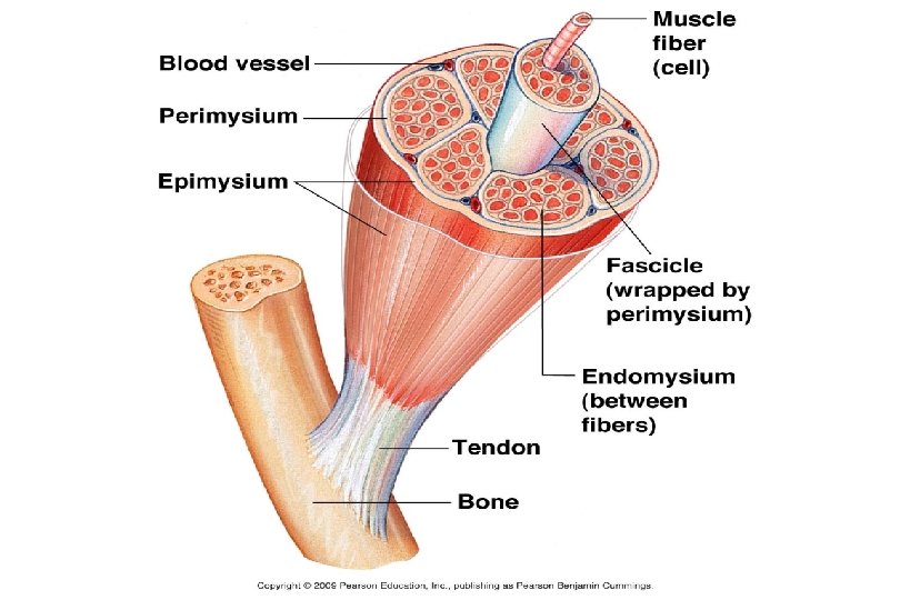
Structure of Skeletal Muscle • entire gross structure covered by connective tissue - epimysium • muscle is made of small bundles called fascicle which are bound by perimysium • each fascicle is made of a bundle of muscle cells or fibers which are surrounded by endomysium (all coverings are continuous extensions)
Muscle Fiber Structure
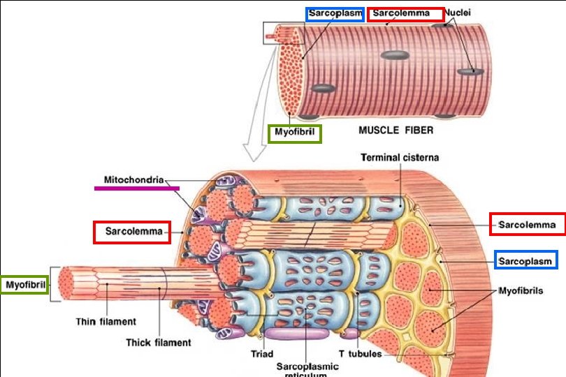
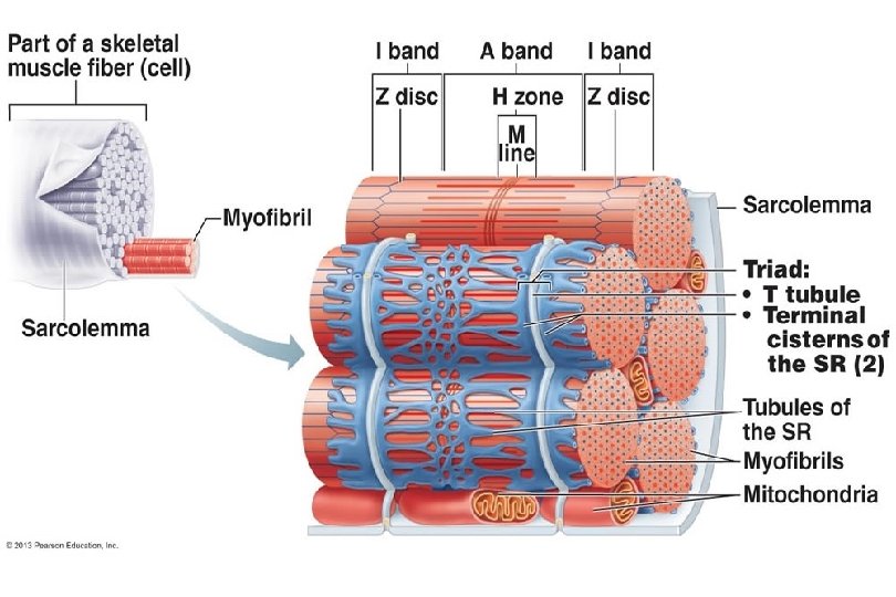
Muscle Fiber Structure • sarcolemma – muscle fiber plasma membrane • sarcoplasm – muscle fiber cytoplasm, contains myoglobin – a red pigment that stores oxygen • Each muscle fiber is made of bundles of rod-shaped structures of myofibrils (organelles). • (100’s to 1000’s per cell)
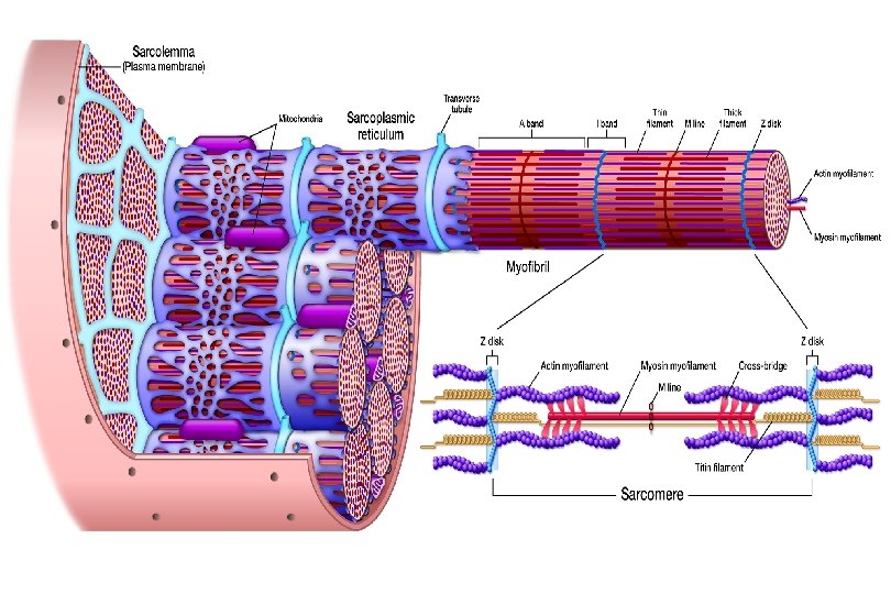
They have repeating pattern of striations called sarcomeres
The banding is caused by an orderly overlapping arrangement of protein filaments - myofilaments
Myofilaments: 1. myosin - thick, extend entire length of A band 2. actin - thin, extend across the I band part way into A band
• Z line - attachment site for actin, ends of sarcomere • I band - light bands, actin only • A band - dark bands, myosin and actin overlap
• • H zone - only visible in relaxed, myosin only M line - sarcomere center
Structure Connective Tissue organ Epimysium surrounds Perimysium tissues Endomysium surrounds cells organelles molecules Gross muscle Fascicle (bundles of cells) Muscle fiber (muscle cell) Myofibril (contracting organelle) Myofilaments Actin – thin filament Myosin – thick filament
Connective Tissue Epimysium Perimysium Endomysium Structure Gross muscle surrounds Fascicle (bundles of cells) organ tissues Muscle fiber cells Myofibril organelles (muscle cell) (contracting organelle) Myofilaments molecules Actin – thin filament Myosin – thick filament
Molecular Structure of Myofilaments
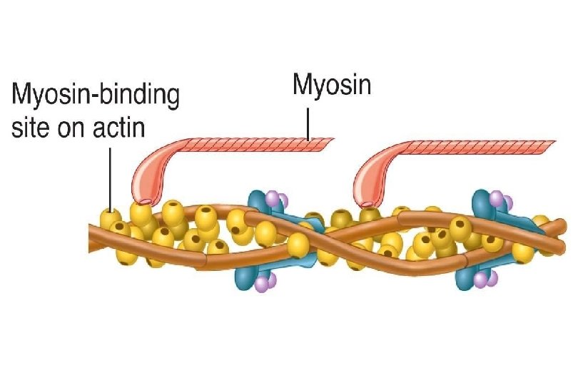
Myosin • rod like tail or axis ending in 2 globular heads - “crossbridges” - heads contain ATPase tail heads (enzyme)
Actin • globular subunits - G actin are found in long strands, like a double strand of twisted beads
Actin • tropomyosin spirals around beads, to add strength and stiffen, thin ribbon • troponin molecules bond actin to tropomyosin, has binding sites that will join with myosin crossbridge Binding sites
Sarcoplasmic Reticulum
Sarcoplasmic Reticulum • smooth endoplasmic reticulum of muscle cells • storage area for Ca ions • At H zones and A - I junctions tubes fuse and form lateral channels - terminal cisternae which feed into transverse tubules (T tubules) tubules at each Z line. • T tubules receive nerve stimuli and provide a pathway for oxygen, glucose, and Ca ions. (because continuous with membrane, nerve impulse allowed deep into muscle)
The End