MUSCLE TISSUE PPT 3 How does a muscle


 stroke. Figure 9. 12, step 3")


- Slides: 61
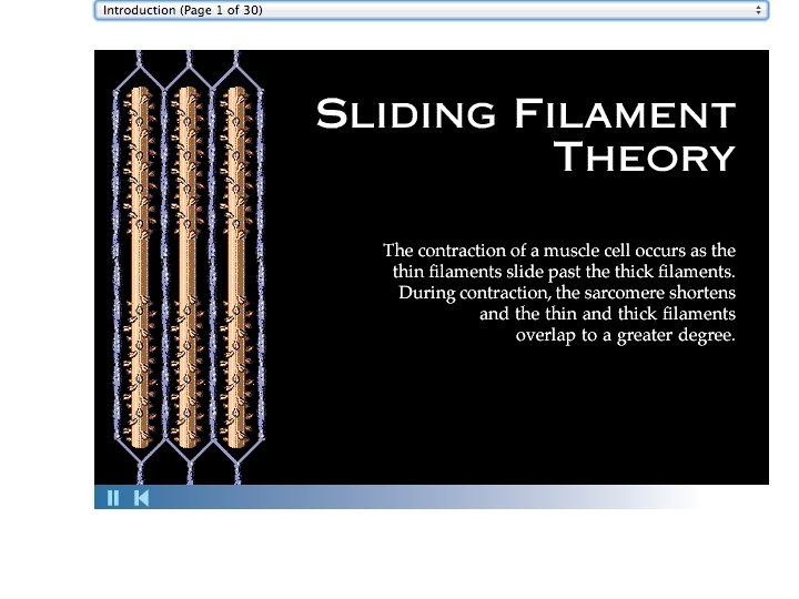
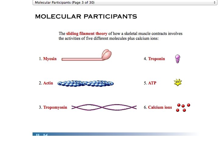
MUSCLE TISSUE PPT 3 • How does a muscle cell contract?
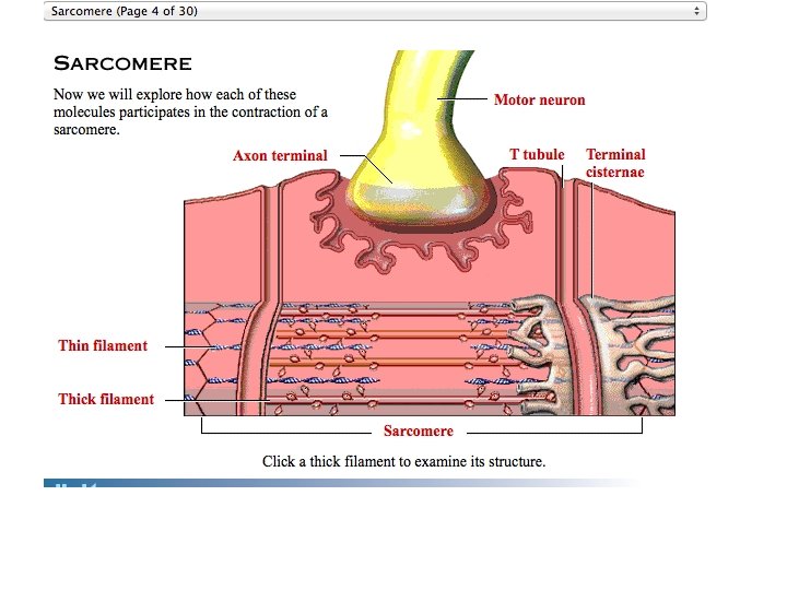
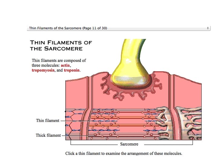
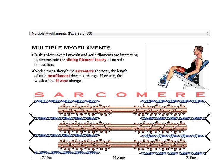
Sarcomeres • A sarcomere is one small part of a myofibril • Sarcomeres are lined up in the myofibril like box cars of a train • There are many myofibrils in a muscle cell
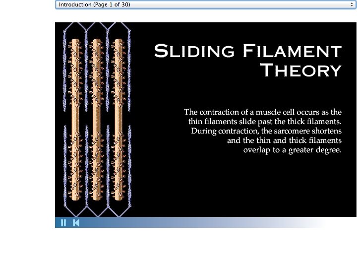
Sarcomeres • Since a sarcomere is one small part of a myofibril 1. When sarcomeres shorten, myofibrils shorten Contraction of the cell 2. When myofibrils shorten, the muscle cell shortens 3. When the muscle cells shorten, the whole muscle shortens
Contraction of the cells leads to contraction of the whole muscle
How does it all work to contract a cell? Each student is a sarcomere. Each row of students is a myofibril Each myofibril is in the cell (room!) Your top arm is a thin filament Your bottom arm is a thick filament Your elbows are the Z discs Touch your elbows to the person next to you and do not lose contact. Start with your own arms totally overlapped. Your top arm is a thick filament Your bottom arm is a thin filament Use your fingers (myosin heads) to walk the top arm along the bottom arm What happens to the length of the myofibril What would happen to the walls if you were attached to them? Contract and expand your myofibril as a team.
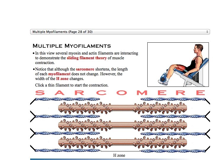
Do the thin and thick filaments shrink?
The action potential continues down the muscle cell, falling into a T tubule. Action potential is generated ACh Terminal cisterna of SR Sarcolemma Ca 2+ Figure 9. 11, step 1
Axon terminal Open Na+ Channel Na+ Synaptic cleft ACh Na+ K+ K+ ++ ++ + + Action potential + + +++ + of d ------ ep o l ar iza tio n ACh Na+ K+ Closed K+ Channel ve Wa The cell is negative inside at rest 1 Sarcoplasm of muscle fiber Figure 9. 9, step 1
Axon terminal Synaptic cleft Open Na+ Channel Na+ + ACh Na+ ++ ++ + K+ Action potential + + +++ + 2 Generation and propagation of the action potential (AP) of d +++++ ep o l ar iza tio n ACh Na+ K+ K+ Closed K+ Channel ve Wa 1 Sarcoplasm of muscle fiber Figure 9. 9, step 2
Axon terminal Synaptic cleft Open Na+ Channel Na+ + ACh iza tio n K+ ++ ++ + K+ Action potential + + +++ + 2 Generation and propagation of the action potential (AP) of d ep o l ar ACh Na+ K+ Na+ Closed K+ Channel ve Wa 1 Local depolarization: generation of the end plate potential on the sarcolemma Sarcoplasm of muscle fiber Figure 9. 9, step 2
Axon terminal Synaptic cleft Open Na+ Channel Na+ + ACh iza tio n K+ ++ ++ + K+ Action potential + + +++ + 2 Generation and propagation of the action potential (AP) of d ep o l ar ACh Na+ K+ Na+ Closed K+ Channel ve Wa 1 Local depolarization: generation of the end plate potential on the sarcolemma Sarcoplasm of muscle fiber Figure 9. 9, step 2
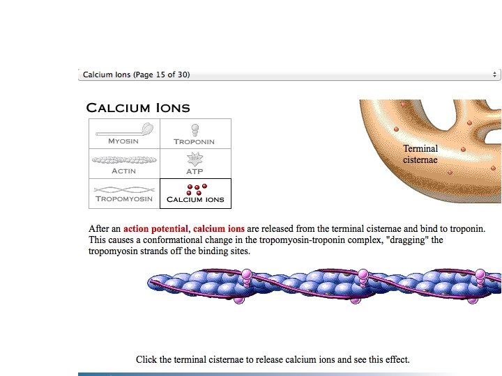
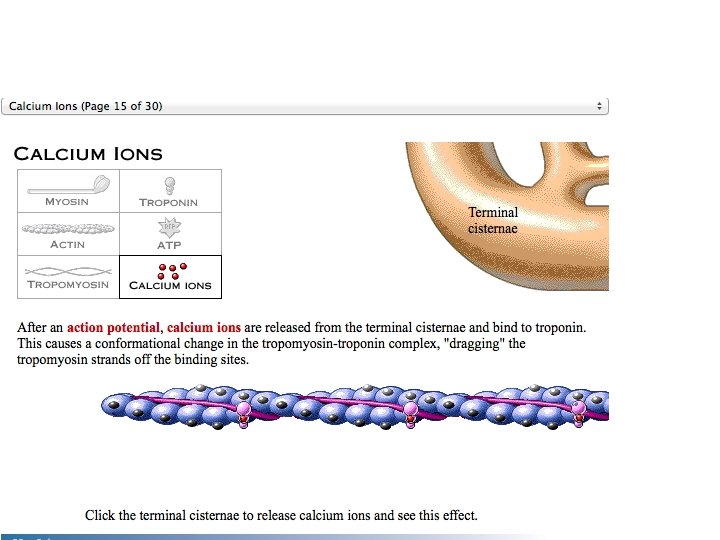
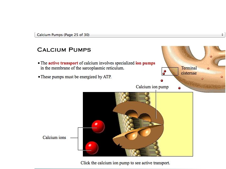
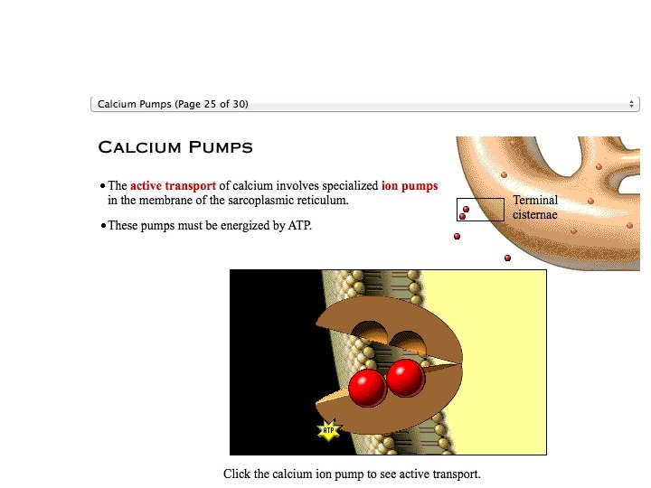
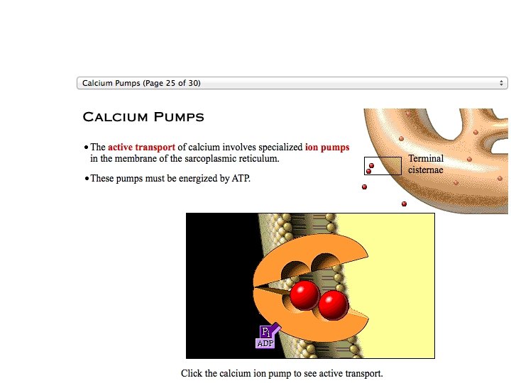
The Action potential is a signal that moves along the sarcolemma and down the T tubules, and then Ca++ is released from the SR into the cytoplasm Steps in E-C Coupling: Voltage-sensitive tubule protein Sarcolemma T tubule Ca 2+ release channel Terminal cisterna of SR Ca 2+ Figure 9. 11, step 3
. T tubule Ca 2+ release channel . Ca 2+ Figure 9. 11, step 4
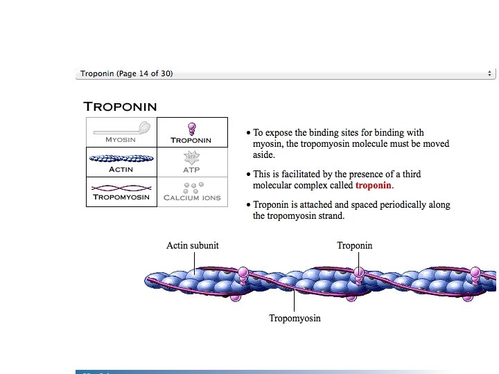
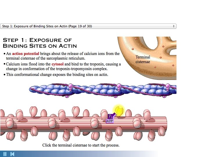
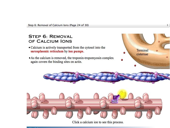
Steps in E-C Coupling: Sarcolemma Voltage-sensitive tubule protein T tubule 1 Action potential is propagated along the sarcolemma and down the T tubules. Ca 2+ release channel 2 Calcium ions are released. Terminal cisterna of SR Ca 2+ Actin Troponin Ca 2+ Tropomyosin blocking active sites Myosin 3 Calcium binds to troponin and removes the blocking action of tropomyosin. Active sites exposed and ready for myosin binding 4 Contraction begins Myosin cross bridge The aftermath Figure 9. 11, step 2
Actin Ca 2+ Troponin Tropomyosin blocking active sites Myosin The aftermath Figure 9. 11, step 5
Actin Ca 2+ Troponin Tropomyosin blocking active sites Myosin 3 Calcium binds to troponin and removes the blocking action of tropomyosin. Active sites exposed and ready for myosin binding The aftermath Figure 9. 11, step 6
Actin Ca 2+ Troponin Tropomyosin blocking active sites Myosin 3 Calcium binds to troponin and removes the blocking action of tropomyosin. Active sites exposed and ready for myosin binding Myosin cross bridge 4 Contraction begins The aftermath Figure 9. 11, step 7
Steps in E-C Coupling: Sarcolemma Voltage-sensitive tubule protein T tubule 1 Action potential is propagated along the sarcolemma and down the T tubules. Ca 2+ release channel 2 Calcium ions are released. Terminal cisterna of SR Ca 2+ Actin Troponin Ca 2+ Tropomyosin blocking active sites Myosin 3 Calcium binds to troponin and removes the blocking action of tropomyosin. Active sites exposed and ready for myosin binding 4 Contraction begins Myosin cross bridge The aftermath Figure 9. 11, step 8
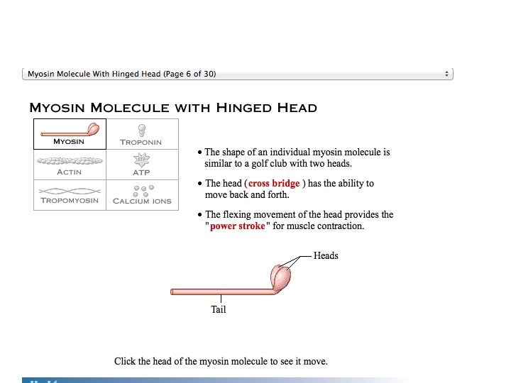
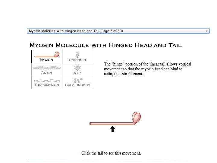
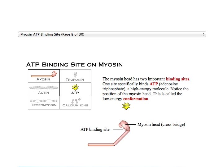
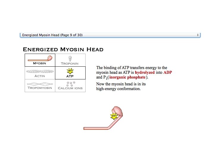
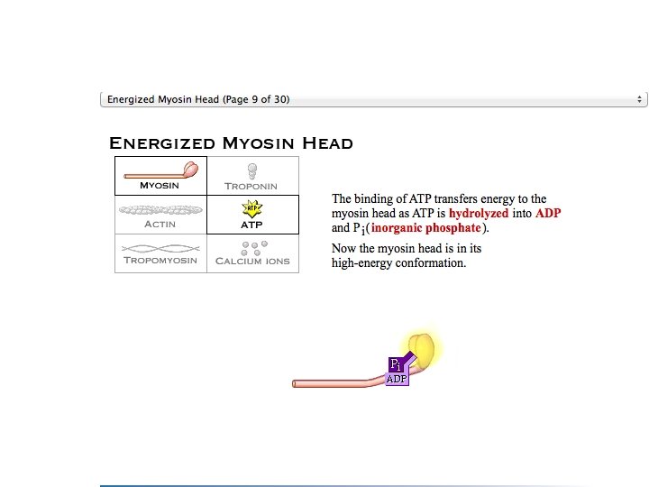
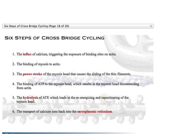
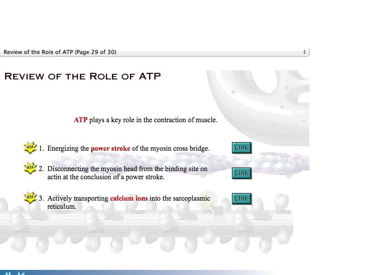
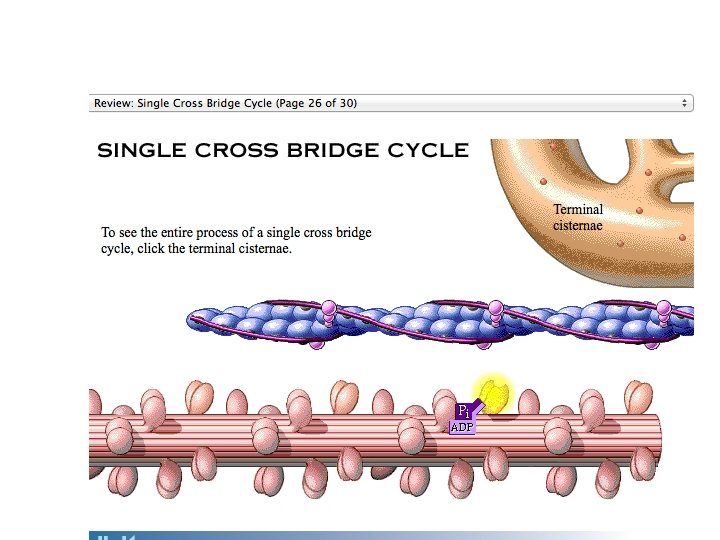
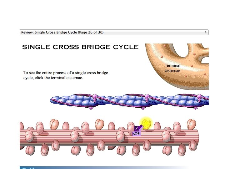
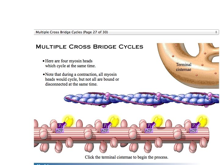
ATP ENERGY Is required for contraction. Thin filament Actin Ca 2+ Myosin cross bridge ADP Pi Thick filament When you run out of ATP you stop moving! Rigor Mortis: After death, no new ATP is made, so the myosin cannot release from action. Myosin Cross bridge formation. 1 ADP Pi Pi ATP hydrolysis 2 The power (working) stroke. 4 Cocking of myosin head. ATP 3 Cross bridge detachment. Figure 9. 12
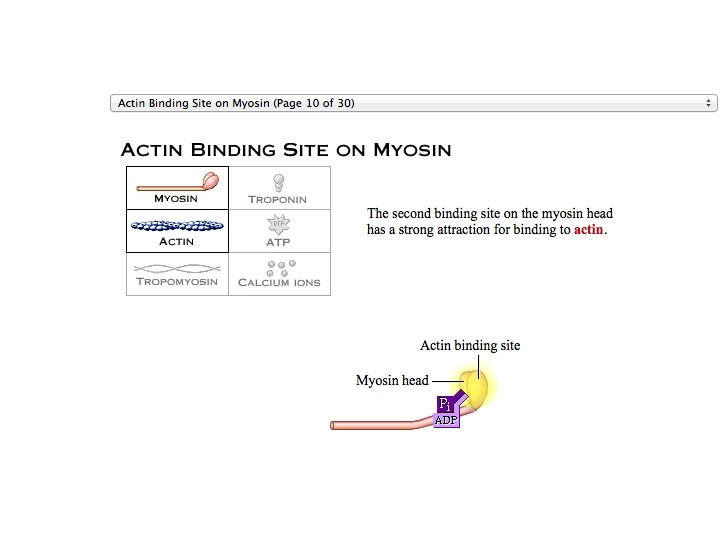
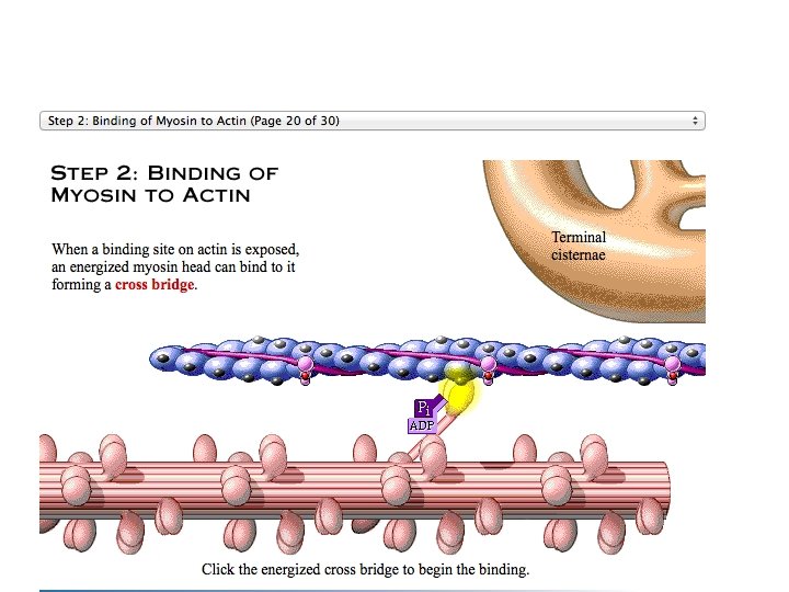
Actin Ca 2+ Myosin cross bridge Thin filament ADP Pi Thick filament Myosin 1 Cross bridge formation. Figure 9. 12, step 1
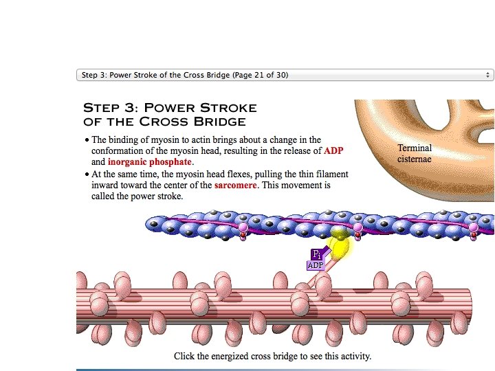
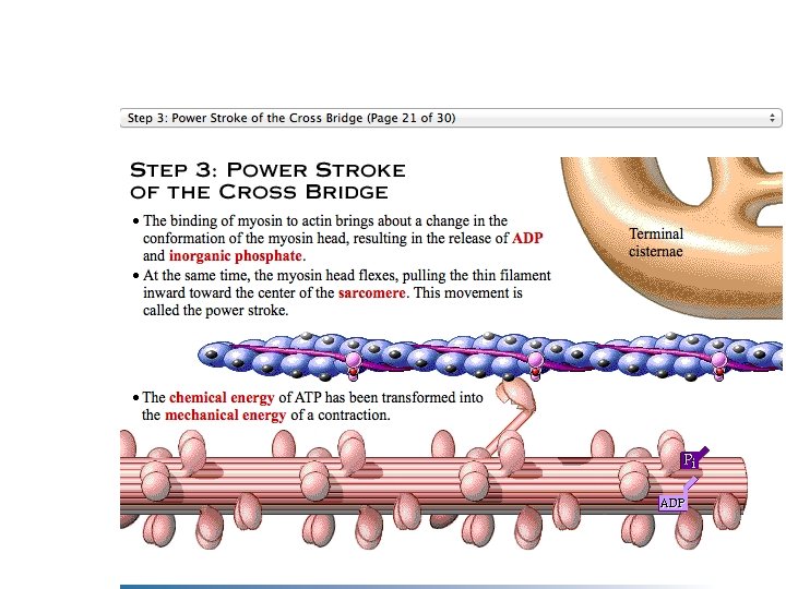
ADP Pi 2 The power (working) stroke. Figure 9. 12, step 3
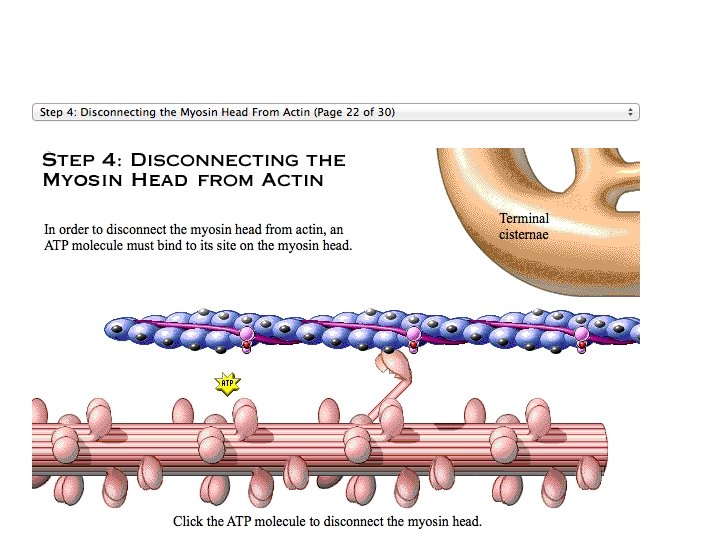
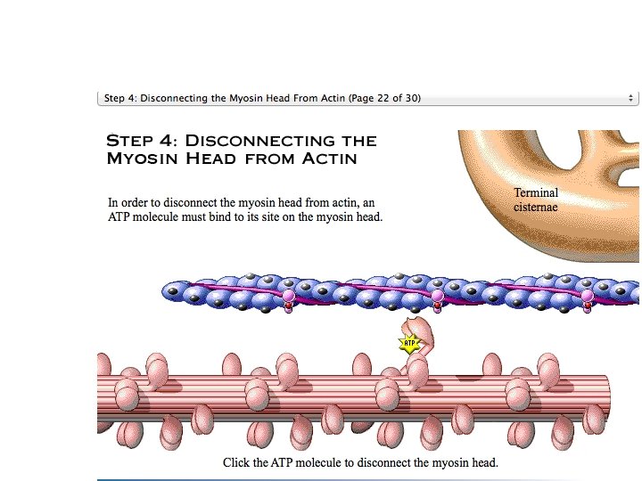
ATP 3 Cross bridge detachment. Figure 9. 12, step 4
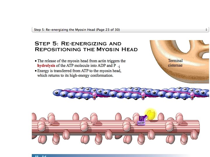
ADP ATP Pi hydrolysis 4 Cocking of myosin head. Figure 9. 12, step 5
Thin filament Actin Ca 2+ Myosin cross bridge ADP Pi Thick filament Myosin Cross bridge formation. 1 ADP Pi Pi ATP hydrolysis 2 The power (working) stroke. 4 Cocking of myosin head. ATP 3 Cross bridge detachment. Figure 9. 12