Muscle Tissue Skeletal Muscle Notes 3 Types of


")


 contains bundles of")


 • Z")


")


 2 Lactic acid 36 -38")


- Slides: 38
Muscle Tissue & Skeletal Muscle Notes
3 Types of Muscle Tissue • Skeletal muscle- striated and voluntary (it is subject to conscious control)
• Smooth muscle- not striated and involuntary (we do not consciously control it) – Lines our digestive system and empties our bladder and bowels
• Cardiac muscle- striated and involuntary – Only found in the heart
Functions of Skeletal Muscle • • Produce movement Maintain posture Support soft tissue Body temperature
THE ANATOMY OF A MUSCLE
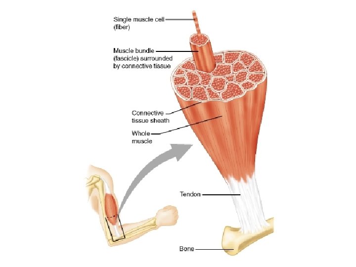
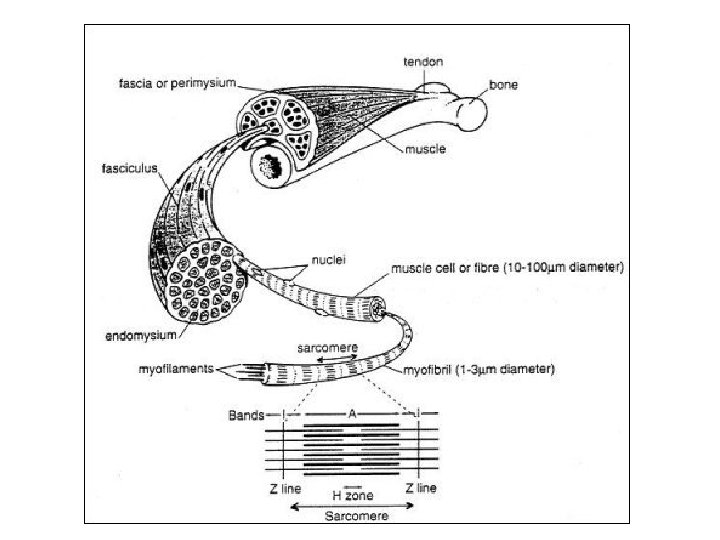
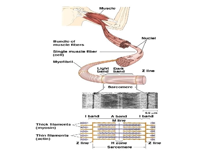
Anatomy of Skeletal Muscle • Each muscle is composed of bundles of muscle fibers. • Each muscle fiber (cell) has many nuclei and is a cluster of myofibrils • Myofibrils contain two types of protein filaments that are arranged in a regular, over-lapping pattern – Myosin – thicker filament – Actin – thinner filament
Sarcomere • The functional unit of contraction in skeletal muscle myofibrils • Located between two Z lines • One end of each actin filament is attached to the Z line • Myosin filaments are located between two actin filaments and overlap them on each end
CW: Color and Label
Muscle Tissue Anatomy • Epimysium - outer covering of muscles • Fascicle - a bundle of muscle fibers
Muscle Tissue Anatomy • Perimysium - each fascicle is covered by the perimysium • Endomysium - thin covering around each muscle fiber - Both perimysium & endomysium contain blood vessels and nerve endings
Muscle Tissue Anatomy • Myofibrils - each muscle fiber (muscle cell) contains bundles of myofibrils
• Sarcomere – tiny contractile units that make up myofibrils - Made of 2 proteins: 1. Thin Filaments – made of actin 2. Thick Filaments – made of myosin
• Sarcomere - what gives skeletal muscle its STRIATIONS • A – BAND - area where actin & myosin overlap • I – BAND - area of actin only
• Z – LINES - where I – Bands attach (actin) • Z – Lines are also the boundaries of the sarcomere
Muscle Contractions
Sliding Filament Theory • The myosin filaments pull the actin filaments towards the center of the sarcomere • This shortens the sarcomere which in turn shortens the muscle
Sliding Filament Theory • 1. A neural impulse is sent to the muscle you want to move. • 2. the neuron releases a neurotransmitter called acetylcholine over the surface of a muscle; the neurotransmitter enters the muscle • 3. Neurotrasmitter sacs release calcium
Sliding Filament Theory • 4. Calcium triggers the binding sites on myosin – myosin are uncovered • 5. Actin fibers attach and move up the binding site – z-lines get closer = contraction • 6. Calcium is reabsorbed – binding sites recover – actin releases – z-line moves apart = relaxation
• Pink is actin • Blue is myosin The myosin pulls the actin toward the center of each sarcomere which in turn shortens or contracts the muscle
All-or-None Response • The shortening of the sarcomere occurs along the entire length of the muscle fiber • The strength of a muscle contraction depends on – How often the individual muscle fibers are stimulated to contract – How many muscle fibers contract within a given muscle
Skeletal Muscle Contractions • Controlled voluntarily by the nervous system • Motor Unit – a motor neuron (nerve cell) and all of the muscle fibers it controls
Energy for Muscle Contractions • Energy for muscle contractions comes from ATP (adenosine triphosphate) • Glucose is converted into ATP by mitochondria during cellular respiration
Two Cellular Respiration Pathways • Aerobic Respiration requires a supply of oxygen in order to take place • Produces the maximum number of ATP molecules (36 -38 ATP’s for each glucose molecule converted) • ATP is used in long continuous exercise (distance running)
Two Cellular Respiration Pathways • Anaerobic Respiration occurs when available oxygen has been depleted • Produces only 2 ATP’s per glucose molecule • Also produces Lactic Acid – causes muscle soreness & fatigue • Typically occurs during short periods of intense exercise
Cellular Respiration Summary (glucose) 2 Lactic acid 36 -38
Oxygen Supply • Oxygen is carried to the muscle cells by red blood cells through the circulatory system
Fatigue • Decrease in the strength of muscle contractions due to repeated stimulation without periods of rest • If continued, muscle will lose ability to contract • Occurs when ATP supply is depleted and oxygen is not replenished fast enough – lactic acid builds up in the muscle fibers
Oxygen Debt & Recovery Period • Oxygen debt – amount of oxygen needed to restore pre-exertion oxygen levels • During recovery (rest) period, oxygen is replenished along and more ATP is produced while lactic acid is broken down
Skeletal Muscles • Attached to bones by tendons • Origin – the end of the muscle attached to the bone that remains stationary during a muscle contraction • Insertion – the end of the muscle attached to the bone that moves during a muscle contraction • The insertion always moves toward the origin
Skeletal Muscles • Attached to the bones of the appendicular skeleton in opposing pairs (flexors and extensors) • Flexors – cause the limb to bend at the joint • Extensors – cause them to straighten
Twitch • Single stimulus • Contraction usually followed by a relaxation stimulus • Involuntary – Lack of K and Na gives you cramps
Tetanus • A state of constant contraction
Cramp • a painful, involuntary muscle contraction, usually caused by fatigue or strain – Causes include: • • Poor blood circulation in the legs Overexertion of the calf muscles while exercising Insufficient stretching before exercise Muscle fatigue Dehydration Magnesium and/or potassium deficiency Malfunctioning nerves (pinched nerve in the neck or back)
Injuries • Strain - an injury to the muscles caused by overstretching • Sprain - an injury to the joints caused by overstretching