Muscle Anatomy and Physiology Muscle Tissue Muscle Tissue


 voluntary muscles")


- Slides: 42
Muscle Anatomy and Physiology
Muscle Tissue
Muscle Tissue • same brief description as tissue chapter • ½ of body mass • able to convert ATP (chemical energy) to mechanical energy
1. Cardiac Muscle • in heart only • striated and branching • involuntary
2. Smooth Muscle • visceral – – – GI tract walls, urinary bladder, bronchi, arrector pili, iris
2. Smooth Muscle • smooth spindle shape • involuntary • contractions are slow and sustained without fatigue
2. Smooth Muscle • cells are surrounded by connective tissue - endomysium • usually arranged in sheets running in different directions
3. Skeletal Muscle • • make up the skeletal muscles striated (banded) voluntary muscles respond rapidly but fatigue easily
Functions of Skeletal Muscle
Functional Characteristics of Skeletal Muscle 1. contractibility - shortens with force 2. excitability - responds 3. extensibility - stretches 4. elasticity - recoils
Function of Muscle 1. Movement 2. Posture maintenance - continuous tiny adjustments 3. Heat generation - by product of metabolism – – nearly ¾ of energy released from ATP escapes as heat skeletal muscle is at least 40% of body mass
Structure of Skeletal Muscle
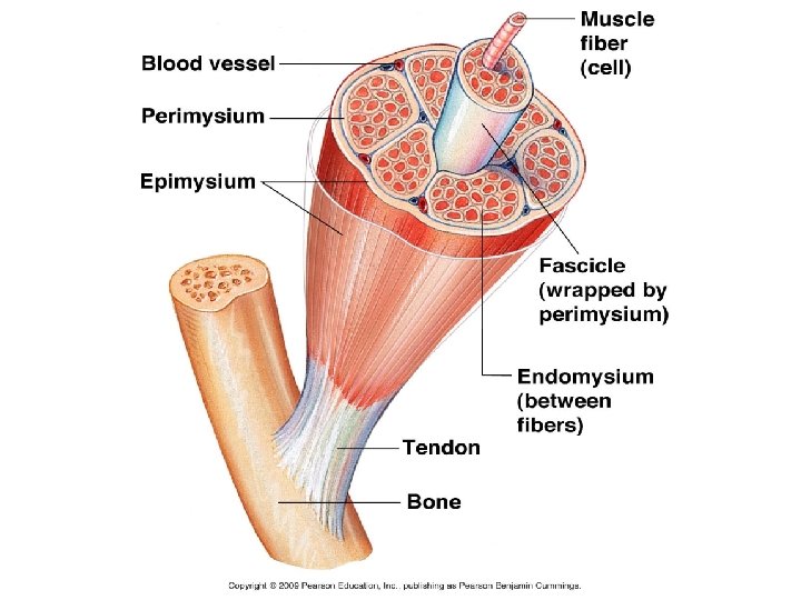
Structure of Skeletal Muscle • entire gross structure covered by connective tissue - epimysium • muscle is made of small bundles called fascicle which are bound by perimysium • each fascicle is made of a bundle of muscle cells or fibers which are surrounded by endomysium (all coverings are continuous extensions)
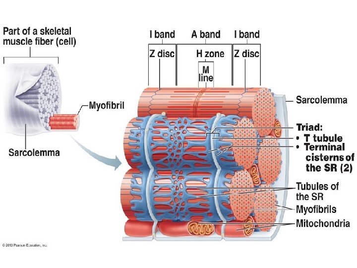
Muscle Fiber Structure
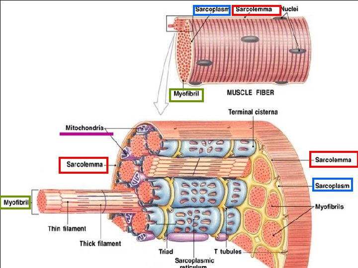
Muscle Fiber Structure • sarcolemma – muscle fiber plasma membrane • sarcoplasm – muscle fiber cytoplasm, contains myoglobin – a red pigment that stores oxygen • Each muscle fiber is made of bundles of rod-shaped structures of myofibrils (organelles). • (100’s to 1000’s per cell)
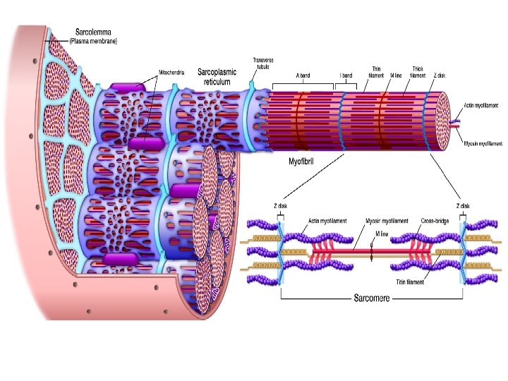
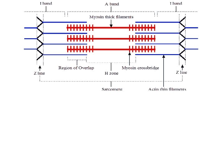
They have repeating pattern of striations called sarcomeres.
The banding is caused by an orderly overlapping arrangement of protein filaments - myofilaments
Myofilaments: 1. myosin - thick, extend entire length of A band 2. actin - thin, extend across the I band part way into A band
• Z line - attachment site for actin, ends of sarcomere • I band - light bands, actin only • A band - dark bands, myosin and actin overlap
• H zone - only visible in relaxed, myosin only • M line - sarcomere center
Structure Connective Tissue Epimysium surrounds organ Gross muscle Perimysium tissues Fascicle Endomysium surrounds cells organelles molecules (bundles of cells) Muscle fiber (muscle cell) Myofibril (contracting organelle) Myofilaments Actin – thin filament Myosin – thick filament
Connective Tissue Structure Epimysium surrounds Gross muscle organ Perimysium surrounds Fascicle tissues Endomysium surrounds (bundles of cells) Muscle fiber (muscle cell) cells Myofibril organelles Myofilaments molecules (contracting organelle) Actin – thin filament Myosin – thick filament
Molecular Structure of Myofilaments
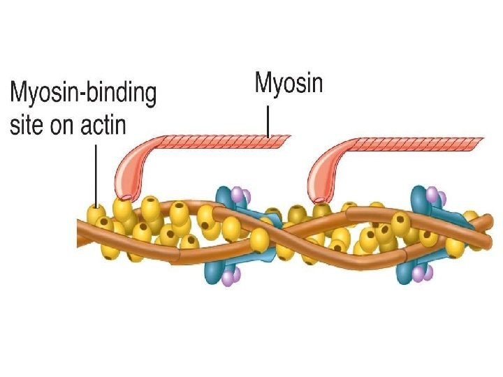
Myosin • tail rod like tail or axis ending in 2 globular heads - “crossbridges” - heads contain ATPase (enzyme) heads
Actin • globular subunits - G actin are found in long strands, like a double strand of twisted beads
Actin • tropomyosin spirals around beads, to add strength and stiffen, thin ribbon • troponin molecules bond actin to tropomyosin, has binding sites that will join with myosin crossbridge Binding sites
Sarcoplasmic Reticulum
Sarcoplasmic Reticulum • smooth endoplasmic reticulum of muscle cells • storage area for Ca ions • At H zones and A - I junctions tubes fuse and form lateral channels - terminal cisternae which feed into transverse tubules (T tubules) tubules at each Z line. • T tubules receive nerve stimuli and provide a pathway for oxygen, glucose, and Ca ions. (because continuous with membrane, nerve impulse allowed deep into muscle)
The End
• Muscles are attached to bones directly or indirectly by extensions of epimysium. indirect - are more common 1. rope like tendons 2. flat aponeurosis direct - a direct connection
Patterns of Arrangement • arrangement of fascicle produce muscles with different shapes and function • most common arrangements are: 1. parallel - strap-like muscles, ex: biceps 2. pennate - short, obliquely attached to central tendon, feather shape, ex: rectus femoris 3. convergent - broad origin converging to a single tendon, ex: pectoralis major 4. circular - circular fibers, control openings and closings, ex: sphincters, orbicularis oris, orbicularis oculi
• They have repeating pattern of striations called sarcomeres. Z line - attachment site for actin, ends of sarcomere I band - light bands, actin only A band - dark bands, myosin and actin overlap H zone - only visible in relaxed, myosin only M line - sarcomere center
• The banding is caused by an orderly overlapping arrangement of protein filaments - myofilaments. myosin - thick, extend entire length of A band actin - thin, extend across the I band part way into A band Z line - disklike protein sheet, anchors actin and connects each myofibril to the next H line - less dense (myosin only) M line - darker, fine strands connect adjacent myosins in this area
Sarcoplasmic Reticulum • smooth endoplasmic reticulum of muscle cells • its connecting tubules lie in spaces between myofibrils and run parallel to them • storage area for Ca ions • At H zones and A - I junctions tubes fuse and form lateral channels - terminal cisternae • which feed into transverse tubules (T tubules) at each Z line. (from penetration of sarcolemma) • T tubules receive nerve stimuli and provide a pathway for oxygen, glucose, and Ca ions. (because continuous with membrane, nerve impulse allowed deep into muscle)