
Histology Part III MUSCLE AND NERVOUS TISSUE THE

- Slides: 22
Histology: Part III MUSCLE AND NERVOUS TISSUE
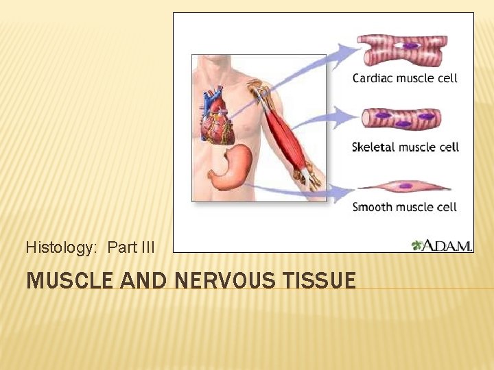
THE 3 MUSCLE TYPES 1. Skeletal – attached to bones of skeleton. 2. Cardiac – the contractile tissue of the heart. 3. Smooth – movement of materials through passageways or ducts.
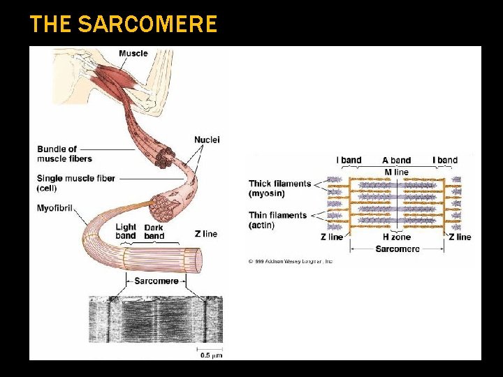
LOCATION AND DESCRIPTION Location/Description 1. Location – throughout the body 2. Description – Specialized Structures a. fibers shorten alongitudinal axis b. sarcoplasm – muscle cell cytoplasm c. sarcolemma – muscle cell membrane SARCOMERE = Functional muscle unit.
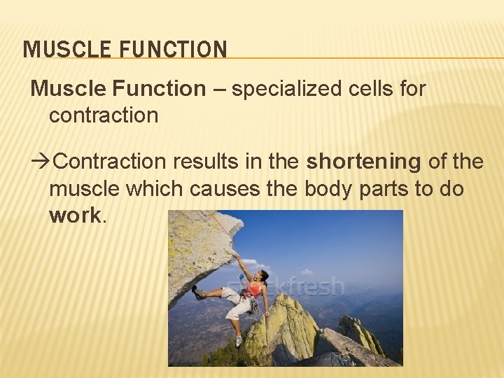
MUSCLE FUNCTION Muscle Function – specialized cells for contraction Contraction results in the shortening of the muscle which causes the body parts to do work.
THE SARCOMERE
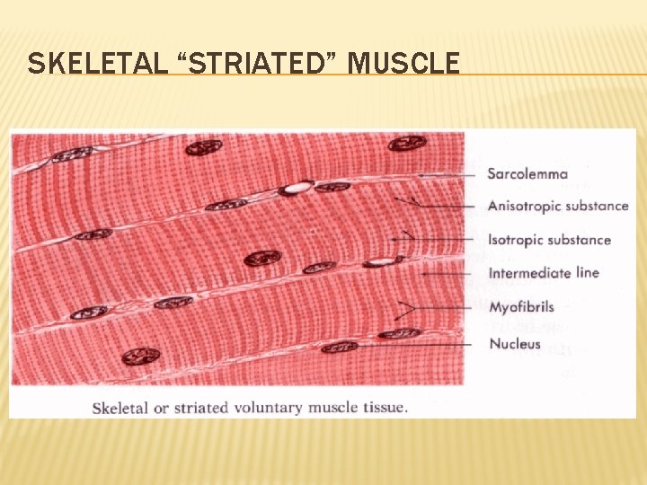
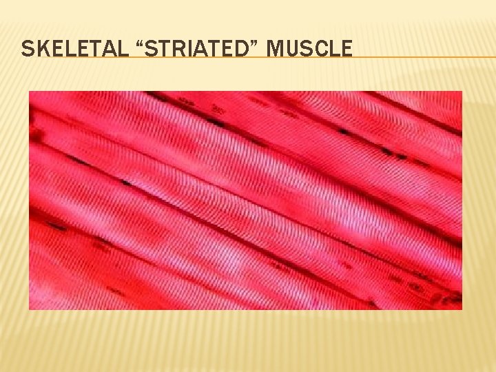
CLASSIFICATION: SKELETAL (STRIATED) § Classification 1: Skeletal Muscle (striated, voluntary muscle) A. Description § § very long fibers (1 ft. ) Multinucleated , sphincter control Shows a banded appearance can’t divide B. Location – all along skeleton C. Function – moves skeleton; guards entrances and exits to digestive, respiratory, and urinary tracts
SKELETAL “STRIATED” MUSCLE
SKELETAL “STRIATED” MUSCLE
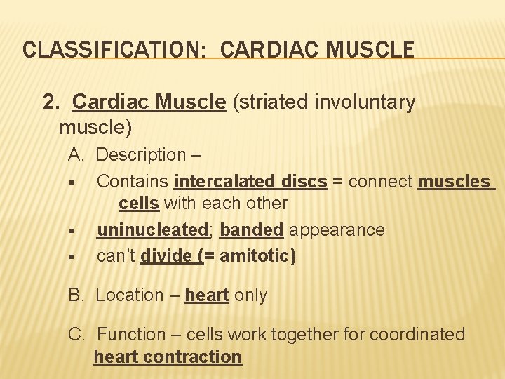
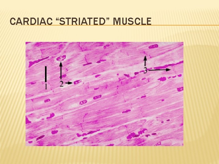
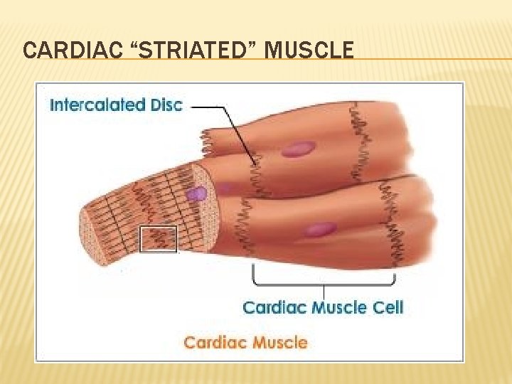
CLASSIFICATION: CARDIAC MUSCLE 2. Cardiac Muscle (striated involuntary muscle) A. Description – § Contains intercalated discs = connect muscles cells with each other § uninucleated; banded appearance § can’t divide (= amitotic) B. Location – heart only C. Function – cells work together for coordinated heart contraction
CARDIAC “STRIATED” MUSCLE
CARDIAC “STRIATED” MUSCLE
CARDIAC “STRIATED” MUSCLE
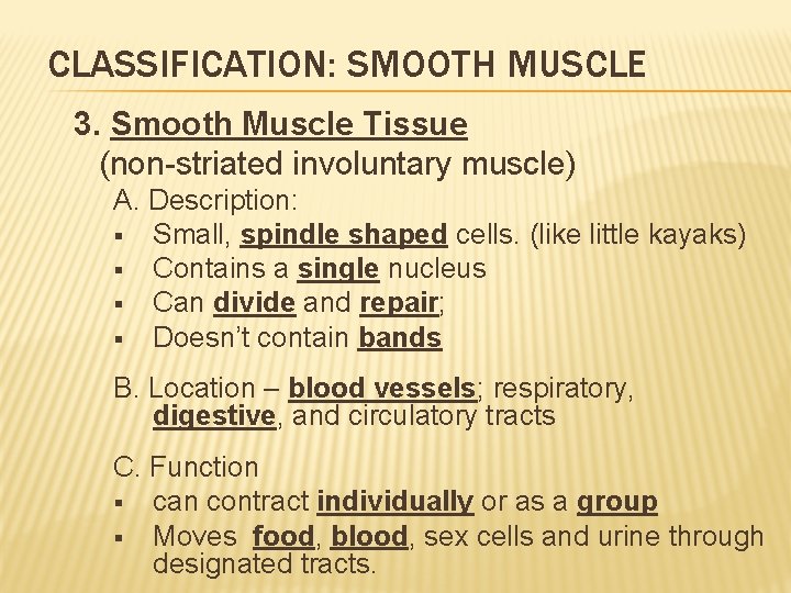
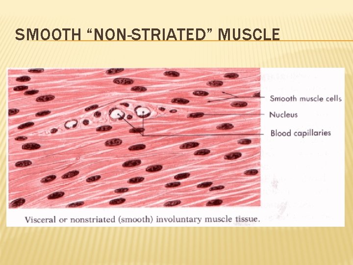
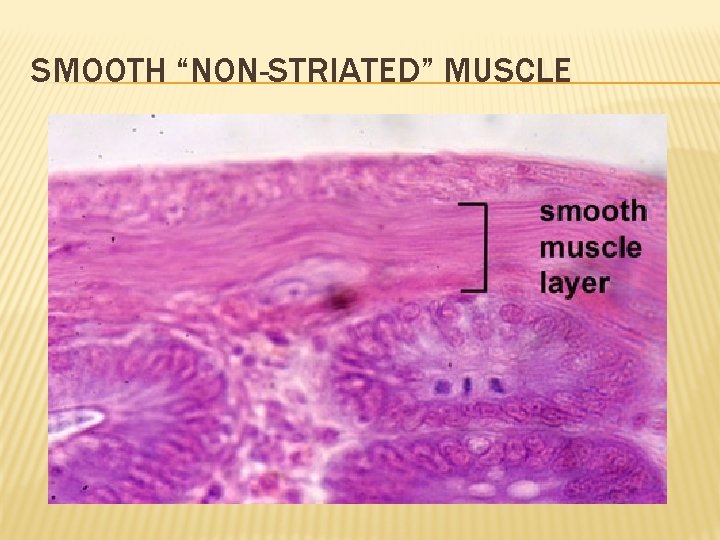
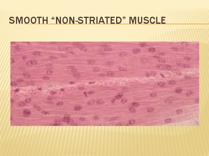
CLASSIFICATION: SMOOTH MUSCLE 3. Smooth Muscle Tissue (non-striated involuntary muscle) A. Description: § Small, spindle shaped cells. (like little kayaks) § Contains a single nucleus § Can divide and repair; § Doesn’t contain bands B. Location – blood vessels; respiratory, digestive, and circulatory tracts C. Function § can contract individually or as a group § Moves food, blood, sex cells and urine through designated tracts.
SMOOTH “NON-STRIATED” MUSCLE
SMOOTH “NON-STRIATED” MUSCLE
SMOOTH “NON-STRIATED” MUSCLE
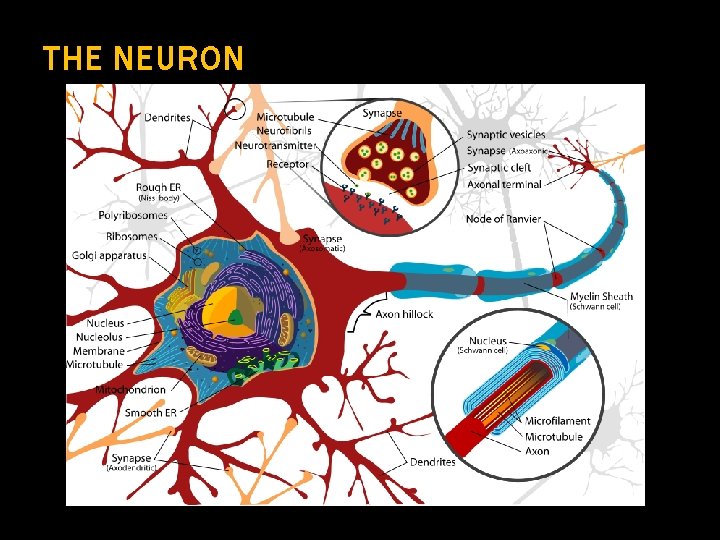
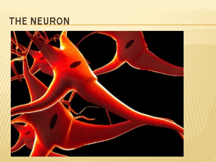
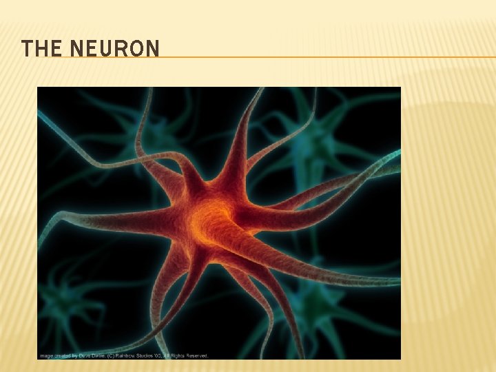
NERVOUS TISSUE A. Location/Description 1. Location – make up 98% brain and spinal cord 2. Description of Neurons § Neurons – longest cells in body (39 inches) § Incapable of dividing (=amitotic) § Cell body contains nucleus and other essential organelles. § Dendrites receive info from the axon terminals of the adjacent neuron. § Axon transmits electrical impulse through cell.
THE NEURON
THE NEURON
THE NEURON
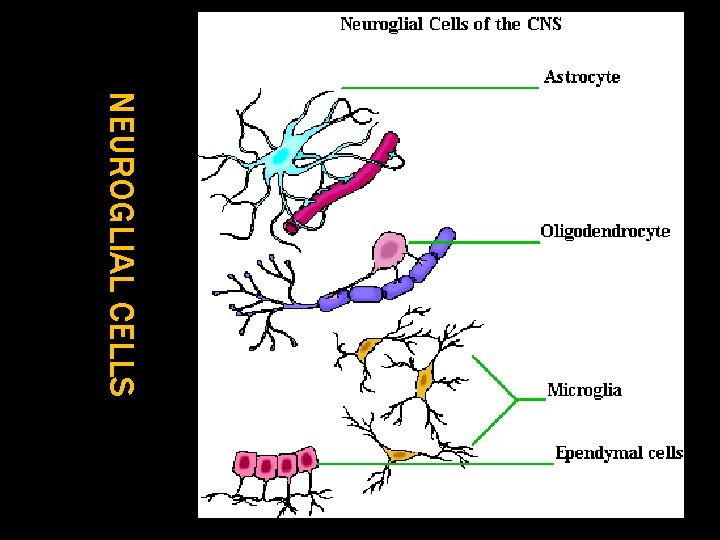
NERVOUS TISSUE A. Location/Description 3. Description of Neuroglial Cells = non-neuronal cells that support neurons in brain and spine. Functions of Neuroglia: a. Surround and stabilize neurons. b. Form myelin that insulates neurons from each other. c. Supply nutrients and oxygen to neurons. d. Destroy pathogens and remove dead neurons.
NEUROGLIAL CELLS