
Chapter 7 General Physiology Copyright 2015 by Saunders
Chapter 7 General Physiology Copyright © 2015 by Saunders, an imprint of Elsevier Inc. All rights reserved.
Lesson 7. 1 Skeletal and Muscular Systems 1. 2. 3. 4. Pronounce, define, and spell the Key Terms. Explain why understanding physiology is important to the dental assistant. Locate the skeletal system; explain its purpose, components, and functions; and describe the signs and symptoms of its common disorders. Locate the muscular system; explain its purpose, components, and functions; and describe the signs and symptoms of its common disorders. Copyright © 2015 by Saunders, an imprint of Elsevier Inc. All rights reserved. 2
Introduction Physiology is the study of how living things function It continues beyond the study of anatomy into how parts work, what they do, and why Copyright © 2015 by Saunders, an imprint of Elsevier Inc. All rights reserved. 3
General Physiology The human body has 10 systems Each system has specific organs within it Each body system performs specific functions In a healthy human being, all 10 systems are functioning Copyright © 2015 by Saunders, an imprint of Elsevier Inc. All rights reserved. 4
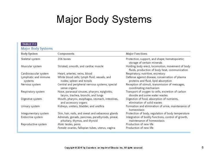
Major Body Systems Copyright © 2015 by Saunders, an imprint of Elsevier Inc. All rights reserved. 5
Skeletal System There are 206 bones in the human body For descriptive purposes, the skeleton is divided into the axial and appendicular skeletal systems Copyright © 2015 by Saunders, an imprint of Elsevier Inc. All rights reserved. 6
Axial Skeleton Consists of the skull, spinal column, ribs, and sternum Its function is to protect the major organs of the nervous, respiratory, and circulatory systems The bones of the skull are of primary importance to the dental assistant Copyright © 2015 by Saunders, an imprint of Elsevier Inc. All rights reserved. 7
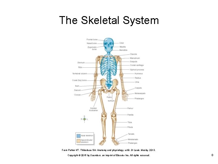
The Skeletal System From Patton KT, Thibodeau GA: Anatomy and physiology, ed 8, St Louis, Mosby, 2013. Copyright © 2015 by Saunders, an imprint of Elsevier Inc. All rights reserved. 8
Appendicular Skeleton Consists of the upper extremities and shoulder girdle plus the lower extremities and pelvic girdle It protects the organs of digestion and reproduction Copyright © 2015 by Saunders, an imprint of Elsevier Inc. All rights reserved. 9
Bone is the hard connective tissue that makes up most of the human skeleton It consists of an organic component (the cells and matrix) and an inorganic (mineral) component Ø Ø The minerals, mainly calcium and phosphate, give rigidity to bone These minerals stored in bone also act as a mineral reservoir to maintain essential blood mineral concentrations in times of inadequate supply in the body Copyright © 2015 by Saunders, an imprint of Elsevier Inc. All rights reserved. 10
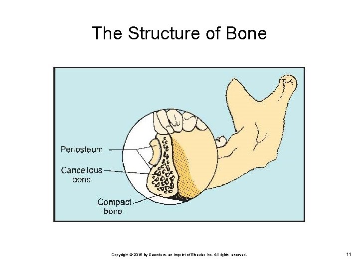
The Structure of Bone Copyright © 2015 by Saunders, an imprint of Elsevier Inc. All rights reserved. 11
The Periosteum A specialized connective tissue covering all bones of the body It is responsible for the life of the bone and is capable of repair Ø Ø The outer layer is a network of dense connective tissue containing blood vessels The inner layer is loose connective tissue containing osteoblasts Copyright © 2015 by Saunders, an imprint of Elsevier Inc. All rights reserved. 12
Kinds of Bone Compact bone Ø Ø Hard, dense, and very strong It forms the outer layer of bone, where it is needed for strength Cancellous bone Ø Ø Lighter in weight, but not as strong as compact bone It is found in the interior of bones Copyright © 2015 by Saunders, an imprint of Elsevier Inc. All rights reserved. 13
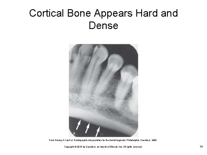
Cortical Bone Appears Hard and Dense From Haring JI, Lind LJ: Radiographic interpretation for the dental hygienist, Philadelphia, Saunders, 1993. Copyright © 2015 by Saunders, an imprint of Elsevier Inc. All rights reserved. 14
Trabeculae Bony spicules in cancellous bone that form a mesh of intercommunicating spaces filled with bone marrow Appear as a weblike structure on radiographs Copyright © 2015 by Saunders, an imprint of Elsevier Inc. All rights reserved. 15
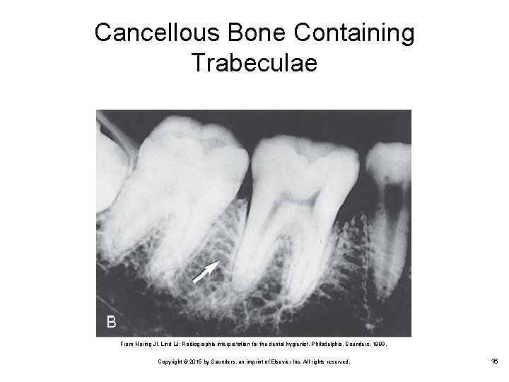
Cancellous Bone Containing Trabeculae From Haring JI, Lind LJ: Radiographic interpretation for the dental hygienist, Philadelphia, Saunders, 1993. Copyright © 2015 by Saunders, an imprint of Elsevier Inc. All rights reserved. 16
Bone Marrow Inside the spaces of cancellous bone are two types of bone marrow: Ø Red marrow • • Ø Filled with blood vessels Makes red and white blood cells and platelets Yellow marrow • • Contains mainly fat cells Found primarily in the shafts of long bones Copyright © 2015 by Saunders, an imprint of Elsevier Inc. All rights reserved. 17
Cartilage It is tough, yet more elastic than bone It is found where bones join together Nonvascular connective tissue In addition to the ends of bone, cartilage gives form to the nose and ears Copyright © 2015 by Saunders, an imprint of Elsevier Inc. All rights reserved. 18
Joints Areas where two bones come together There are three basic types of joints Ø Ø Fibrous: Do not move; example: the sutures of the skull Cartilaginous: Made of connective tissue and cartilage • Ø An example is the joints between the bones of the vertebrae Synovial: The movable joints, account for most of the joints in the body Copyright © 2015 by Saunders, an imprint of Elsevier Inc. All rights reserved. 19
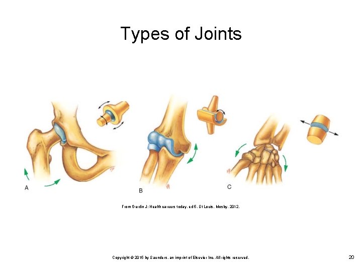
Types of Joints From Gerdin J: Health careers today, ed 5, St Louis, Mosby, 2012. Copyright © 2015 by Saunders, an imprint of Elsevier Inc. All rights reserved. 20
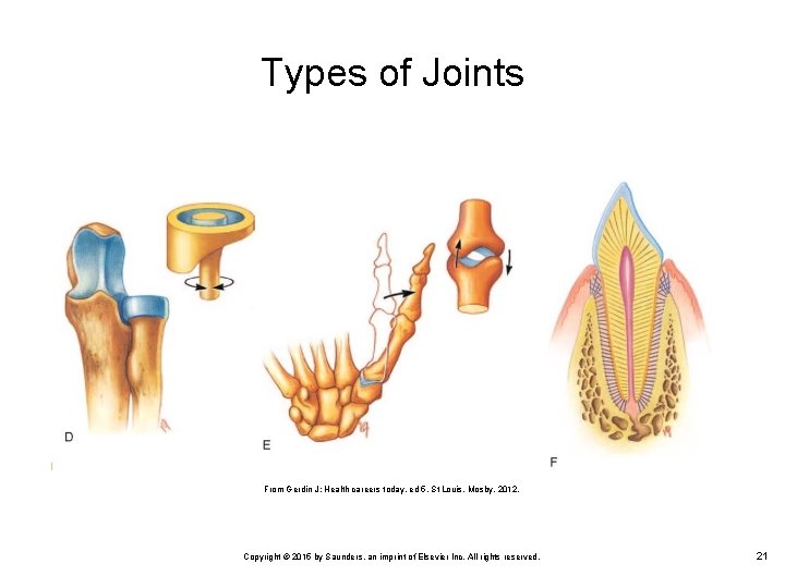
Types of Joints From Gerdin J: Health careers today, ed 5, St Louis, Mosby, 2012. Copyright © 2015 by Saunders, an imprint of Elsevier Inc. All rights reserved. 21
Muscular System Gives us the ability to stand, walk, run, jump, move our eyes, smile, and frown It is composed of more than 600 individual muscles For muscles to make the body move, they must work together Each muscle consists of muscle tissue, connective tissue, nerve tissue, and vascular (blood) tissue Copyright © 2015 by Saunders, an imprint of Elsevier Inc. All rights reserved. 22
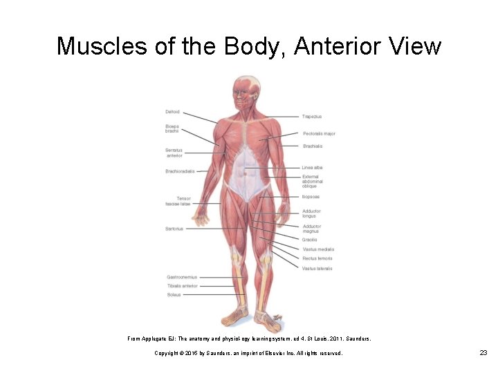
Muscles of the Body, Anterior View From Applegate EJ: The anatomy and physiol ogy learning system, ed 4, St Louis, 2011, Saunders. Copyright © 2015 by Saunders, an imprint of Elsevier Inc. All rights reserved. 23
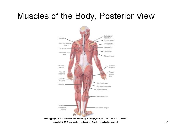
Muscles of the Body, Posterior View From Applegate EJ: The anatomy and physiol ogy learning system, ed 4, St Louis, 2011, Saunders. Copyright © 2015 by Saunders, an imprint of Elsevier Inc. All rights reserved. 24
Types of Muscles Striated muscle Ø Ø Ø Smooth muscle Ø So named because dark and light bands in the muscle fibers yield a striped, or striated, appearance Is known as skeletal or voluntary muscle Attached to the bones of the skeleton; makes bodily motion possible Moves the internal organs, such as the digestive tract, blood vessels, and secretion ducts leading from glands Cardiac muscle Copyright © 2015 by Saunders, an imprint of Elsevier Inc. All rights reserved. 25
Smooth Muscle Involuntary muscle is under the control of the autonomic nervous system and is not controlled voluntarily Visceral muscle is so named because it is found in the visceral (internal) organs, except the heart It is also found in hollow structures such as the digestive and urinary tracts Copyright © 2015 by Saunders, an imprint of Elsevier Inc. All rights reserved. 26
Cardiac Muscle Striated in appearance but resembles smooth muscle in action Cardiac muscle forms most of the walls of the heart, and contraction of this muscle results in the heartbeat It combines the power of striated voluntary muscles with the steady reliability of smooth, involuntary ones Copyright © 2015 by Saunders, an imprint of Elsevier Inc. All rights reserved. 27
Muscle Function Contraction is the tightening of a muscle, during which it becomes shorter and thicker Relaxation occurs when a muscle returns to its original form or shape Ø Ø The muscles of the body are arranged in opposing pairs so that when one contracts, the other relaxes It is these contrasting actions that make motion possible Copyright © 2015 by Saunders, an imprint of Elsevier Inc. All rights reserved. 28
Muscle Function (Cont. ) Muscle origin Ø Ø The place where the muscle begins (originates) This is the more fixed attachment, the end of the muscle that is closer to the midline of the body, or both Muscle insertion Ø Ø The place where the muscle ends (inserts) It is the more movable end, the portion of the muscle that is farther from the midline of the body, or both Copyright © 2015 by Saunders, an imprint of Elsevier Inc. All rights reserved. 29
Lesson 7. 2 Cardiovascular and Nervous Systems 5. 6. Locate the cardiovascular system; explain its purpose, components, and functions; and describe the signs and symptoms of its common disorders. Locate the nervous system; explain its purpose, components, and functions; and describe the signs and symptoms of its common disorders. Copyright © 2015 by Saunders, an imprint of Elsevier Inc. All rights reserved. 30
Cardiovascular System Consists of the: Ø Ø Ø Circulatory system Heart Lymphatic system These systems provide life sustaining capabilities for the survival of body cells and tissues Copyright © 2015 by Saunders, an imprint of Elsevier Inc. All rights reserved. 31
Circulatory System The two primary functions of the circulatory system are: Ø Ø Transporting oxygen and nutrients to the cells and transporting carbon dioxide and waste products from the cells, as well as transporting hormones and antibodies throughout the body Regulating body temperature and chemical stability Copyright © 2015 by Saunders, an imprint of Elsevier Inc. All rights reserved. 32
Heart Each day the heart pumps 4000 gallons of blood at a speed of 40 mph through 70, 000 miles of vessels The heart is a hollow muscle with four chambers Heart size varies from individual to individual but is generally the same as a closed fist Copyright © 2015 by Saunders, an imprint of Elsevier Inc. All rights reserved. 33
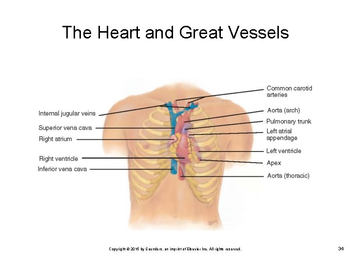
The Heart and Great Vessels Copyright © 2015 by Saunders, an imprint of Elsevier Inc. All rights reserved. 34
The Pericardium The heart is enclosed in a double walled membranous sac known as the pericardium Pericardial fluid between the layers prevents friction when the heart beats Copyright © 2015 by Saunders, an imprint of Elsevier Inc. All rights reserved. 35
Heart Chambers The heart functions as a double pump: Ø Ø The right side pumps blood to the lungs The left side sends blood to the rest of the body The coronary vessels supply blood to the heart muscle Each side is subdivided into an upper and a lower chamber Copyright © 2015 by Saunders, an imprint of Elsevier Inc. All rights reserved. 36
Heart Valves One way valves prevent the backflow of blood and separate the chambers of the heart by opening and closing with each heartbeat Ø Ø The tricuspid valve is found between the right atrium and the right ventricle The mitral valve has two cusps and lies between the left atrium and the left ventricle Two semilunar valves have three crescent shaped flaps Ø Ø Pulmonary semilunar valve allows blood to flow from the right ventricle into the pulmonary artery Aortic semilunar valve allows blood flow from the left ventricle into the aorta Copyright © 2015 by Saunders, an imprint of Elsevier Inc. All rights reserved. 37
Blood Flow Through the Heart The right atrium receives blood from the superior and inferior venae cavae Blood flows from the right atrium into the right ventricle The right ventricle receives blood from the right atrium and pumps it into the pulmonary artery, which carries it to the lungs The left atrium receives oxygenated blood from the lungs through the four pulmonary veins Ø Blood flows from here into the left ventricle The left ventricle receives blood from the left atrium Ø Blood then goes to the aorta, and is pumped to all parts of the body, except the lungs Copyright © 2015 by Saunders, an imprint of Elsevier Inc. All rights reserved. 38
Blood Vessels Arteries are large blood vessels that carry blood away from the heart to all regions of the body Capillaries are a system of microscopic vessels that connect the arterial and venous systems Veins form a low pressure collecting system to return the waste filled blood to the heart Copyright © 2015 by Saunders, an imprint of Elsevier Inc. All rights reserved. 39
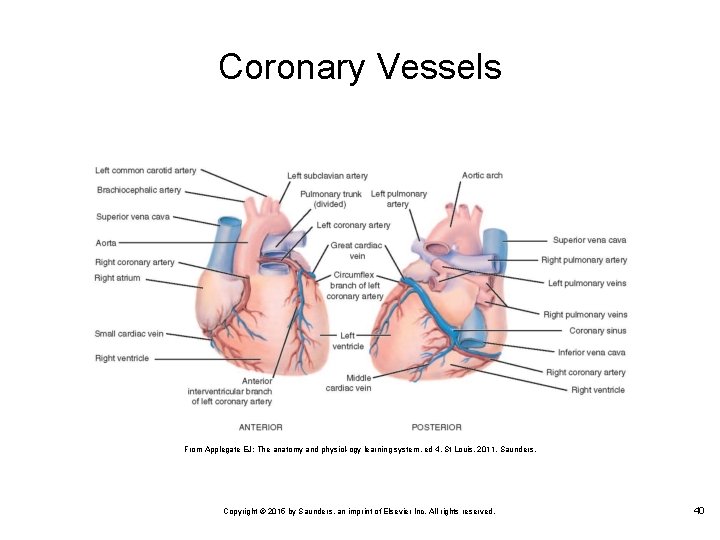
Coronary Vessels From Applegate EJ: The anatomy and physiol ogy learning system, ed 4, St Louis, 2011, Saunders. Copyright © 2015 by Saunders, an imprint of Elsevier Inc. All rights reserved. 40
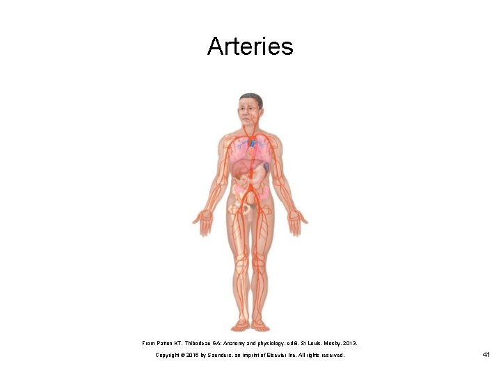
Arteries From Patton KT, Thibodeau GA: Anatomy and physiology, ed 8, St Louis, Mosby, 2013. Copyright © 2015 by Saunders, an imprint of Elsevier Inc. All rights reserved. 41
Blood and Blood Cells Plasma is a straw colored fluid that transports nutrients, hormones, and waste products Plasma is 91% water Red blood cells contain the blood protein hemoglobin, which plays an essential role in oxygen transport White blood cells, also known as leukocytes, have the primary function of fighting disease in the body Copyright © 2015 by Saunders, an imprint of Elsevier Inc. All rights reserved. 42
Blood Typing and Rh Factor The safe administration of blood from donor to recipient requires typing and cross matching Blood typing is based on the antigens and antibodies found in the blood The most important classifications are A, AB, B, and O Patients who receive blood that is incompatible with their own may experience a serious and possibly fatal reaction Copyright © 2015 by Saunders, an imprint of Elsevier Inc. All rights reserved. 43
Blood Typing and Rh Factor (Cont. ) The Rh factor is an additional antigen that is present on the surfaces of red blood cells in some individuals A person whose blood contains the factor is Rh positive A person whose blood does not contain the factor is Rh negative Anti Rh antibodies are not naturally found in plasma as they are in blood types, but they do develop if the patient is exposed to the Rh factor Copyright © 2015 by Saunders, an imprint of Elsevier Inc. All rights reserved. 44
Lymphatic System The structures of the lymphatic system: Ø Ø Lymph vessels Lymph nodes Lymph fluid Lymphoid organs Drainage vessels absorb excess protein from tissues and return it to the bloodstream Lymphoid organs contribute to the immune system to assist with destruction of harmful microorganisms Copyright © 2015 by Saunders, an imprint of Elsevier Inc. All rights reserved. 45
Lymph Vessels Lymph capillaries are thin walled tubes that carry lymph from the tissue spaces to the larger lymphatic vessels Ø Ø Similar to veins, lymphatic vessels have valves that prevent the backflow of fluid Lymph fluid always flows toward the thoracic cavity, where it empties into veins in the upper thoracic region Specialized lymph vessels, called lacteals, are located in the small intestine Ø Lacteals aid in the absorption of fats from the small intestine into the bloodstream Copyright © 2015 by Saunders, an imprint of Elsevier Inc. All rights reserved. 46
Lymph Nodes Small round or oval structures that are located in the lymph vessels They fight disease by producing antibodies; this is part of the immune reaction Major lymph node sites of the body include: Ø Ø Cervical (in the neck) Axillary (under the arms) Inguinal (in the lower abdomen) Lymph fluid Copyright © 2015 by Saunders, an imprint of Elsevier Inc. All rights reserved. 47
Lymphoid Organs Tonsils Ø Ø Masses of lymphatic tissue that are located in the upper portions of the nose and throat, where they form a protective ring of lymphatic tissue Nasopharyngeal tonsils are found in the nasopharynx Palatine tonsils are located in the oropharynx between the anterior and posterior pillars of fauces Lingual tonsils are located on the back of the tongue Copyright © 2015 by Saunders, an imprint of Elsevier Inc. All rights reserved. 48
Lymphoid Organs (Cont. ) Spleen Ø Ø Produces lymphocytes and monocytes, which are important components of the immune system It also filters microorganisms and other debris not destroyed by the lymphatic system Copyright © 2015 by Saunders, an imprint of Elsevier Inc. All rights reserved. 49
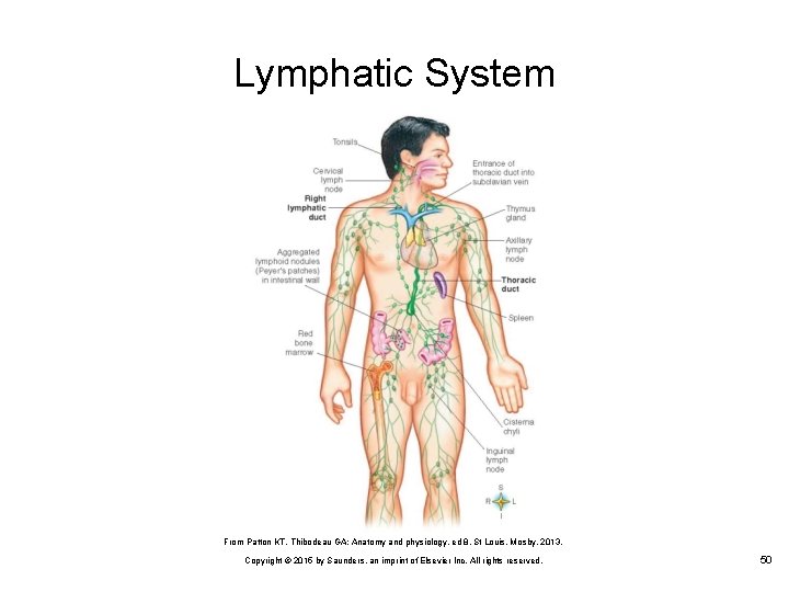
Lymphatic System From Patton KT, Thibodeau GA: Anatomy and physiology, ed 8, St Louis, Mosby, 2013. Copyright © 2015 by Saunders, an imprint of Elsevier Inc. All rights reserved. 50
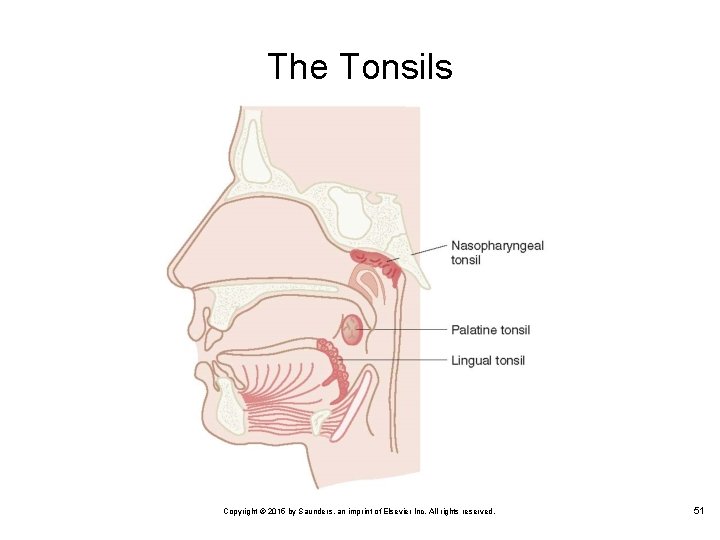
The Tonsils Copyright © 2015 by Saunders, an imprint of Elsevier Inc. All rights reserved. 51
Nervous System The nervous system is the communication system of the body Instructions and directions are sent out by this system to the various organs in the body The nervous system can be compared to computer communications, with the brain and spinal cord as the main computer and the nerves as the cables that carry messages to and from this center Copyright © 2015 by Saunders, an imprint of Elsevier Inc. All rights reserved. 52
Nervous System (Cont. ) Central nervous system (CNS) Ø The brain and the spinal cord Peripheral nervous system (PNS) Ø The cranial nerves and the spinal nerves Copyright © 2015 by Saunders, an imprint of Elsevier Inc. All rights reserved. 53
Neurons The basic function of neurons is to direct communication or nerve impulses Neurons have the properties of excitability, the ability to respond to a stimulus, and conductivity, which refers to transmission of an impulse There are three types of neurons: Ø Ø Ø Sensory neurons Motor neurons Associative neurons Copyright © 2015 by Saunders, an imprint of Elsevier Inc. All rights reserved. 54
Neurons (Cont. ) A synapse is the space between two neurons or between a neuron and a receptor organ A neurotransmitter is a chemical substance that allows the impulse to jump across the synapse from one neuron to another Nerves covered with myelin are referred to as white matter Nerves that do not have the protective myelin sheath make up the gray matter of the brain and spinal cord Copyright © 2015 by Saunders, an imprint of Elsevier Inc. All rights reserved. 55
Central Nervous System Brain Ø The brain is the primary center for the regulation and coordination of the body’s activities, and each part of the brain controls different aspects of body function Spinal cord Ø The spinal cord carries all of the nerves that affect the limbs and lower part of the body and is the pathway for impulses going to and from the brain Copyright © 2015 by Saunders, an imprint of Elsevier Inc. All rights reserved. 56
Peripheral Nervous System The autonomic nervous system controls unconscious activities such as breathing, heart rate, body temperature, blood pressure, and pupil size The somatic nervous system controls conscious activities Copyright © 2015 by Saunders, an imprint of Elsevier Inc. All rights reserved. 57
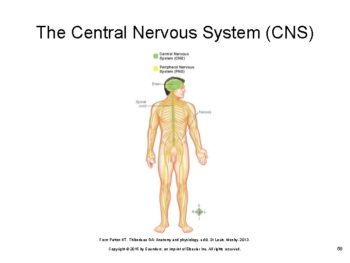
The Central Nervous System (CNS) From Patton KT, Thibodeau GA: Anatomy and physiology, ed 8, St Louis, Mosby, 2013. Copyright © 2015 by Saunders, an imprint of Elsevier Inc. All rights reserved. 58
Lesson 7. 3 Respiratory, Digestive, Endocrine, Urinary, Integumentary, and Reproductive Systems 7. 8. 9. Locate the respiratory system; explain its purpose, components, and functions; and describe the signs and symptoms of its common disorders. Locate the digestive system; explain its purpose, components, and functions; and describe the signs and symptoms of its common disorders. Locate the endocrine system; explain its purpose, components and functions; and describe the signs and symptoms of its common disorders. Copyright © 2015 by Saunders, an imprint of Elsevier Inc. All rights reserved. 59
Lesson 7. 3 Respiratory, Digestive, Endocrine, Urinary, Integumentary, and Reproductive Systems 10. 11. 12. 13. Locate the urinary system; explain its purpose, components, and functions; and describe the signs and symptoms of its common disorders. Locate the integumentary system; explain its purpose, components, and functions; and describe the signs and symptoms of its common disorders. Locate the reproductive system; explain its purpose, components, and functions; and describe the signs and symptoms of its common disorders. Give examples of conditions that require interaction among body systems. Copyright © 2015 by Saunders, an imprint of Elsevier Inc. All rights reserved. 60
Respiratory System Delivers oxygen to the millions of cells in the body and transports the waste product carbon dioxide out of the body The respiratory system consists of the: Ø Ø Ø Ø Nose Paranasal sinuses Pharynx Epiglottis Larynx Trachea Alveoli Lungs Copyright © 2015 by Saunders, an imprint of Elsevier Inc. All rights reserved. 61
Nose Air enters the body through the nostrils of the nose and passes through the nasal cavity Ø The nose is divided by a wall of cartilage called the nasal septum The nose and the respiratory system are lined with mucous membrane, a specialized form of epithelial tissue Incoming air is filtered by the cilia, which are thin hairs that are attached to the mucous membrane just inside the nostrils Copyright © 2015 by Saunders, an imprint of Elsevier Inc. All rights reserved. 62
Pharynx After passing through the nasal cavity, air reaches the pharynx, which is commonly known as the throat There are three divisions of the pharynx: Ø Ø Ø The nasopharynx is located behind the nose and above the soft palate The oropharynx extends from the soft palate above to the level of the epiglottis below The laryngopharynx extends from the level of the epiglottis above to the larynx below Copyright © 2015 by Saunders, an imprint of Elsevier Inc. All rights reserved. 63
Epiglottis The oropharynx and the laryngopharynx serve as a common passageway for food from the mouth and air from the nose During swallowing, the epiglottis acts as a lid and covers the larynx so that food does not enter the lungs Copyright © 2015 by Saunders, an imprint of Elsevier Inc. All rights reserved. 64
Larynx Also known as the voice box, it contains the vocal bands, which make speech possible The larynx is protected and is held open by a series of cartilaginous structures The largest cartilage forms the prominent projection in front of the neck, called the “Adam’s apple” Copyright © 2015 by Saunders, an imprint of Elsevier Inc. All rights reserved. 65
Trachea Air passes from the larynx to the trachea The trachea extends from the neck into the chest, directly in front of the esophagus It is protected and held open by a series of C shaped cartilaginous rings Copyright © 2015 by Saunders, an imprint of Elsevier Inc. All rights reserved. 66
Lungs The trachea divides into two branches called bronchi Ø Each bronchus leads to a lung, where it divides and subdivides into increasingly smaller branches; bronchioles are the smallest of these branches Alveoli are tiny grapelike clusters found at the end of each bronchiole Ø Exchange of gases between the lungs and the blood takes place in the alveoli Copyright © 2015 by Saunders, an imprint of Elsevier Inc. All rights reserved. 67
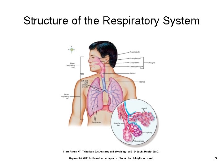
Structure of the Respiratory System From Patton KT, Thibodeau GA: Anatomy and physiology, ed 8, St Louis, Mosby, 2013. Copyright © 2015 by Saunders, an imprint of Elsevier Inc. All rights reserved. 68
Digestive System The role of the digestive system is to provide the body with the nutrients, water, and electrolytes necessary for life The digestive system functions under involuntary control Copyright © 2015 by Saunders, an imprint of Elsevier Inc. All rights reserved. 69
Digestive Process Ingestion: Occurs when food is taken into the mouth Digestion: Begins in the mouth as food is masticated with saliva and then swallowed Movement: The chewed food is subjected to peristalsis, the rhythmic wavelike contractions that move the food through the digestive tract Absorption: The nutritional elements in the gastrointestinal tract pass through the lining of the gastrointestinal tract and into the bloodstream Elimination: In the large intestine, the solid by products of digestion are eliminated from the body in feces Copyright © 2015 by Saunders, an imprint of Elsevier Inc. All rights reserved. 70
Structures of the Digestive System The major structures of the digestive system are the: Ø Ø Ø Ø Mouth Pharynx Esophagus Stomach Small intestine Large intestine Liver, gallbladder, and pancreas Copyright © 2015 by Saunders, an imprint of Elsevier Inc. All rights reserved. 71
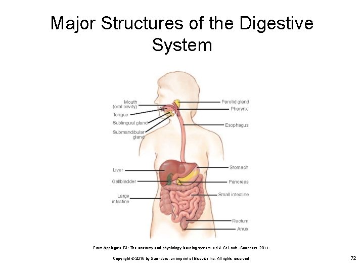
Major Structures of the Digestive System From Applegate EJ: The anatomy and physiology learning system, ed 4, St Louis, Saunders, 2011. Copyright © 2015 by Saunders, an imprint of Elsevier Inc. All rights reserved. 72
Endocrine System The endocrine system consists of ductless glands and tissues that help regulate the body’s activities The glands secrete hormones directly into the blood These hormones affect metabolism, growth, and development The hormones are secreted directly into the bloodstream, not through a duct Copyright © 2015 by Saunders, an imprint of Elsevier Inc. All rights reserved. 73
Endocrine Glands Thyroid gland Parathyroid gland Ovaries Testes Pituitary gland Pancreas Adrenal medulla Copyright © 2015 by Saunders, an imprint of Elsevier Inc. All rights reserved. 74
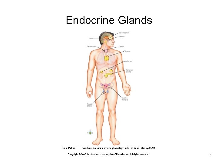
Endocrine Glands From Patton KT, Thibodeau GA: Anatomy and physiology, ed 8, St Louis, Mosby, 2013. Copyright © 2015 by Saunders, an imprint of Elsevier Inc. All rights reserved. 75
Urinary System The urinary system is also known as the excretory system Its principal function is to maintain fluid volume and composition of the body fluids Waste products leave the body in the form of urine, and the needed substances are returned to the blood Copyright © 2015 by Saunders, an imprint of Elsevier Inc. All rights reserved. 76
Organs of the Urinary System The kidneys are the site of formation of urine, which carries waste materials away from the blood The ureters transport the urine from the kidney The bladder stores urine until it can be disposed of The urethra is the conduit through which the bladder is emptied during the process of urination Copyright © 2015 by Saunders, an imprint of Elsevier Inc. All rights reserved. 77
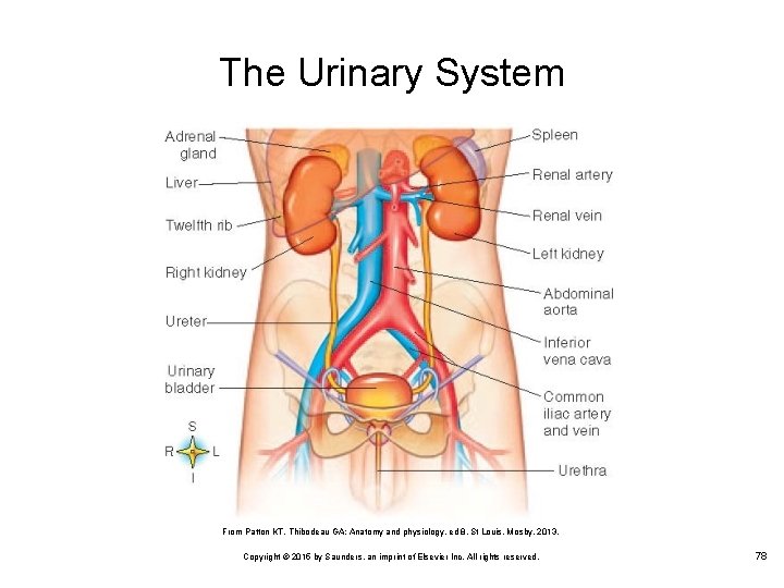
The Urinary System From Patton KT, Thibodeau GA: Anatomy and physiology, ed 8, St Louis, Mosby, 2013. Copyright © 2015 by Saunders, an imprint of Elsevier Inc. All rights reserved. 78
Integumentary System The skin is the body’s first line of defense against disease Functions of the skin system: Ø Ø Ø Helps regulate body temperature Provides a barrier that prevents bacteria from entering the body Excretes liquids and salts Provides sensitivity to touch Uses ultraviolet rays from the sun to convert chemicals into vitamin D necessary for absorption of calcium Copyright © 2015 by Saunders, an imprint of Elsevier Inc. All rights reserved. 79
Skin Structures The skin is composed of different layers, including: Ø Epidermis • Ø Dermis • Ø The outer layer of the skin; has no blood supply of its own Contains many free nerve endings and receptors, which allow for detection of touch, temperature, and pain Subcutaneous fat • A layer of loose connective tissue that anchors the skin to underlying organs; insulates the body against heat loss and cushions underlying organs Copyright © 2015 by Saunders, an imprint of Elsevier Inc. All rights reserved. 80
Skin Appendages Hair Ø Nails Ø Enclosed in a follicle and consists of a root and a shaft Contains a root and a body Glands Ø Ø Ø Sebaceous glands Sudoriferous glands Apocrine sweat glands Copyright © 2015 by Saunders, an imprint of Elsevier Inc. All rights reserved. 81
Female Reproductive System External Ø Ø Mons pubis Labia majora and minora Vulva Clitoris Internal Ø Ø Ovaries Fallopian tubes Uterus Vagina Copyright © 2015 by Saunders, an imprint of Elsevier Inc. All rights reserved. 82
Male Reproductive System Produces and transports sperm In the male, several organs serve as parts of both the urinary tract and reproductive system This system consists of the: Ø Ø Excretory ducts Testes Prostate Seminal vesicles Copyright © 2015 by Saunders, an imprint of Elsevier Inc. All rights reserved. 83
Interaction Among the 10 Body Systems Body systems do not operate independently Exert important effects on each other Something happening to one system tends to affect other systems Copyright © 2015 by Saunders, an imprint of Elsevier Inc. All rights reserved. 84

Questions? Copyright © 2015 by Saunders, an imprint of Elsevier Inc. All rights reserved. 85
- Slides: 85