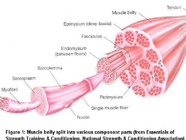
Skeletal Muscle Tissue Hierarchy Muscle belly fasciculus myofiber

Skeletal Muscle Tissue
 → fasciculus → myofiber (cell) → myofibril → myofilaments")
Hierarchy Muscle (belly) → fasciculus → myofiber (cell) → myofibril → myofilaments
 Junction Where nerves meet the muscle to give message")
Myoneural (neuromuscular) Junction Where nerves meet the muscle to give message

Color Code: Anatomy of Muscle Tendon-connects muscle to periostium b/c. belly is surrounded by epimysium d. fasciculus-bundle of fibers surrounded by perimysium e. Myofibers-surrounded by endomysium f/g. myofibril- made up of myofilaments a.


Muscle Size Ø The number of myofibers doesn’t change a. Atrophy: decrease in size due to lack of use in extended atrophy, the fiber will actually die. If you don’t use it, it will shut down!
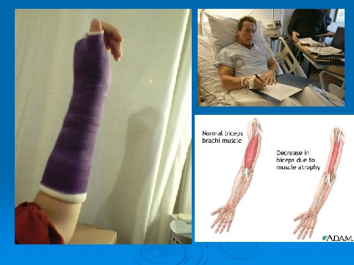

Atrophy can be reversed unless it is permanent damage!!!!!

Hypertrophy Ø Increase in number and size of myofibrils

Testosterone Ø Stimulates hypertrophy Steroid-synthetic testosterone hormone to artificially build muscle. Do boys and girls have it? Yes-boys produce 20 -30 more than females!!

Ø Maintenance of muscle mass and strength Ø Maintenance of bone density and strength Ø Mental and physical energy Ø Excessive testosterone in males can lead to an increased risk of prostate cancer


Anatomy of Myofiber Sarcolemma: Cell membrane 2. Sarcoplasm: muscle cytoplasm, contains myoglobin & mitochondria, glycogen-stored glucose. 1.
: surrounds myofibrils stores calcium ions Ca ++ (important for muscle")
3. Sarcoplasmic Reticulum (SR): surrounds myofibrils stores calcium ions Ca ++ (important for muscle contraction) a. Cisternae: calcium storage area b. T Tubules: transverse tubules, connect sarcolemma to sarcoplasm.


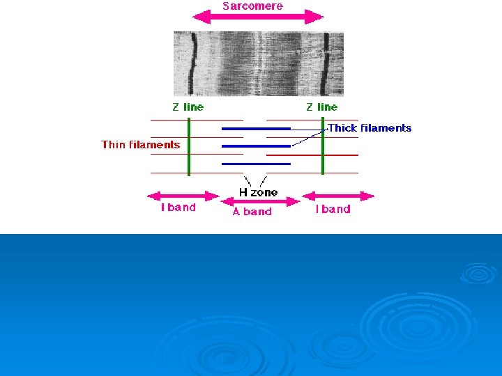
Anatomy of Myofibril Ø Responsible for contraction Ø 2 myofilaments: actin- the thin one myosin- the thick one Ø Sarcomeres: functional unit of contraction; area between 2 z-lines Ø 10, 000 or more sarcomeres along each myofiber.

g. I Band: Light region, actin only f. A Band: dark region, both actin and myosin h. H Zone: center of the A band, myosin only

j. Z line: dark zig-zag line in center of I band k. Actin: thin myofilament They have active sites (handles) if calcium is present.

l. Myosin: thick myofilament m. Cross Bridges: part of myosin, attach to actin in the presence of calcium

Sliding Filament Theory Actin filaments slide past myosin 2. Z lines get closer together 3. Sarcomere shortens-neither actin or myosin get shorter. 1.
- Slides: 23