Muscle Tissue Xing Wenying Introduction n Components l


- Slides: 40

Muscle Tissue Xing Wenying 邢文英

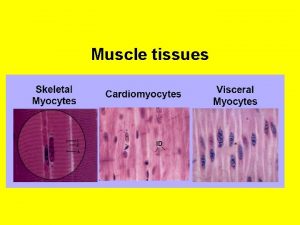
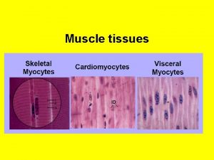
Introduction n Components: l Muscle cells(muscle fibers) • Elongated, thread-like, containing myofilaments and being contractile • cell membrane → sarcolemma; cytoplasm → sarcoplasm; SER → sarcoplasmic reticulum l CT: contain blood vessels, lymphatics and nerves.

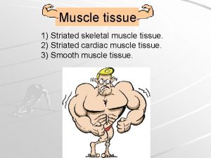
n classification : According to the structure and function structure striated unstriated

Skeletal muscle

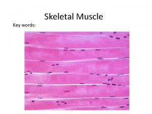
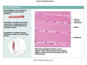
n Histological features of skeletal muscle fibers l l l Long cylindrical, 1 -40 mm long, 10 -100 um in diameter Multinucleate, ovoid nuclei, just under the sarcolemma Cross-striations: alternating dark and light bands

n Histological features of skeletal muscle fibers l Dark bands--A bands: H band, M line Light bands--I bands: Z line

sarcomere: • The sarcomere is the basic contractile unit of striated muscle. It is the segment of a myofibril between two adjacent Z line. • 1 sacromere = 1/2 I band+1 A band+1/2 I band l

l Myofibrils: Sarcoplasm is filled with long cylindrical filamentous bundles called myofibrils.

n Ultrastructure of skeletal muscle fibers Myofibril Sarcoplasmic reticulum Mitochondrium Transverse tubule

l myofibrils and myofilaments • Myofibrils are composed of bundles of myofilaments. • Thick filaments and thin filaments.


Thick filaments ü 1. 5μm long and 10 nm in diameter ü Occupy A band ü Made up of myosin molecules: --rods: overlap --heads: cross bridges, have ATPase activity.


Thin filaments 1 um long, 5 nm in diameter ü situated in both ending ü sides of each sarcomere; Run between and parallel to thick filaments; One end attached to the Z line, the other is free at H band ü composed of actin, tropomyosin, troponin

Thin filaments A. Actin: binding site for myosin.

Thin filaments B. Tropomyosin

Thin filaments C. Troponin: is a complex of three sub-units. Tn T(troponin T) Tn C(troponin C) Tn I(troponin I)


Arrangement ü I band -- only thin filaments ü A band -- both thick and thin filaments ü H band -- only thick filaments ü Z line -- anchor for thin filaments ü M line – fixation of thick filaments

l Transverse tubule (T tubule) u Definition: A transverse distributed tubular system formed by Sarcolemma invaginating into sarcoplasm, encircling each myofibrils. u Location: A-I junctional part u Function: Rapidly conduct impulses for contraction to every myofibrils

l Transverse tubule (T tubule) Sarcolemma with opening to T-tubule T tubule

l Sarcoplasmic reticulum (SR) u Definition: A longitudinal distributed tubular system formed by SER, encircling each myofibril between 2 adjacent T tubules. u Terminal cisternae: Ends of L tubule dilate and fuse to form flattened sac.

l Sarcoplasmic reticulum (SR) Triad: 1 T tubule + 2 terminal cisternae u Function: store and release calcium ions regulating concentration of Ca 2+ within sarcoplasm u

l Other organelles and inclusion Mitochondria Glycogen lipid droplet providing the energy necessary for the reactions involved in contraction.

n Mechanism of contraction -- sliding filament hypothesis The thin filaments slide past the thick filaments and insert further into the A band. u I band sarcomere become shorter, H band shortens or disappears, whereas A band remains the same length. u Ca 2+ and ATP play an important role. u


Cardiac muscle l Mainly distributed in the wall of heart; l Has more connective tissue and capillaries; l Some specialized as Purkinje fibers.

n Microstructure of cardiac muscle l Short cylindrical, 80 -150 um long, 10 -20 um in diameter, with branches, associated with each other; 1 or 2, ovoid, centrally-located pale-staining nuclei; l Exhibit cross striations, but less distinct; l

l Intercalated disk: unique characteristic

n Ultrastructure of cardiac muscle l Myofibril have different diameter, the boundary of myofibril is not very clear. l T tubules are larger, located at Z-line level.

n Ultrastructure of cardiac muscle l Sarcoplasmic reticulum is not well-developed, forms less and smaller terminal cisternae, so Diads are common consisting of 1 T tubule and 1 terminal cisternae on one side.

n Ultrastructure of cardiac muscle Intercalated discs • Specialized cell junctions at Z lines. l

n Ultrastructure of cardiac muscle l rich in mitochondria, glycogen, pigment.

Smooth muscle Seen in blood vessels and hollow viscera, arranged in layers.

n Structure of Smooth Muscle fiber


Summary n n n The structure of skeletal muscle. The structure of cardiac muscle. The structure of smooth muscle. Mechanism of contraction -- sliding filament hypothesis

Summary n The structure of cardiac muscle.

Summary n The structure of smooth muscle.

Homework Ø Review the characteristics of skeletal muscle and cardiac muscle. Ø Prepare for nerve tissue.
 Eric xing
 Eric xing
 Eric xing
 Liudong xing
 Eric p. xing
 Qing wen nin gui xing
 Pid xing
 Bakit mahalaga ang kasanayan sa pagsulat ng bionote
 Nǐ xìng shénme
 Hou pei chen
 Nǐ guì xìng
 Cmu machine learning
 Eric xing cmu
 Eric xing
 Jaringan epitel dapat ditemukan di
 Skeletal muscle tone
 Sarcomere
 Cardiac muscle
 Asynchronous recruitment of motor units
 Muscle tissue ppt
 Classification of muscle tissue
 Skeletal muscle
 Muscles and muscle tissue chapter 9
 Chapter 10 muscle tissue
 Epithelium location
 Muscle
 Cross section of skeletal muscle
 Muscle tissue
 Summation anatomy
 Muscle tissue parts
 Cardiac muscle tissue
 Axial muscles
 Types of muscle
 Tissue that connects muscle to bone
 Muscle
 Define myocele
 Skeletal muscle tissue structure
 Similar image
 Muscle tissue parts
 Cardiac muscle tissue parts
 Types of muscle tissue


