MUSCLE TISSUE Muscle tissue facilitates movement of the


- Slides: 19

MUSCLE TISSUE

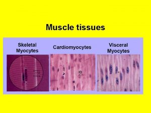
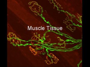
§ Muscle tissue facilitates movement of the animal by contraction of individual muscle cells (referred to as muscle fibers). § § Three types of muscle fibers occur in animals: skeletal (striated), smooth and cardiac

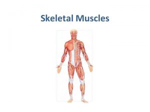
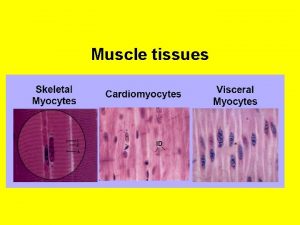
TYPES OF MUSCLE TISSUE Muscle occurs in three distinct types: § Skeletal muscle tissue attaches to bones (via tendons) for voluntary movement: This is the type of muscle that you eat if you eat meat. § Smooth muscle tissue contains spindle shaped cells with a single nucleus: it lines the gut, blood vessels and glands its operation is involuntary. § Cardiac (heart) muscle is composed of short, striated cells that can function in units due to the contracting signal that passes from cell to cell by way of the gap junctions.

Skeletal muscle tissue attaches to bones (via tendons) for voluntary movement

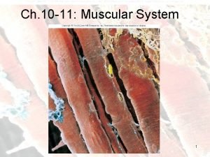
Cellular organization of skeletal muscle fibers § Unlike most tissues, skeletal muscle does not consist of individual cells. Rather it is formed from huge, striated (actin and myosin) multinucleated, long cells (fibers), which are often bundled together to form a “muscle”. § The muscle fibers develop by fusion of many individual embryonic cells called myoblasts. § Each individual skeletal muscle fiber extends over much of the length of the muscle in which it resides (up to many centimeters), with a uniform diameter that is typically around 50 µm.

Microscopic features of the muscle fiber § The unique contractile cytoplasm of muscle fibers is called sarcoplasm (sarco = flesh). § The contractile machinery is concentrated into myofibrils (myo = muscle), long narrow structures (1 -2 µm in diameter) which extend the length of the fiber and form the bulk of the sarcoplasm. § itochondria (once called sarcosomes) and § sarcoplasmic reticulum (a highly specialized form of endoplasmic reticulum) surround each myofibril. § The plasma membrane of muscle fibers is sometimes called the sarcolemma. § The many nuclei within each fiber are displaced from the center of the fiber by the specialized sarcoplasm and are located adjacent to the sarcolemma (except in special cases, like the intrafusal fibers of muscle spindles, and regenerated fibers).

Skeletal Muscle Identification: Teased or l. s. section shows distinct, very large, straight fibers. Looks more like hair than any other tissue. Also has distinct striations. § Features to Know: striations (1) composed of dark Abands and light I-bands; nuclei (2) pushed to edge of fiber; sarcolemna (plasma membrane surrounding fiber). Where Located: skeletal muscles; under voluntary control.

Skeletal Muscle - also called volutnary or striated muscle. The striations (stripes) are due to a very orginized pattern of proteins found within the cells. § The black arrow is pointing to a nucleus. § the entire length of the fiber may include hundreds nuclei.

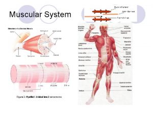
Striations (regularly spaced bands) § Each myofibril is marked by striations (alternating light and dark bands) within the fibers along its length. Dark and light bands are composed of myosin and actin, respectively. § In relaxed muscle, a single repeat of this pattern is about 2 µm long (the length varies with degree of contraction) and is called a sarcomere. § Because the striations on adjacent myofibrils are usually aligned, the entire muscle fiber appears uniformly striated.

Sarcomere § In each sarcomere, the broad band which appears dark in standard histological procedures is called the A-band. This band indicates the location of thick filaments (myosin); it is darkest where thick and thin filaments overlap. § The broad light band between the dark bands is the I-band. The Iband indicates the location where thin filaments (actin) extend beyond the thick filaments. § A distinct dark line running down the middle of the I-band is the Z-line, where thin filaments are attached end to end. Where thick filaments are attached end-to-end in the center of the A-band is the M-line. § When muscle is stretched, an H-band appears along the middle of the A-band, between the free ends of the thin filaments. From Blue Histology (Copyright Lutz Slomianka 1998 -2004) The image should be animated, if you watch patiently. )

Skeletal Muscle fibers, Cross Section § Any particular cross section of a muscle fiber may reveal one or a few nuclei, or none, but the entire length of the fiber may include hundreds.

The structural organization of skeletal muscle cross section, slide at 100 X Individual muscle fibers (4) are bundled into fascicles (5). Surrounding individual fibers, fascicles, and the entire muscle are layers of connective tissue (1 -3). § Features to Know: connective tissue sheaths: epimysium (1), perimysium (2), & endomysium (3); muscle fiber with nuclei (6) along edges; fascicle (5). You may see

Skeletal Muscle cross section at 400 x

Smooth Muscle - also called involuntary or visceral muscle. Smooth muscle is stronger than skeletal muscle. If you ever had stomach cramps you know how strong smooth muscle can be - a better example is labor (for those of you who have experienced or seen child birth) - the uterus has a large quantity of smooth muscle that works to "birth" a baby. § Muscle cells are packed tightly together (no gaps between cells) and usually not distinct. § These cells function in involuntary movements and/or autonomic responses (such as breathing, secretion, ejaculation, birth, and certain reflexes). These fibers are components of structures in the digestive system, reproductive tract, and blood vessels.

Cellular organization of Smooth muscle fibers lack the banding, although actin and myosin still occur. Smooth muscle fibers are spindle shaped cells that form masses. § Nuclei may or may not be visible. Note lack of striations.

Smooth muscle fibers are components of structures in the wall of the digestive system, Inner Circular, Outer Longitudinal of smooth muscle fibers

Smooth muscle fibers are components of structures in the blood vessels. § Smooth Muscle in Arterial Wall

Cardiac (heart) muscle is composed of short, striated cells § Note faint striations across fibers. Fibers distinct, typically with numerous small gaps between them. § Features to Know: nuclei (1), intercalated disk (2).

Like skeletal muscle, cartiac muscle is striated, but cardiac muscle also contains structures called intercalated disks (blue arrow) that work to produce the rhythmic contractions of the heart. Intercalated disks are found only in cardiac muscle. § And fibers in cardiac muscle are much smaller & branched.
 Accurate cutting facilitates sewing and improve garment
 Gandhi philosophy of wealth management
 Accounting treatment of departmental accounts
 Movement vs non movement area
 Axial in dance
 Embrasure hooks
 Fulcrum line definition
 Tissue ward movement
 Fixator muscles
 Isotonic vs isometric contraction examples
 Origin of superior oblique
 Intercostal muscles
 Jaringan epitel dapat ditemukan di
 Cardiac muscle tissue
 Cardiac muscle tissue parts
 Muscle tissue parts
 Types of muscle
 Muscles and muscle tissue chapter 9
 Muscle and nervous tissue
 Skeletal muscle tissue structure


