DNA DISCOVERY OF STRUCTURE OF DNA The structure


- Slides: 40
DNA �DISCOVERY OF STRUCTURE OF DNA The structure of DNA double helix and how it was discovered. Chargaff, Watson and Crick, and Wilkins and Franklin worked to solve this question.
Introduction Today the DNA double helix is most ionoic of all biological molecules. Untill 1950 s the double helix structure of DNA remained a mystry. In this article we will briefly described the work of all the major scientists on DNA.
Chargaff rules CHARGAFF RULES. ON the key piece of information related to the structure of DNA came from AUASTRIAN biochemist Erwin Chargaff. He analyzed the DNA of different species determining its composition of A, T, C and G bases
He made several key observations. • 1; , A, T , C and G were not found in equal quantities. • 2; The amounts of the bases varied among species but not between individuals of same species. • 3; The amount of A always equaled the amount of T , and the amount of C always equaled the amount of G.
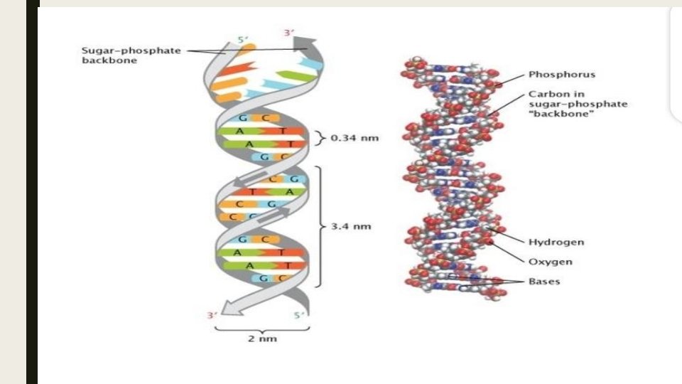
WATSON AND CRICK, S model of DNA The structure of DNA as represented in WATSON and CRICK, S model is DOUBLE STRANDED ANTIPARALLEL RIGHT HANDED HELIX NITROGENOUS BASES found inside Form HYDROGEN BOND PAIRS that hold the DNA strands together.
ANTIPARALLEL ORIENTATION Double stranded DNA is an antparallel molecule that mean it is composed of two strandas that run alongside each other but point in opposite. In a double stranded DNA molecule the 5 end (PHOSPHATE BEARING) of one strand aligns with the 3 end (HYDROXYL BEARING END) of its partner and vice versa.
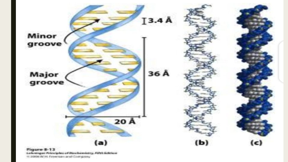
Right hand helix RIGHT HANDED HELIX In Watson and Crick, s model the two strands of DNA twist around each other to form a right handed helix. All helics have a handness which is property that describes how there grooves oriented in space.
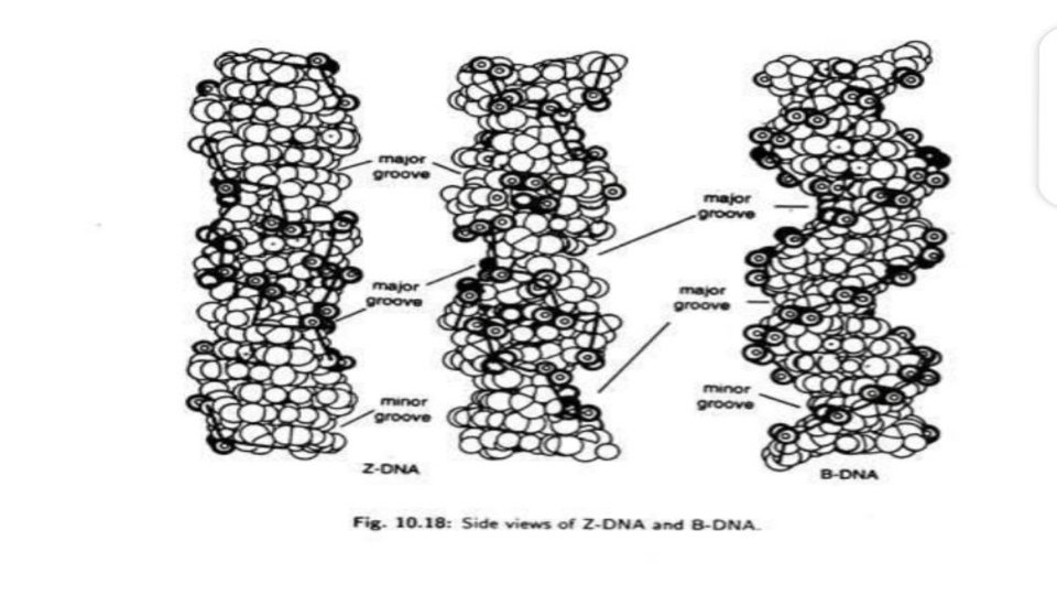
The twisting of the DNA and the geometry of the bases creates a wider gap (called the major groove) and the narrower gap( called the minor groove). These grooves run along the length of molecule. These grooves are important building sites for proteins that maintain DNA and regulate gene activity.
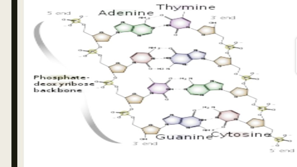
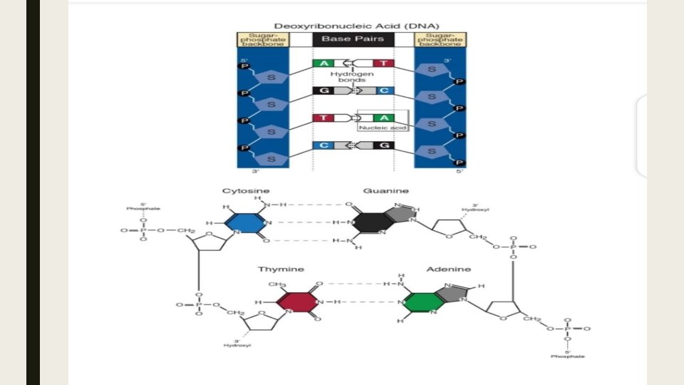
Base pairing BASE PAIRNING In Watson and Crick, s model the two strandas of double helix are held together by hydrogen bonds between nitrogenous bases on opposite strands. Each pair of bases lies flat , forming a rung on the ladder of DNA molecule. Base pairs are not made up of just any combination of bases. Instead if there is an A found on one strand it must pair with T on other strand vice versa. Similarly G pair with C. Two hydrogen bonds formed between A And T and threee between G and C.
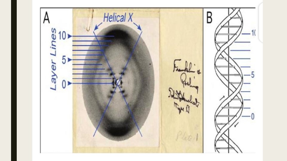
ROSALIND FRANKLIN Franklin discovered the structure of DNA. He obtained excellent X-ray diffraction photographs of DNA.
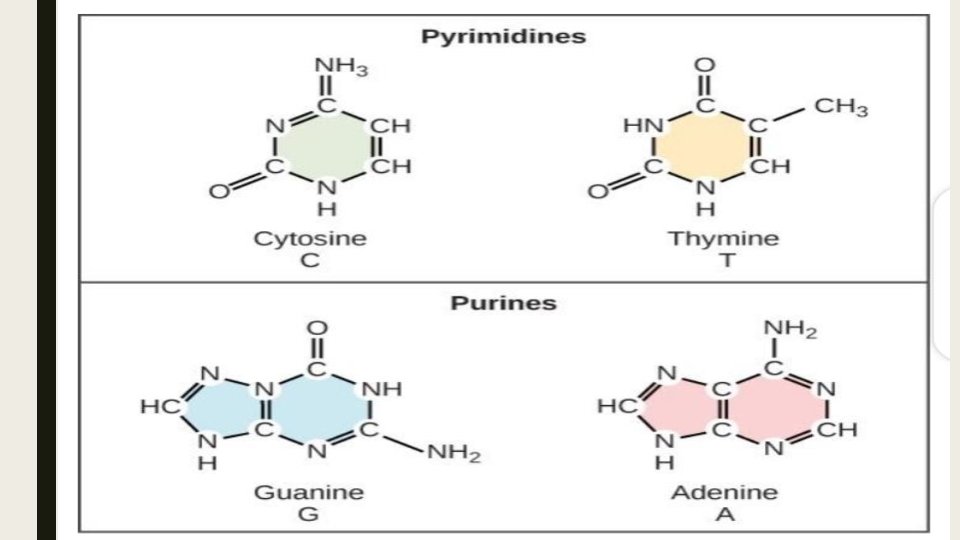
CHEMICAL NATURE OF DNA • CHEMICAL NATURE OF DNA Hydrolysis of DNA showed that it is composed of: • phosphoric acid , a phosphate group • A 5 Carbon sugar , as deoxyribose sugar • the purine bases , cytosine and thymine • the pyrimide bases cytosine and thymine
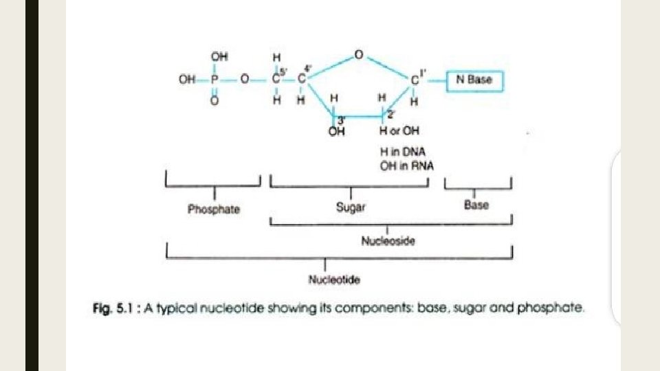
DNA is a Polymer ■ DNA is a polymer. A polymer is a long chain like molecule , comprising of numerous individual units called monomers linked together in series. The monomer in a DNA is nucleotide.
NUCLEOTIDES ■ Each nucleotide is composed of ■ A sugar ■ A nitrogenous base ■ A phosphate group ■ The nitrogenous base is linked to sugar.
THE SUGAR COMPONENT OF DNA ■ In DNA the sugar component is pentose sugar, deoxyribose. It can exist in chain form or ring form. It contains hydroxyl group attached with carbon atom number two with a hydrogen group
■ THE NITROGENOUS BASES ■ These are complex single ring pyrimides and double ring purines
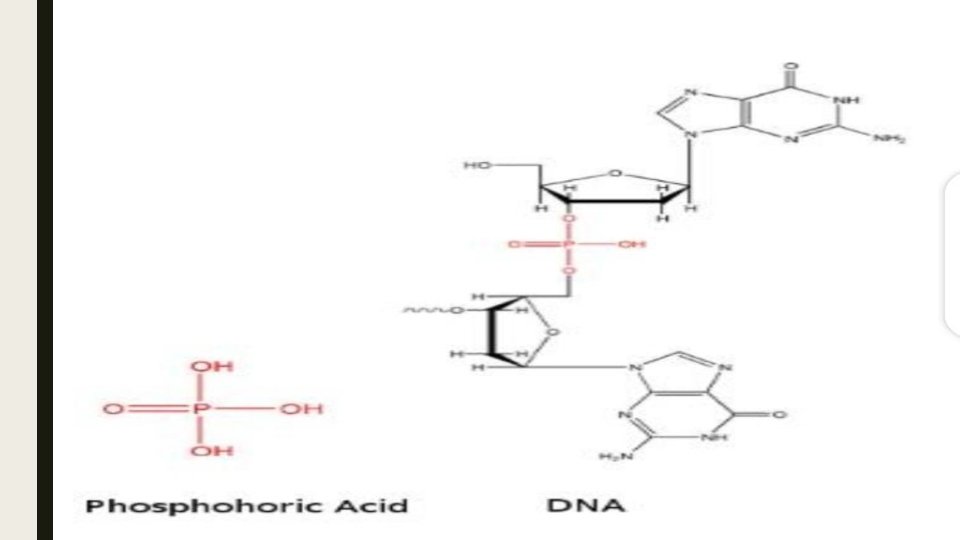
. ■ THE PHOSPHORIC ACID COMPONENT ■ A nucleoside is converted into a nucleotide by attachment of phosohoric group to a 5 C of sugar.
. ■ ALTERNATIVE FORMS OF DNA ■ DNA exist in many forms such as:
B DNA ■ The form OF DNA described above is called B DNA. It is a right-handed helix. It turns in clockwise manner when viewed down its axis. The bases are stacked almost exactly perpendicular to the main axis with about ten base pairs per turn.
A DNA ■ If the water contents increase to about 75% the A form DNA will occur. In this form the bases are titled with regard to the axis and there are more base pairs per turn as compared to B DNA.
Z DNA ■ gar –phosphate backbone forms a zigzag structure. The Z DNA looks like B DNA in which each base was rotated 180 degree , resulting in a zigzag left-handed structure. Z DNA is thought to be involved in regulation gene expression in eukaryotes.
RNA ■ It is abbreviation of ribonucleic acid. ■ It is complexed compound of high molecular weight that functions in cellular protein synthesis and replaces DNA. As a carrier of genetic codes in some viruses.
■ NITROGENOUS BASES: ■ The nitrogenous bases in RNA are ■ ADENINE ■ GUANINE ■ CYTOSINE ■ URACIL
■ �SUGAR ■ Ribose sugar present in RNA. ■ �TYPES OF RNA: e ■ There are three types of RNA
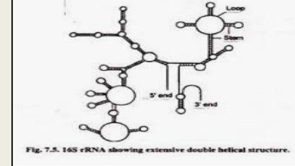
RIBOSOMAL RNA ■ It is largest of RNA molecules and usually constitute about 80% of all RNA in the cells. The molecule is composed of thousands of nucleotides consisting of a single strand with regions in which base pairing result in double helix projecting on either side. ■ It is produced inside the nucleus. It takes part in formation of messenger RNA and translational RNA
MESSENGER RNA ■ It is produced in nucleus from the coded instruction in the DNA and then passes into the cytoplasm where it becomes associated with the ribosomes. It carries chemical information from DNA OF gene to ribosome for protein synthesis. ■ Molecules of messenger RNA are 75 -3000 nucleotides long and are not folded in any special way.
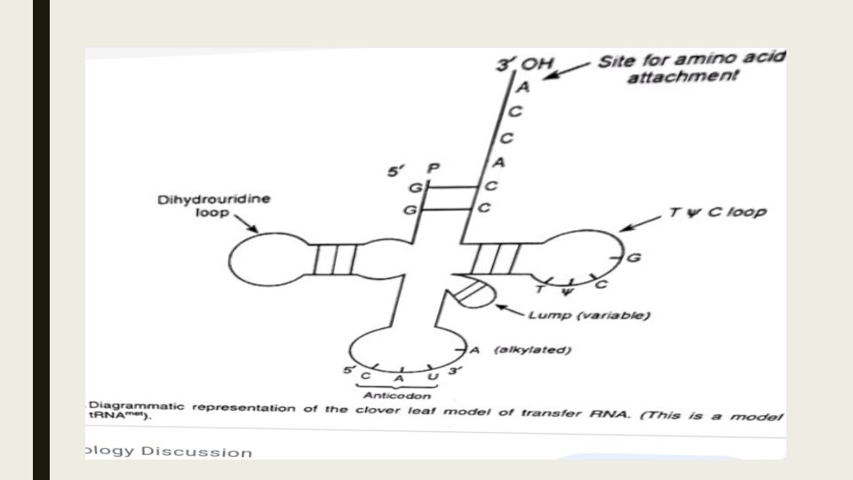
TRANSFER RNA ■ It is smallest class of RNA molecules and consisting of 75 -90 nucleotides. It carries amino acids to ribosomes during protein synthesis each organism synthesis a number of different t. RNAs each in multiple copies. All cells have atleast 20 different kinds of t. RNAs molecules. A t RNA molecules is nearly similar in bacteria and eukaryotes.
CLOVER LEAF MODEL OF t. RNA ■ More is known about the secondary structure of this molecule then any other RNA. All t. RNAs have a two –dimensional clover leaf model because it can be folded into a base pair structure like a clover leaf. ■ Each molecule is held together by a precise base pairing arrangement between different parts of RNA D loop strands each of the base paired region is twisted to form a double helix but when the variable loop molecule is flattened out it has a clover leaf shaped
■Thank you