Stomach and Duodenum Dr Othman Tajoury MD MRCSEng
, FEBS(Euro. Board) Supervisor of clinical")


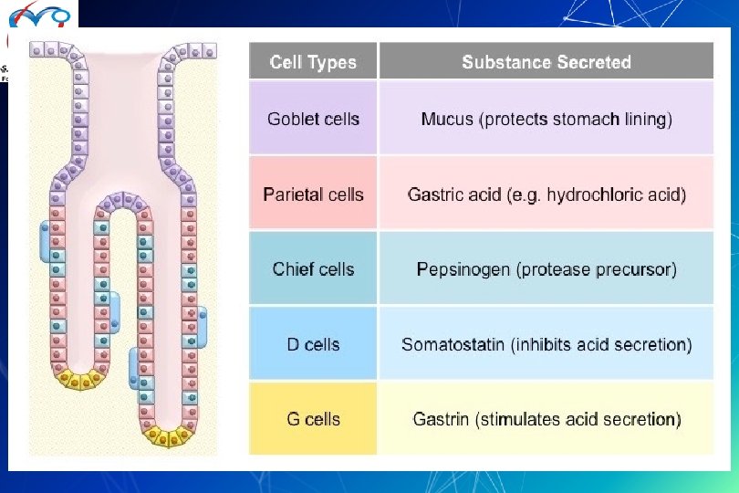
: • Location: Body • Function: Histamine production •")
 1 - Gastritis 2 - Mallory-Weiss Syndrome 3 - Gastric Polyps")
 • Widespread inflammation of the mucosa. • Causes:")


 • <")

 •")


 Serological tests ; 1 - H. pylori Ig. G antibodies")


, anywhere in stomach.")
 2. Perforation 3. Malignancy")


 mucosa-associated lymphoid tissue 3")


 is")

 • Duodenal Polyps")


. • If")
 •")

 • Gastric outlet")


. ▰ Some")


 Syndrome (gastrinoma)")


- Slides: 80
Stomach and Duodenum Dr. Othman Tajoury MD, MRCS(Eng. ), FEBS(Euro. Board) Supervisor of clinical phase LIMU
Learning Objectives • To understand the anatomy and pathophysiology of the • • stomach in relation to disease To be able to decide the appropriate techniques in the investigation and treat peptic ulcer disease and its complications To understand the critical importance of gastritis and Helicobacter pylori in upper gastrointestinal disease To be able to recognise the presentation of gastric cancer To know about the causes of duodenal obstruction and the presentation of duodenal tumours
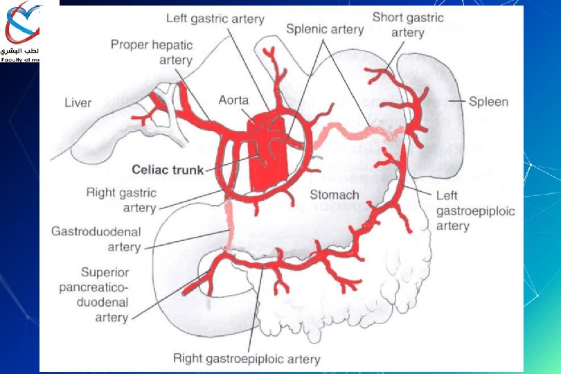
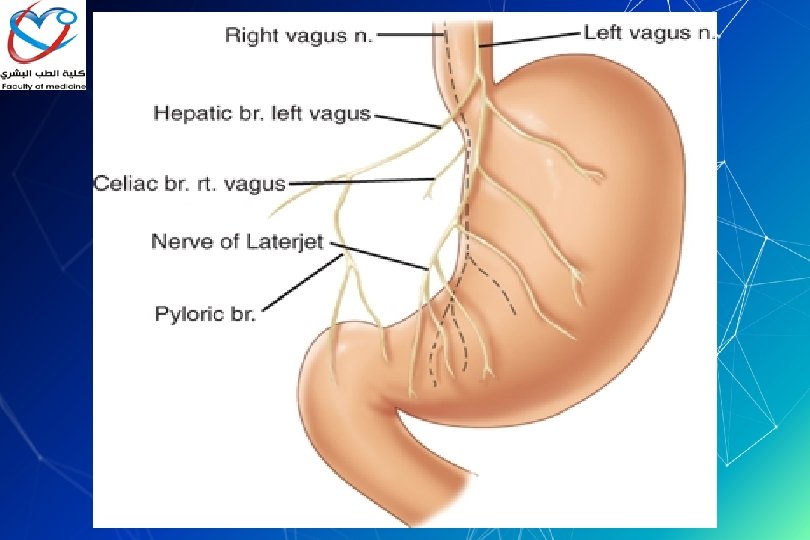
Anatomy
Cell Types • Parietal: • Location: Body • Function: secrete acid and intrinsic factor (production of hydrogen ions to form hydrochloric acid) • Chief: • Location: Body • Function: produce pepsinogen I pepsin & digestive protease • Goblet: • Location: Body, Antrum • Function: mucus production
Cell Types • Entero. Chromaffin-Like (ECL): • Location: Body • Function: Histamine production • G cells: • Location: Antrum • Function: Gastrin production • D cells: • Location: Body, Antrum • Function: produce Somatostatin • Endocrine cells in the duodenum produce cholecystokinin and secretin.
Benign Gastric Disease(s) 1 - Gastritis 2 - Mallory-Weiss Syndrome 3 - Gastric Polyps 4 - Bezoars 5 - Gastric (peptic) Ulcer Disease
1 - Gastritis (acute or stress) • Widespread inflammation of the mucosa. • Causes: H. pylori, NSAIDS, physiologic stress.
Types of gastritis Type A gastritis: autoimmune, antibodies to the parietal cells. Type B gastritis: Helicobacter-associated gastritis. Reflux gastritis: common after gastric surgery Erosive gastritis: NSAIDs and alcohol are common causes. Stress gastritis: B/O reduction in the blood supply to superficial mucosa of the stomach. ▰ Ménétrier’s disease: gross hypertrophy of the gastric mucosal folds, premalignant. ▰ Lymphocytic gastritis: rare ▰ ▰ ▰
Gastritis Ø S&S: • Nausea, emesis, hematemesis, melena, hematochezia, etc. • Treatment: Ø Prevention, removal of offending agent. Ø Gastric decompression and support. ØAcid suppression (PPI), Ø Gastrectomy in severe cases
2 - Mallory-Weiss Syndrome
3 - Gastric Polyps • There are two types: a- Hyperplastic b- Adenomatous A- Hyperplastic polyps: • More common • Usually benign B- Adenomatous: • Precancerous
Gastric Polyps • Adenomatous polyps: • Greater risk of malignancy 40% (Precancerous) • < 0. 5 cm in diameter = very low risk. • > 1. 5 cm in diameter = very high risk. • Once a polyp is diagnosed, should be removed, 2. 5% recurrence rate
Peutz-Jegher’s Syndrome Autosomal dominant genetic disorder characterized by the development of benign hamartomatous polyps in the gastrointestinal tract and hyper pigmented macules on the lips and oral mucosa
4 - Bezoars • Phytobezoars = indigestible plant material (e. g. , cellulose) • Trichobezoars = an extreme form of hairball.
5 - Gastric Ulcers
Pathogenesis of ulcer • Increase acid production ▰ Lack of protection of the mucosa • 85%-95% of PU pts are colonized with H. pylori.
Helicobacter pylori (H. pylori) Serological tests ; 1 - H. pylori Ig. G antibodies (sensitivity and specificity 80% to 95% ) 2 - Breath test; the patient is given either 13 C- or 14 Clabeled urea to drink. H. pylori metabolizes the urea rapidly, and the labeled carbon is absorbed. then measured as CO 2 in the patient’s expired breath to determine whether H. pylori is present (sensitivity and specificity 94% to 98%)
3 - Upper esophagogastroduodenal endoscopy; biopsy is obtained and the diagnosis of H. pylori can be made by several methods: • The biopsy urease test; it provides rapid testing at the time of biopsy. • Histologic identification of organisms - considered the gold standard of diagnostic tests. • Culture of biopsy specimens for H. pylori, when antimicrobial susceptibility testing is desired. • Treatment: Triple therapy; amoxicillin, tetracycline (not to be used for children <12 yrs. ), metronidazole, or clarithromycin, plus proton pump inhibitor.
Gastric ulcers
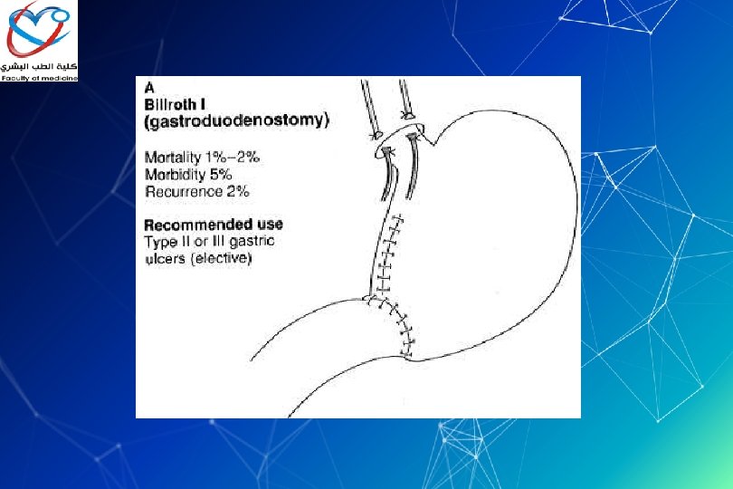
Type I -Occur at the incisura. -Not associated with hyperacidity -Associated with Blood group “A”
Type II • A combination of 2 ulcers, one in the stomach, the other in the duodenum. • Usually occur in hypersecretory states. • Associated with Blood group“O”.
Type III • Located pre-pyloric. • Associated with hypersecretion. • Type “O” blood association. • Typically 2 -3 cm from pylorus, can be multi.
Type IV • Occur high on lesser curve near the GE junction. • Not associated with hypersecretion. • Usually result from defective mucosal defense.
Type V ▰ Medication-induced (NSAID/ acetylsalicylic acid), anywhere in stomach.
Complications of gastric ulcer 1. Bleeding; (penetration) 2. Perforation 3. Malignancy
Treatment of gastric ulcer Ø Ø ▰ ▰ Medical - Prevention, triple therapy, ppi. Surgical - in few cases Primary goals ; Excision of the ulcer and Reduction of acid/pepsin output Secondary goals ; Minimize bile reflux and gastric stasis
Bleeding gastric ulcer : operative procedures ▰ ▰ Endoscopic management with biobsy Simple oversewing with biopsy Gastric resection including ulcer (Gastrectomy) Excision of ulcer with TV and drainage without TV and drainage?
Perforated gastric ulcer : operative procedures ▰ Biopsy and simple closure ▰ Gastric resection
Malignant Gastric Disease 1 • Adenocarcinoma 2 • Lymphoma (MALToma) mucosa-associated lymphoid tissue 3 • Gastrointestinal Stromal Tumor (GIST)
1 -Gastric Adenocarcinoma
Gastric Adenocarcinoma • Adenocarcinoma accounts for 95% of all gastric cancers. • US and Europe = low risk areas • Asia (Japan/China), Russia, Chile = high risk areas.
Risk Factors • H. pylori infection. • Pernicious anemia. • Achlorhydria. • Chronic gastritis. • H/o caustic injury (corrosive ingestion) • Adenomatous polyps.
Ca stomach Classification • 4 Macroscopic types: Cauliflower, Ulcerative, Infiltrating, Colloid. § 3 Microscopic: Adenocarcinoma 95%, SCC 4%. Anaplastic 1% • DIO classification: - Diffuse - Intestinal - Otherwise
Diffuse ▰ On top of atrophic gastritis. • undifferentiated with classic signet-ring cells. • Patients are usually younger. • Associated with ABO blood group “A”. • Spreads via lymphatics and local extension.
Intestinal ▰ On top of intestinal metaplasia& Biliary gastritis • Well differentiated with glandular elements. • More common form in high incidence areas. • Patients are usually older male. • Hematogenous spread.
Gastric Adenocarcinoma. Investigations : OGD , CT , EUS , PET , Laparoscopy. . Treatment : Depend on site of tumor (Total , distal or proximal Gastrectomy + LN dis section)
2 -Gastric Lymphoma
Gastric Lymphoma • Stomach is the primary site for 2/3 of all GI lymphomas. • Predominately non-Hodgkin’s variant. • Symptoms are similar to Adeno-ca. • Dx: via biopsy (endoscopic vs. open). • If Dx is made prior to surgery, do lymphoma work-up. • Treatment: chemotherapy vs. surgery.
3 -Gastrointestinal stromal tumors GIST
GIST • Formerly called leiomyomas or leiomyosarcomas. • Stomach is the most common site. • Can be “benign” or “malignant”. • Malignant - direct invasion, mitotic • Work-up: CT , EUSS, etc
Treatment: - Surgical Resection w/ margins. - Gleevec : - Gleevec (imatinib mesylate) is a type of drug called a tyrosine kinase inhibitor (TKI). - Targeted drug therapy is different from traditional chemotherapy in that it is very selective
Duodenum • 4 parts • Produces many enzymes • D 2: posterolateral insertion of ampulla. • Becomes jejunum at the angle of Treitz
Benign Duodenal Disease • Duodenal Ulcer Disease(Uncomplicated or Complicated) • Duodenal Polyps
Uncomplicated Ulcer • In contrast to gastric ulcers, duodenal ulcers rarely harbor malignancy.
Uncomplicated Ulcer Important risk factors: • H. pylori , NSAIDs , tobacco. • Treatment : mainly medical; ( triple therapy) instead of surgical.
Presentation • Burning, gnawing pain in the epigastrium. • it may radiate to the back. • Exacerbated by fasting. • Relief gained from meds and eating.
Evaluation/Testing • Non-invasive H. pylori testing. (Serum antibodies, breath urease, fecal antigen). • If (+) test = H. pylori treatment (triple therapy). • If primary treatment fails then do upper endoscopy. • OGD allows for visualization, biopsy, evaluation for alternate diagnosis.
Treatment • Non-operative • Remove all ulcerogenic agents. • Start acid suppression (PPI) • Eradicate H. pylori.
Treatment • H. pylori therapy: First Line: • Amoxicillin and Clarithromycin or Clarithromycin and Flagyl (+) PPI • Treat for 7 – 14 days. • Recheck for H. pylori after treatment. • Continue acid suppression until ulcer is healed.
Complicated Ulcer • 4 major manifestations: • Perforation • Bleeding (penetration) • Gastric outlet obstruction • Intractable
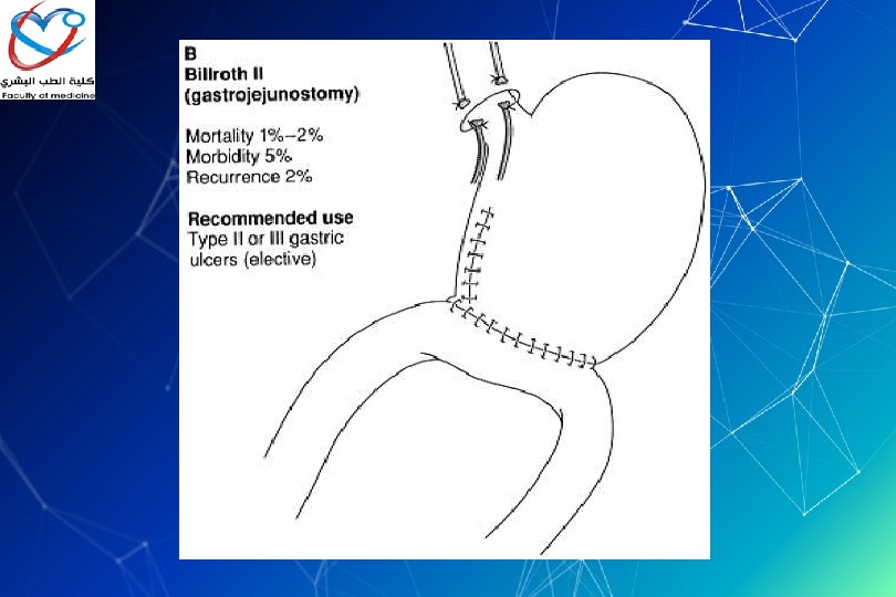
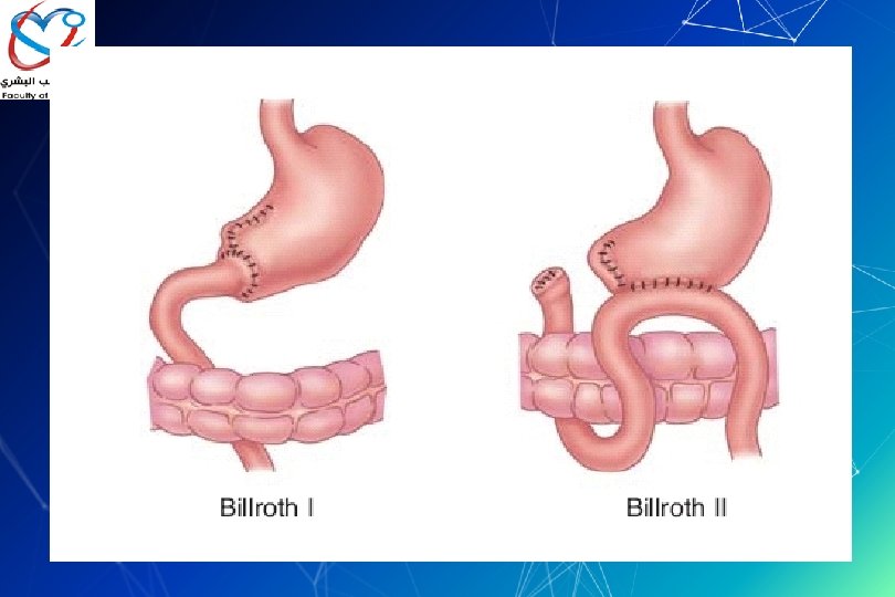
Treatment: Perforation • Typical presentation of acute abdomen. Ø Resuscitation. Ø Operation: 1. Graham patch (omentoplasty). 2. +/- (Antrectomy with drainage) + (vagotomy).
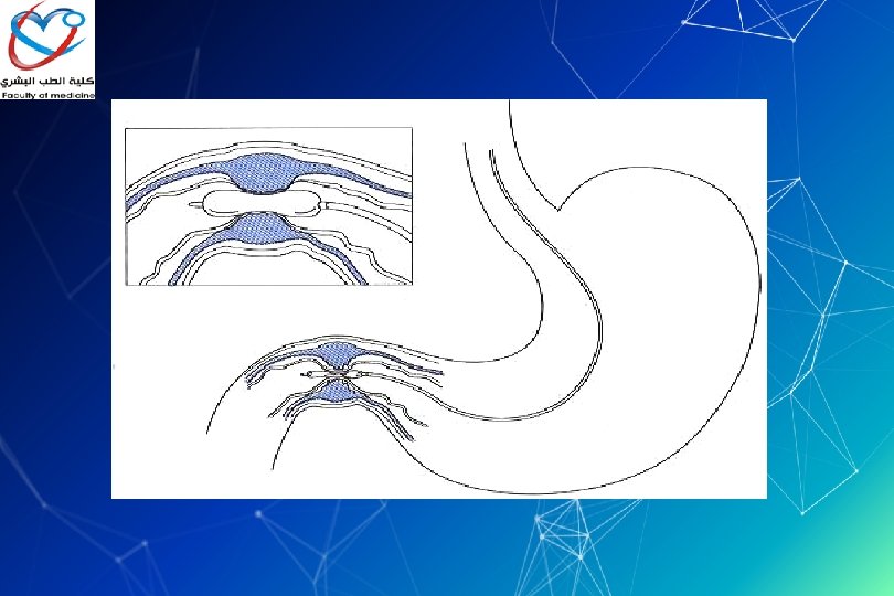
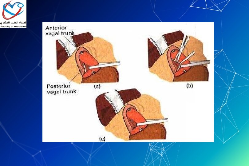
Bleeding Treatment: • Resuscitation. • Upper endoscopy with local treatment. • High-risk stigmata : active bleeding, visible vessel, fresh clot. • Angiography with embolization. • Surgical intervention. Suturing bleeding vessel + (TV) Truncal vagotomy and pyloroplasty PP or HSV
Bleeding Indications for operative intervention: 1. Massive hemorrhage leading to shock 2. Prolonged blood loss requiring continuing transfusion 3. Recurrent bleeding during medical therapy or after endoscopic therapy 4. Recurrent bleeding requiring hospitalization 5. Rare blood groups
Outlet obstruction • NPO, NG, Rehydration, Correct electrolytes. • Some resolve (swelling). ▰ Some Endoscopic dilatation • Some Surgery: -Remove obstruction (Antrectomy with drainage) -Bypass obstruction (Gastroenterostomy).
Intractability ▰ Ulceration persists at 3 months despite active drug therapy ▰ Ulcers recur within 1 year of initial healing despite maintenance therapy ▰ The ulcer disease is characterized by cycles of prolonged activity with brief or absent remissions
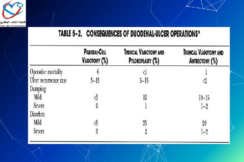
Intractability • Medical failure – look for other causes like, ZE syndrome, non-compliance, etc. • Surgery - HSV - TV + Antrectomy - TV + PP
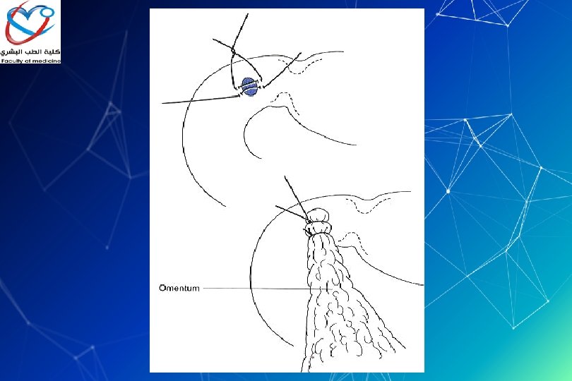
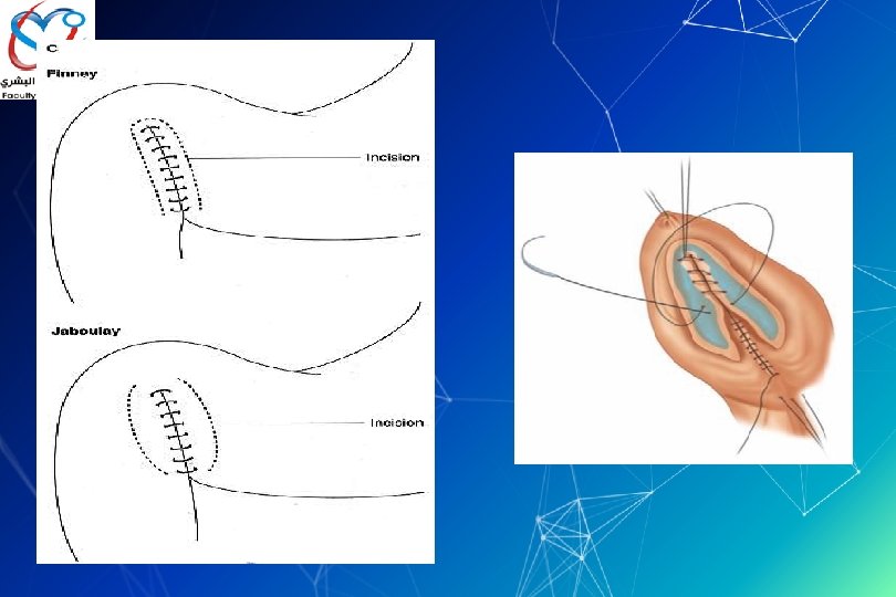
Drainage procedures : pyloroplasty ▰ ▰ ▰ Heineke-Mikulicz Finney Jaboulay
Heineke-Mikulicz pyloroplasty
Duodenal polyps Typically arise as part of a familial disorder. • FAP. - Autosomal dominant - High malignant potential • Peutz-Jegher’s syndrome.
Malignant Duodenal Disease • Adenocarcinoma • Lymphoma ▰ Zolliger-Ellison (ZE) Syndrome (gastrinoma)
Adenocarcinoma • Duodenum is the most common site for small bowel adenocarcinoma. • ~ 2/3 are peri-ampullary (D 2). • Only treatment is resection (if possible). • Double bypass if unresectable. • (+) LN’s = < 15%, 5 year survival.
Adenocarcinoma
Zollinger–Ellison syndrome • Rare disease. • Disease is a direct result of gastrinoma. • 2/3 of gastrinomas are in the “triangle” • Can be sporadic or hereditary. • Strong assoc. with MEN-I (3 p’s) Pituitary, Parathyroid, Pancreas. • Treatment: 1 - PPI 2 -resection (if possible).
Gastrinoma - Tumor in the pancreas or duodenum that secretes excess of gastrin leading to ulceration in the duodenum, stomach and the small intestine. - There is hypersecretion of HCl. - Gastrinoma triangle; region in between three points: - Superior- Body of Gallbladder. - Inferior-junction of the second and third portion of duodenum. - Medial- junction of the neck and body of the pancreas.
Lymphoma • Rare, most small bowel lymphomas are ileal. • Similar presentation to adenocarcinoma. • Diagnose the same as gastric lymphoma. • Resection if resectable. • Chemotherapy and radiation.
Lymphoma
Thank you