Pancreas Anatomy Physiology Investigations Congenital anomalies Dr Amit


, joins the CBD to empty into")
 Drains superior portion of head of the Pancreas Empties")


")
 and acute pancreatitis (right). Evidence of parenchymal edema")


- Slides: 30
Pancreas: Anatomy, Physiology, Investigations Congenital anomalies Dr Amit Gupta Associate Professor Dept of Surgery
Anatomy Lies transversely in the retroperitoneal space, between the duodenum on the right and the spleen on the left Related anteriorly to the omental bursa above, the greater sac below, and the transverse mesocolon Fixed organ
level of the L 2 vertebra From the duodenal C loop to the hilum of the spleen Divided into four parts
Cross-sectional relation of the pancreas to other abdominal structures
Embryology
Pancreatic Ducts Main pancreatic duct (duct of Wirsung), joins the CBD to empty into the duodenum at the ampulla of Vater Diameter -- 2 to 4 mm Pressure --15 to 30 mm Hg
Lesser duct (duct of Santorini) Drains superior portion of head of the Pancreas Empties separately into the 2 nd part of duodenum through the minor papilla
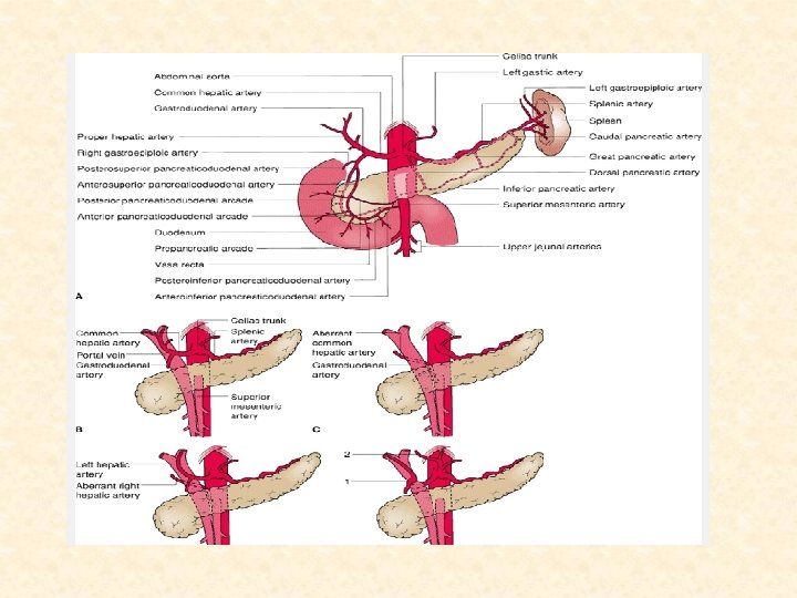
Arterial Supply Celiac Superior mesenteric artery Splenic artery
Venous Drainage Suprapancreatic portal vein Retropancreatic portal vein Splenic veins Infrapancreatic SMV
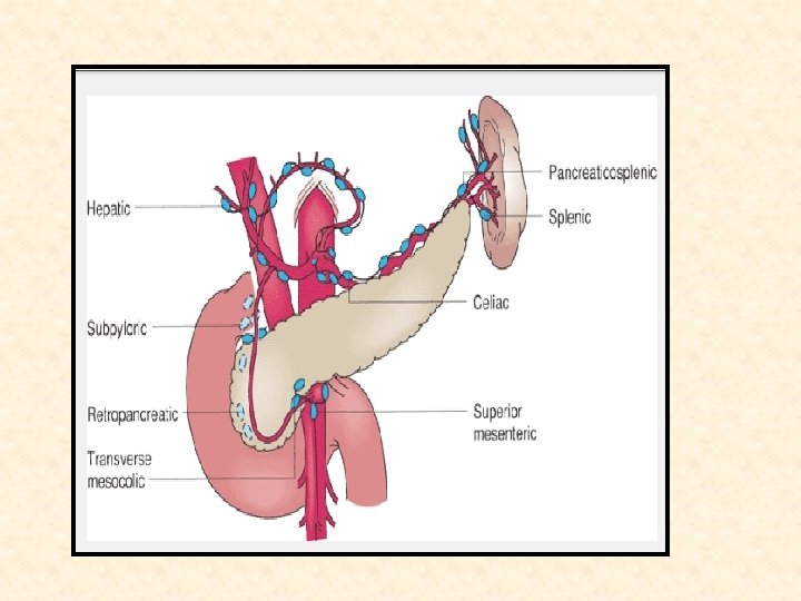
Lymphatic Drainage Drain into five main nodal groups • Superior nodes drain the upper half of the head of the pancreas • Anterior lymphatic drain to the prepyloric and infrapyloric nodes • Inferior group of nodes drain to the superior mesenteric and periaortic nodes • Posterior pancreaticoduodenal lymph nodes drain into right periaortic nodes • Splenic group of nodes drain into the interceliomesenteric lymph nodes
The absence of a peritoneal barrier on the posterior surface of the pancreas results in direct communication of the intrapancreatic lymphatics with retroperitoneal tissues, and this contributes to the high incidence of recurrence after presumably curative resections of pancreatic cancer
Histology Exocrine Structure Acinar cells secrete the enzymes responsible for digestion Ductular network carry the exocrine secretions into the duodenum Constitute 80% to 90% of the pancreatic mass
Endocrine Structure Islets of Langerhans responsible for the secretion of hormones that control glucose homeostasis Contains alpha (A)-- glucagon beta (B)-- insulin delta (D)-- somatostatin pancreatic polypeptide (PP) or F cells– PP Accounts for 2% of the pancreatic mass
Pancreatic Exocrine Enzymes
Pancreatic Endocrine Enzymes
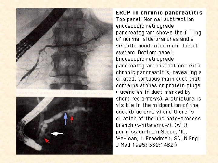
Investigations • Serum Markers Amylase, lipase, trypsinogen, and elastase CA 19. 9 (Tumor Marker) • Lundh meal test • • • Ultrasound EUS CECT Scan MRCP ERCP
Ultrasound image of normal pancreas (left) and acute pancreatitis (right). Evidence of parenchymal edema and peripancreatic fluid accumulation is seen
Non necrotizing acute pancreatitis
Congenital Anomalies
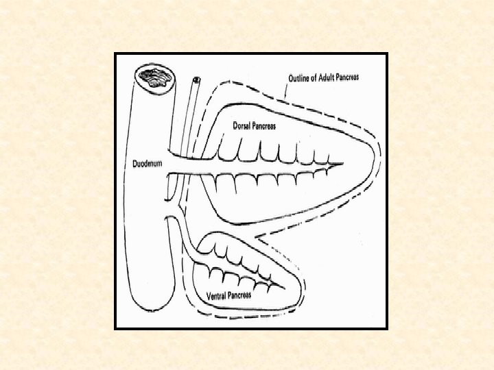
The complex process by which the dorsal and ventral pancreatic primordia fuse during pancreatic development gives rise to congenital variations
Agenesis Very rarely, the pancreas may be totally absent The homeodomain transcription factor IPF 1 (PDX 1) is critical for the development of the pancreas, and germ line (inherited) homozygous mutations in the IPF 1 gene on chromosome 13 q 12. 1 have been reported in a patient with pancreatic agenesis
Pancreas divisum Most common clinically significant Incidence of 3% to 10% Caused by a failure of the fetal duct systems of the dorsal and ventral pancreatic primordia to fuse
Cystic fibrosis v Autosomal recessive v More in Caucasians v Incidence 1 in 2500 live births in U. K. v Defect is mutation in CFTR gene on Ch 7 v High levels of Na 2+, Cl 2 - ions in sweat
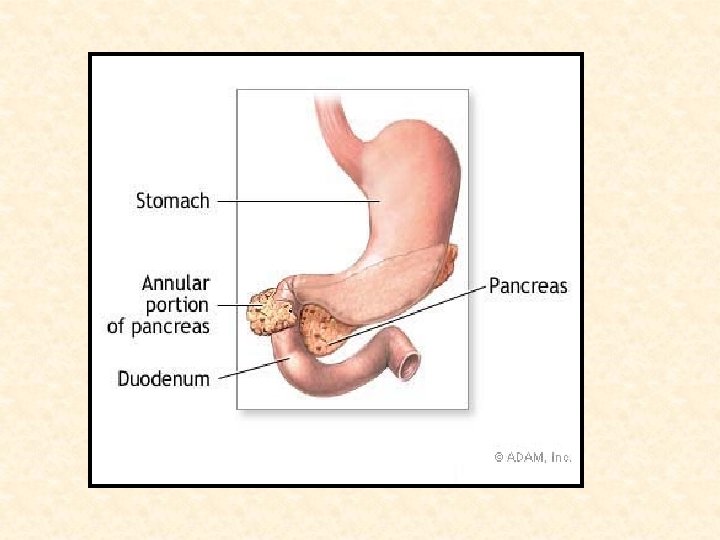
Annular pancreas Develops when one portion of the ventral pancreatic primordium becomes fixed, while the other portion of this primordium is drawn around the 2 nd or 3 rd duodenum Can be associated with congenital duodenal atresia/ stenosis Common in children with Down’s syndrome Present with signs and symptoms of duodenal obstruction such as gastric distention and vomiting Tx- Duodenoduodenostomy
Ectopic pancreas Found in about 2% autopsy specimen Sites for ectopia are stomach and duodenum, followed by the jejunum, Meckel diverticula, and ileum May cause pain from localized inflammation, or, rarely, may incite mucosal bleeding Approximately 2% of islet cell tumors arise in ectopic pancreatic tissue.