Investigations in Stomach and Duodenum Dr Bina Ravi

Investigations in Stomach and Duodenum Dr Bina Ravi MS, MNAMS, FICS LHMC, New Delhi

Investigations • • Functional Bacteriological Biochemical Morphological

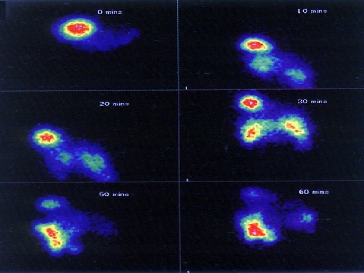
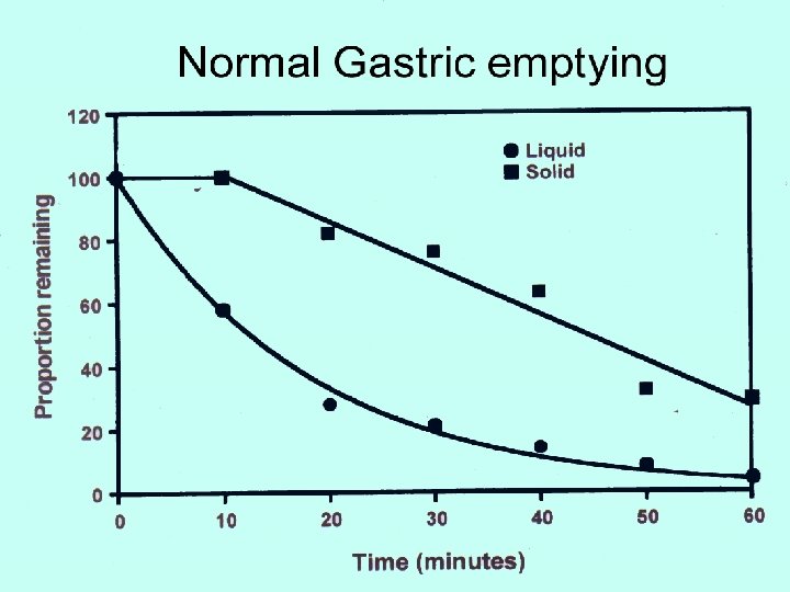
Functional • Gastric Secretory test → p. H monitoring, BAO, MAO, Plasma gastrin studies • Gastric Emptying studies → Ba meal, Bile reflux (HIDA) , Gastric barostat, MRI motility

Recurrent Ulcer • Stomal ulcer • Incomplete vagotomy – Gastric analysis, Sham feeding – Cephalic phase • Retained antrum – ZES – BAO + MAO, Secretin stimulation • Post operative NSAIDs use

ZES • • Duodenal tumors – 50% BAO – > 15 meq / hr N. Fasting Gastrin - < 150 pg/ml Gastrin provocative test – Secretin / Ca IV - > 200 pg/ ml at 15 mts • Endoscopy, EUS, CT, Laparoscopy • SASI, Octreoscan – Sensitivity / Specificity > 75%

Bacteriological • H. pylori • Invasive →Endo. Bx, histology, culture, 80 -95% Sensitivity, 95100%Specificity • Non invasive →Serologic, urea breath test, fecal H. pylori antigen test • H. pylori Negative → screen for Aspirin, NSAIDS levels in blood and urine

Morphological • Bleeding • Acute - DU, GU, Varix, Mallory Weiss, vascular ectasia – Dieulafoys disease • Chronic - DU, GU, Tumor- stomach, periampullary

Biochemical • Tumor Markers • CEA ▲ in 1/3 patients ≈ stage • CEA + Ca 19 -9 or CA 50 ↑ sensitivity

CA Stomach : significance of tumor marker • • • β HCG CA 125 CEA alpha fetoprotein CA 19 -9, tissue staining for C - erb B 2

CA 125, β HCG • Pre-op indicator of Ø aggression Ø tumor burden § Prognostic “Botet”

Diagnosis • • Auto-fluorescence Endoscopic Ultrasound Optical Coherence Tomography Virtual Biopsy

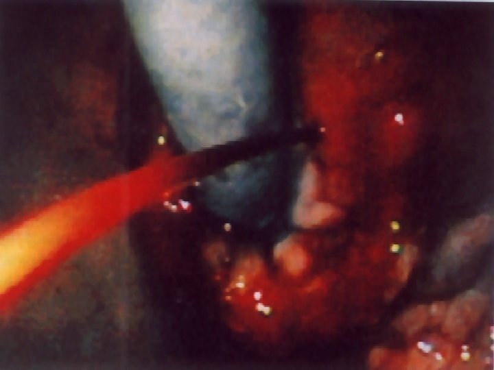
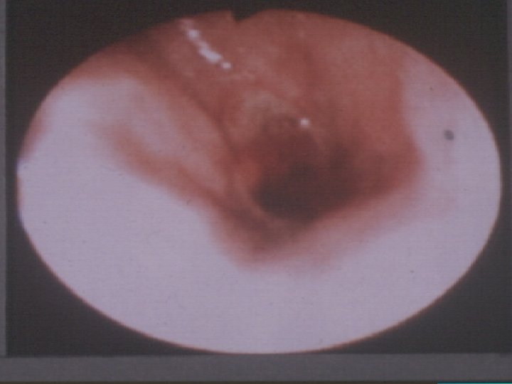
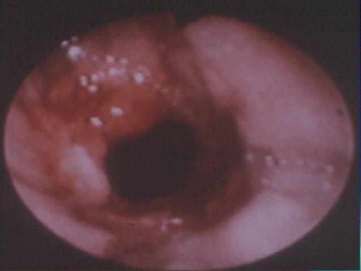
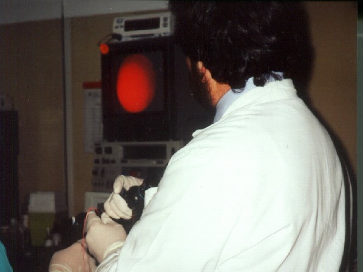
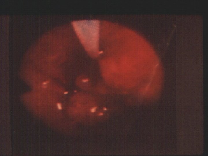
Endoscopy • Size, location, morphology of lesion • Mucosal abnormality, bleeding • Proximal and distal spread of tumor • Distensibility

Endoscopy • Abnormal motility ► SM infiltration, extramural extension – vagal infiltration • Bx – 6 – 10 – 90% accuracy • Early Ca → 0. 1% indigocarmine dye test

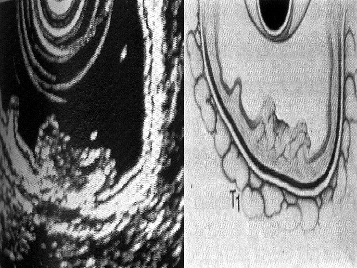
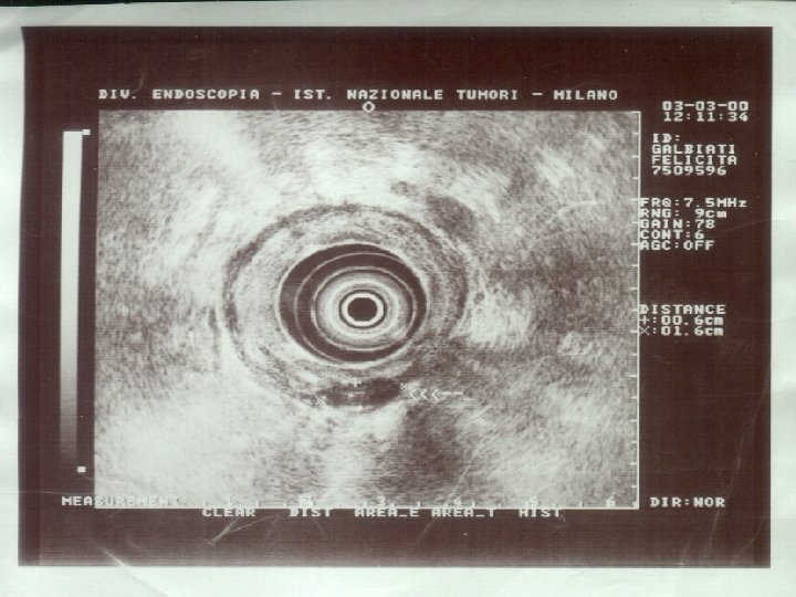
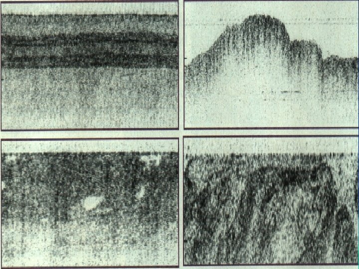
EUS • Good for T & N • Not good for M • Radial probes – 7. 5 or 12 MHz better for Biopsy

T 1

T 2

T 3
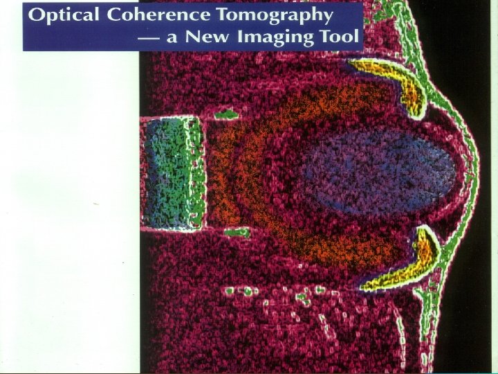

OCT / Virtual Biopsy • Optical coherence tomography • Beyond routine endoscopy • Differentiates - benign and malignant, mucosal dysplasias
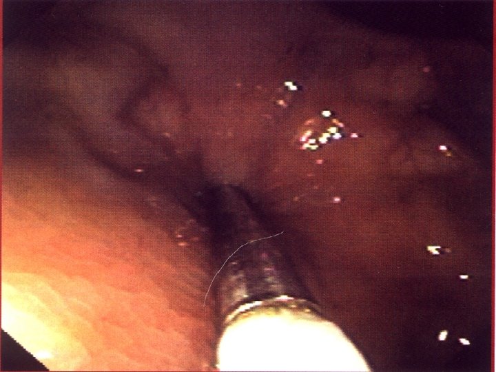

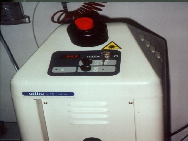
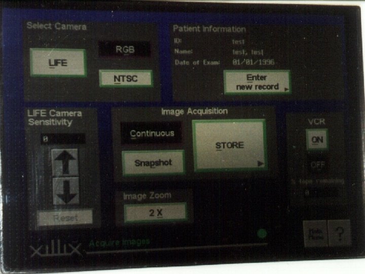
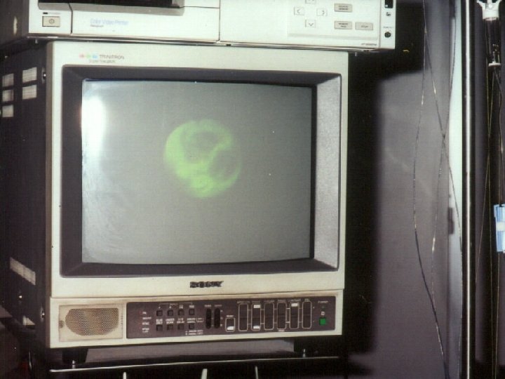
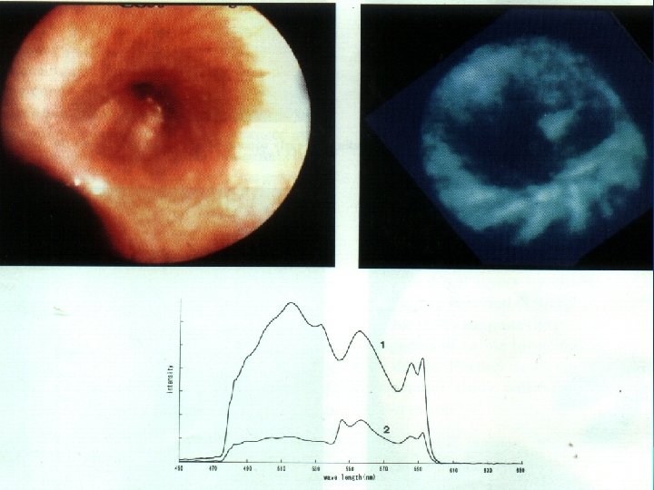
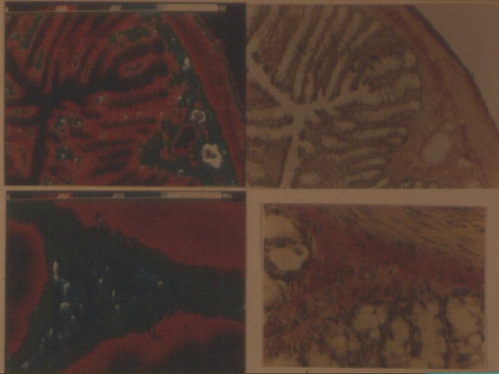
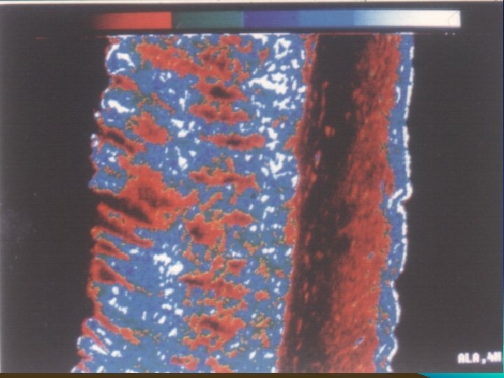
LIFE • Light Induced Fluorescence Endoscopy • Early detection of dysplasias and superficial malignant lesions, in situ Ca

Contrast Radiography • Motility – Ba meal, hypotonic duodenography • Structural changes • Diagnostic accuracy: – Single – 80% – Double – 90%

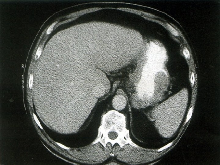
Computed Tomography • Abdomen and chest • Lateral extension, Systemic mets- 75% • Triphasic spiral CT – T stage, stomach filled with water Tako et al 1998 – Adv gastric Ca – 82% Early Ca – 15%

CT – T Staging • • Gastric distension Does not differentiate T 1 and T 2 T 3 stranding in perigastric fat Does not differentiate transmural and perigastric lymphadenopathy • Accuracy 80 – 88% in Advanced disease

CT – N Staging • Size – no predictor of involvement • > 8 mm sensitivity – 48%, specificity 93% • Identifies distal nodes (not seen on EUS) • No of involved nodes N 1 1 -6 RLN according to current TNM classification

CT – M Staging • Liver mets – thin collimation, overlapping slides, dual phase imaging • 75 – 80 % mets detected • Small volume ascites – EUS and CT

Conventional US • Good clinical evidence of liver mets • When treatment options are limited – before palliation • Used in conjunction with or, alternative to MRI – indeterminate lesions on CT

MRI • T assessment – No evidence that MRI better than CT • For identification of indeterminate lesions • IV contrast allergy • Endoluminal MR – experimental only and no advantage over EUS
 18 F • Preferrential accumulation of PEG in tumour •")
PET • FDG (fluorodeoxyglucose) 18 F • Preferrential accumulation of PEG in tumour • Sensitivity 60%, specificity 100%, Accuracy 94% • Detects 20% missed mets on CT • Differentiates: malignancy from inflammation

PET+CT Combo

PET

Laparoscopy • Peritoneal Disease M 1 – CT, EUS, Small volume ascites • Routine use after CT / EUS before radical surgery • Additional information than CT • Complementary to CT / EUS • Accuracy 84%

Laparoscopy US Probes • III dimension in US – detects unsuspected liver and lymphnode metastases • Eliminates need for laparotomy

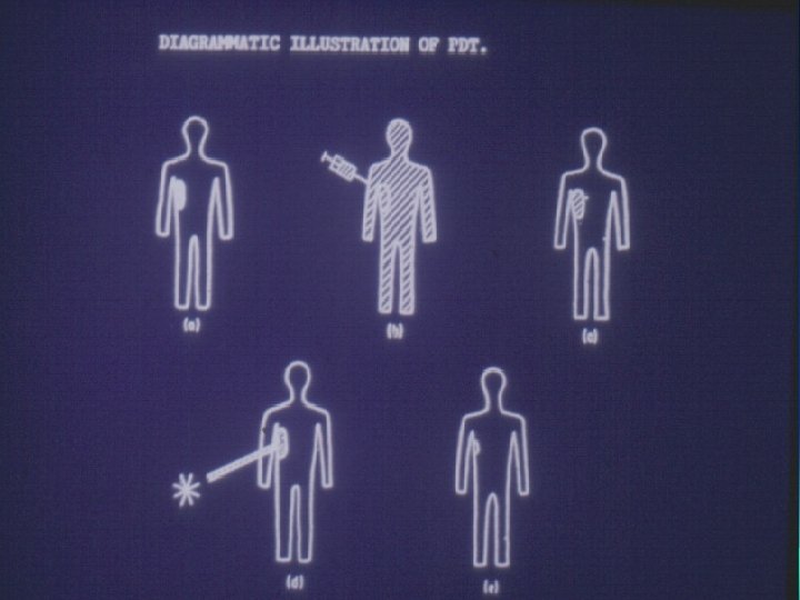
Mechanism of Photo-toxicity • Release of singlet oxygen • S phase cells more vulnerable than G phase cells

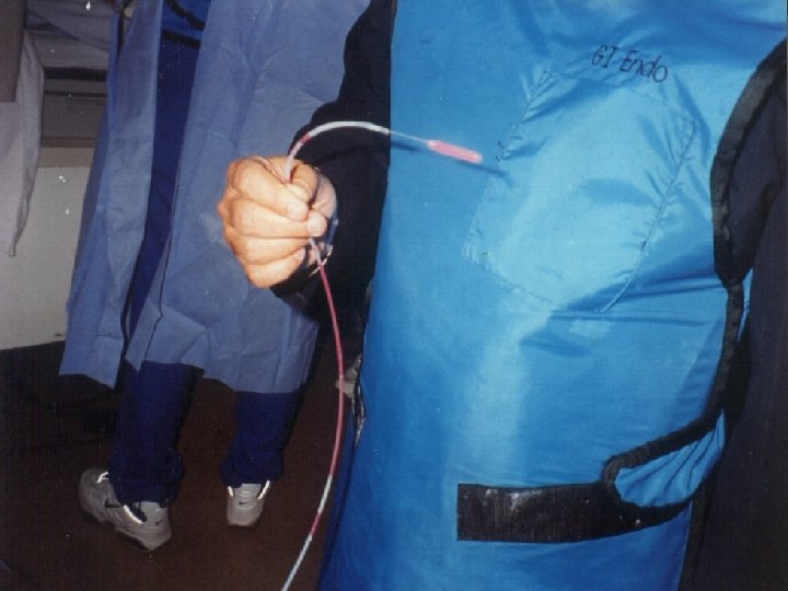

Inside Story – Wonder Pill • Pill with a camera – M 2 A • Pictures taken at 2 frames per second • Microchip in camera with 8 hour battery • Receiver in the belt • Ambulatory endoscopic monitoring

Camera Pill

Summary • H. Pylori – Non invasive, invasive • Gastric acid, Gastrin secretory studies • Tumor markers – CEA, CA 19 -9, • EUS – Early T & N stages • CT – distant metastasis

Summary • OCT – virtual biopsy, LIFE – new methods • PET – Non-invasive, physiological and biological measurement, better tissue differentiation
- Slides: 59