Development Of Pancreas And Small Intestine Embryology 436

Development Of Pancreas And Small Intestine ﴾ ﴿ ﺍ ﺍ ﺍﻹﻧﺴﺎﻥ ﺍ ﻳﺍ ﻳﺍ Embryology 436 M E D I C I N E KING SAUD UNIVERSITY Important Dr. notes Explanation - We recommend you to study anatomy of pancreas and small intestine.

O B J E C T I V E S - At the end of the lecture, the students should be able to : - Describe the development of the duodenum. - Describe the development of the pancreas. - Describe the development of the small intestine. Identify the congenital anomalies of the small intestine : o Congenital omphalocele. o Umbilical hernia. o Meckel’s diverticulum.

Development Of The Duodenum: Useful video - Stages in the development of duodenum, liver, biliary ducts and pancreas (pic. A-D). - Early in the 4 th week, the duodenum develops from the endoderm of primordial gut* of : 1 -Caudal ( )ﺗﺤﺖ part of foregut 2 -Cranial( )ﻓﻮﻕ part of midgut 3 -Splanchnic mesoderm**. Midgut is (Distal part ) Forgut is(Proximal part) -The junction*** of the 2 parts of the gut lies just below or distal to the origin of bile duct (pic, C -D). 5 th week 4 th week liver 5 th week *It’s an intra-embryonic endoderm That Divided into: Foregut, midgut, hindgut **give smooth muscle 6 th week ***it’s the junction between the two part of duodenum

Cont. . -The duodenal loop: 1 -The duodenal loop is formed and projected ventrally, forming a Cshaped loop. 2 -The duodenal loop is rotated with the stomach to the right. (90) degrees 3 - It comes to lie on the posterior abdominal wall retroperitoneally* with the developing pancreas. *except the first small part of duodenum. During 5 th and 6 th weeks, the lumen of the duodenum is temporarily obliterated because of proliferation of its epithelial cells. Normally degeneration of epithelial cells occurs the duodenum normally becomes recanalized by the end of the embryonic period**( end of 8 th week) ﺍﻟﺪﻳﻮﺩﻳﻨﻢ ﻋﺒﺎﺭﺓ ﻋﻦ ﺃﻨﺒﻮﺏ؛ ﻟﺬﻟﻚ ﻓﻲ ﺍﻷﺴﺒﻮﻉ ﺍﻟﺨﺎﻣﺲ ﻭﺍﻟﺴﺎﺩﺱ ﺍﻷﻨﺒﻮﺏ ﻳﺘﺴﺪ ﻭﻳﺘﻘﻔﻞ : ﺍﻟﺨﻼﺻﺔ ** the period from fertilization to the end of 8 th week - Fetal period: from 9 th week to birth ﺑﻌﺪﻳﻦ ﺍﻟﺨﻼﻳﺎ ﺗﺒﺪﺃ ﺗﺘآﻜﻞ ﻭﻣﻊ ﻧﻬﺎﻳﺔ ﺍﻷﺴﺒﻮﻉ ﺍﻟﺜﺎﻣﻦ ، ﻣﻦ ﺍﻟﻮﺳﻂ ﻣﻮﻗﺘﺎ ﺑﺴﺒﺐ ﺗﻜﺎﺛﺮ ﺍﻹﺑﻴﺜﻴﻠﻴﺎﻝ ﺳﻠﺰ ﻳﺮﺟﻊ ﺍﻷﻨﺒﻮﺏ ﻳﻔﺘﺢ ﻣﻦ ﺟﺪﻳﺪ

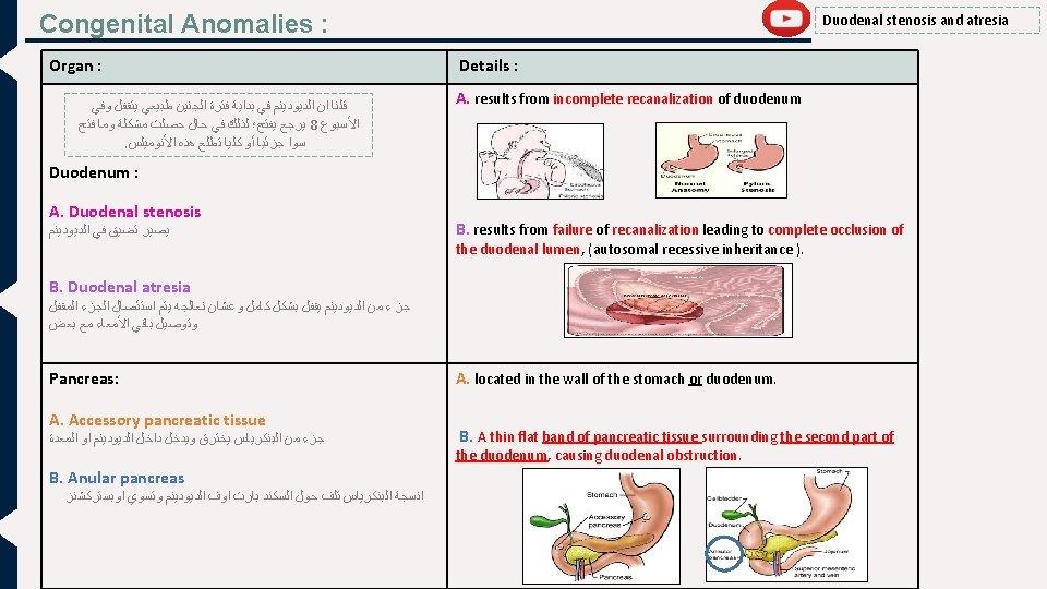
Development Of Pancreas: - The pancreas develops from 2 buds arising from the endoderm of the caudal part of foregut: 1 - ventral pancreatic bud : which develops from the proximal end of hepatic diverticulum (forms the liver and gall bladder). 2 - dorsal pancreatic bud : which develops from dorsal wall of duodenum slightly cranial to the ventral bud. - But Most of pancreas is derived from the dorsal pancreatic bud. ( )ﻻﻧﻬﺎ ﺍﻻﻛﺒﺮ - When the duodenum rotates to the right and becomes C-shaped, the ventral pancreatic bud moves dorsally to lie below and behind the dorsal bud. - Later the 2 buds fused together and lying in the dorsal mesentery. *

Development Of Pancreas: This slide is very important A ventral pancreatic bud A dorsal pancreatic bud (larger ) develops from: Proximal end of hepatic diverticulum (forms the liver and gall bladder). dorsal wall of duodenum slightly cranial to the ventral bud the bud forms: 1 -Uncinate process. 2 -(Inferior part of head of pancreas). Upper part of head, Neck, Body and Tail of pancreas. (so, ventral bud give us small part) (so, dorsal bud give us a lot of parts) pancreatic ducts: They are two ducts open in duodenum The main pancreatic duct ( is derived from ) ﺍﻟﺴﻬﻤﻴﻦ ﺍﻻﺻﻔﺮ ﻭ ﺍﻟﺰﻫﺮﻱ ﻳﻨﺪﻣﺠﻮﺍ ﻣﻊ ﺑﻌﺾ ﻭﻳﻌﻄﻮﻧﺎ ﺍﻟﻤﻴﻦ ﺑﻴﻨﻜﺮﻳﺎﺗﻚ ﺩﻛﺖ The accessory pancreatic duct ( is derived from ) - Before The duct of the ventral bud. The distal part of duct of dorsal bud. ___ Proximal part of duct of dorsal bud. The parenchyma of pancreas is derived from the endoderm of pancreatic buds. Pancreatic islets* develops from parenchmatous pancreatic tissue Insulin secretion begins at 5 th month of pregnancy *important* *its function is to secrete insulin And we can found islet a lot in the tail part of pancreatic tissue After

Development Of Small Intestine: Derivatives of cranial part of the midgut loop : the caudal part of midgut loop: Derivatives of caudal part of foregut : 1. 2. 3. 4. 1. Distal part of the duodenum Jejunum Upper part of the ileum. ( ﻟﻮ ﺗﺨﻴﻠﻨﺎ ﺍﻷﻤﻌﺎﺀ ﻭﺍﻋﺘﺒﺮﻧﺎﻩ ﺗﺴﻠﺴﻞ ﻳﺴﻬﻞ )ﺣﻔﻈﻬﺎ Lower portion of ileum. Cecum and appendix. Ascending colon proximal 2/3 of transverse colon. ** Caudal Foregut * ﻣﻮ ﻣﺮﺓ ﺗﻬﻤﻨﺎ ﻓﻲ ﻫﺬﻩ ﺍﻟﻤﺤﺎﺿﺮﺓ proximal part of duodenum Cranial Midgut Caudal Midgut - So, the small intestine is developed from : ﺍﻏﻠﺐ ﺍﻟﺴﻤﻮﻝ ﺍﻧﺘﻴﺴﺘﺎﻧﺖ ﻣﻦ ﺍﻟﻤﻴﺪ ﻗﺖ Caudal part of foregut. All midgut. 1. 2. Cranial Midgut (distal) Very helpful summarizing pic - Midgut is supplied by superior mesenteric artey (artery of midgut). *** * Don’t forget that also from caudal foregut (proximal part) ** 2, 3, 4 are parts of large intestine, The rest 1/3 of transvers colon developed from hindgut. ***very important; SMA sometimes called artery of midgut. - Stages of development of small intestine : 1. 2. 3. 4. 5. Preherniation stage. Stage of physiological umbilical hernia****. stage of rotation of midgut loop. Stage of reduction of umbilical hernia. Stage of fixation of various parts of intestine **** Hernia is a condition in which part of an organ is displaced and protrudes through the wall of the cavity containing it.
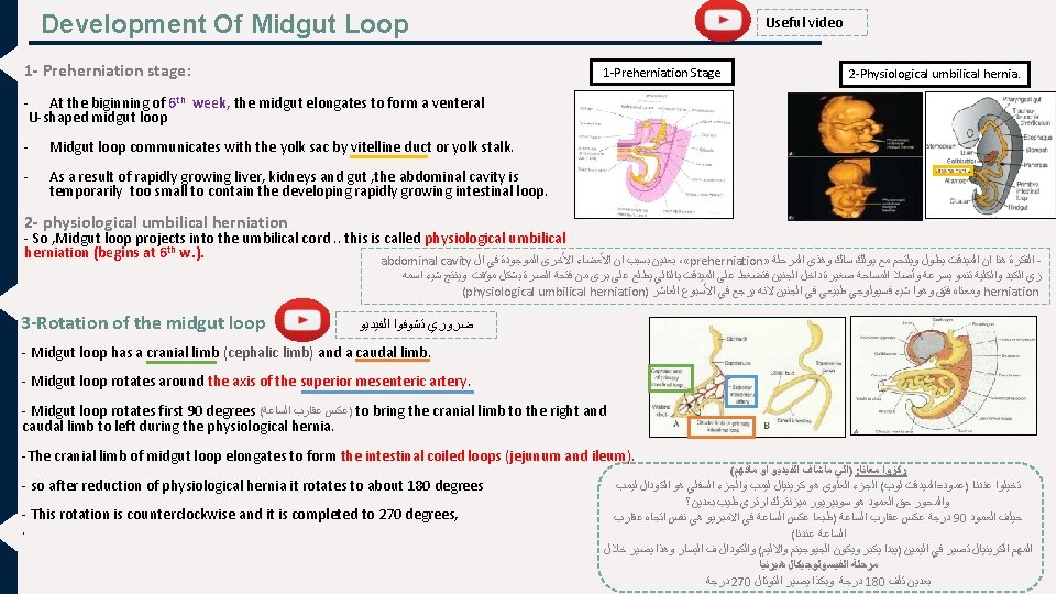

Cont. . 4 -Return of the midgut to abdomen - During 10 th week, the intestines return to the abdomen due to regression of liver and kidneys + expansion of abdominal cavity. It is called reduction of physiological midgut hernia. In 10 th week !! ﻣﻬﻢ ﻫﺬﺍ ﺍﻟﻮﻗﺖ - Rotation is completed and the coiled intestinal loops lie in their final position in the left side. - The caecum at first lies below the liver (A), but later it descends to lie in the right iliac fossa (B) 5 - Fixation of various parts of intestine - The mesentry of jejunoileal loops is at first continuous with that of the ascending colon. - When the mesentry of ascending colon fuses with the posterior abdominal wall , the mesentry of small intestine becomes fan-shaped and acquires a new line of attachment that passes from duodenojejunal junction to the ileocecal junction. ØThe enlarged colon presses the duodenum and pancreas against the posterior abdominal wall. ( C & F) ØMost of duodenal mesentery is absorbed, so most of duodenum except (for about the first 2. 5 cm derived from foregut(upper part of duodenum)) and pancreas become retroperitoneal. ( C & F ) Intestine prior fixation Intestine after fixation

Congenital Anomalies : Organ : Small intestine : important to know The hernial sac is covered by (in every anomalies) A. Congenital Omphalocele This slide is very important Details : • It is a persistence of herniation of abdominal contents into proximal part of umbilical cord due to failure of reduction of physiological hernia to abdominal cavity at 10 th week. • It is accompanied by small abdominal cavity. ﻣﺎﺭﺟﻊ ﺍﻟﻠﻮﺏ ﻓﻲ ﺍﻷﺴﺒﻮﻉ ﺍﻟﻌﺎﺷﺮ ﻭﻋﺸﺎﻥ ﺍﻻﺑﺪﻭﻣﻨﺎﻝ ﻛﺎﻓﺘﻲ ﺻﻐﻴﺮ ﻣﺎﻛﺒﺮ B. Congenital Umbilical Hernia • Herniation of intestines occurs in 1 of 5000 births – herniation of liver and intestines occurs in 1 of 10, 000 births. • The hernial sac is covered by the epithelium of the umbilical cord or the amnion. • Immediate surgical repair is required • The intestines return to abdominal cavity at 10 th week, but herniate through an imperfectly closed umbilicus. ﺍﻻﻧﺘﺴﺘﺎﻧﺖ ﺭﺟﻌﺖ ﻣﻜﺎﻧﻬﺎ ﺑﺲ ﺍﻟﻔﺘﺤﺔ ﻣﺎﺗﻘﻔﻠﺖ • It is a common type of hernia. • The herniated contents are usually the greater omentum and small intestine. • The hernial sac is covered by skin and subcutaneous tissue. • It protrudes during crying , straining or coughing and can be easily reduced through fibrous ring at umbilicus. ﺍﺫﺍ ﺿﻐﻄﻨﺎ ﻋﻠﻰ ﻣﻜﺎﻥ ﺍﻟﻔﺘﺎﻕ ﺗﺪﺧﻞ ﺍﻷﻤﻌﺎﺀ ﺟﻮﺍ ﺑﺪﻭﻥ ﻣﺎﻳﺘﺄﻠﻢ ﺍﻟﻄﻔﻞ ﻟﻜﻦ ﺗﺮﺟﻊ ﺗﻄﻠﻊ ﺍﺫﺍ ﻋﻜﺲ ﺍﻻﻧﻮﻣﻴﻠﻴﺲ ﺍﻟﻲ ﺑﻌﺪﻫﺎ. ﺍﻟﻄﻔﻞ ﺑﻜﻰ ﺍﻭ ﻛﺢ ﺍﻭ ﺑﺬﻝ ﺟﻬﺪ • Surgery is performed at age of 3 -5 years. So it’s not an emergency as the one above

Congenital Anomalies : Organ : This slide is very important Details : • It is one of the most common anomalies of the digestive tract, present in about 2% -4% of people, more common in males. • It is a small pouch from the ileum, and may contain small patches of gastric and pancreatic tissues causing ulceration, bleeding or even perforation. • It is the remnant of proximal part non-obliterated part of yolk stalk (or vitelline duct). Normally, viteline obliterate and disappear , but in this anomalie will not disappear • It arises from antimesenteric border of ileum, 1/2 meter from ileocecal junction. C. ileal (Meckel’s) Diverticulum • It is sometimes becomes inflammed and causes symptoms that mimic appendicitis. ﺍﻟﺰﺍﺋﺪﺓ to ﺍﻟﺘﻬﺎﺏ ﺗﺴﻮﻱ ﻧﻔﺲ by ﺗﻠﺘﻬﺐ ﻟﻤﺎ • 1 - It may be connected the ﺍﻋﺮﺍﺽ umbilucus a fibrous cord (vetline ligament), 2 - or the middle portion forms a cyst 3 - or may remain patent forming the fistula - so, faecal matter is carried through the duct into umbilicus ﻳﺨﺮﺝ ﻣﻦ ﺍﻝ feces ﻭﺍﻟﺨﻄﻮﺭﺓ ﻫﻨﺎ ﺍﻧﻮ ﻣﻤﻜﻦ ﺍﻝ • Yolk stalk: A narrow tube present in the early embryo that connects the midgut of the embryo to the yolk sac outside the embryo through the umbilical opening , It is usually obliterated, but a remnant of it may persist: most commonly as a finger -like protrusion from the small intestine known as Meckelis diverticulum. (girls slids only)

Summary Organ Duodenum Pancreas Midgut loop Period 4 th week Event - Develops from the endoderm of primordial gut - The lumen of the duodenum is temporarily obliterated. 5 th & 6 th week End of 8 th week 5 th month 6 th week - Degeneration of epithelial cells occurs. - Duodenum normally becomes recanalized. - Insulin secretion begins. - Elongates to form a venteral U-shaped midgut loop. - Communicates with the yolk sac by vitelline duct or yolk stalk. - Projects into the umbilical cord (physiological umbilical herniation). 10 th week - Return to the abdomen (reduction of physiological midgut hernia).

Summary Congenital Anomaly Cause And Details Duodenal stenosis Incomplete recanalization of duodenum. Duodenal atresia Failure of recanalization of duodenum. Accessory pancreatic tissue Located in the wall of the stomach or duodenum. Anular pancreas A thin flat band of pancreatic tissue surrounding the second part of the duodenum, causing duodenal obstruction. Congenital Omphalocele Failure of reduction of physiological hernia to abdominal cavity at 10 th week. Congenital Umbilical Hernia Imperfectly closed umbilicus. Ileal (Meckel’s) Diverticulum Remnant of proximal part non-obliterated part of yolk stalk (or vitelline duct). Sometimes become inflamed and can produce pain.

MCQ’s 1. Which part of the pancreas the ventral pancreatic bud forms ? A-Upper part of the head B-Lower part of the head C-Body D-Tail 2. Which artery the midgut loop rotates around its axis ? A-Splenic artery B-Inferior mesenteric artery C- Superior mesenteric artery D-Celiac trunk 3. The cranial limb of midgut loop gives rise : A- The liver B-The pancreas C-The stomach D-The jejunum and ilum 4. The umbilical hernia is: A- Uncommon type. B- Resulting from imperfect closed umbilicus. C- Covered by the epithelium of umbilical cord. D-Not be easily reduced at the umbilicus. 6. The Meckel’s diverticulum : A- Is a duodenal pouch. B- Arises from the mesenteric border of the ileum. C- Is a remnant of the proximal nonobliterated part of yolk stalk. D- Is a physiological hernia of intestine. C-An abdominal wall defect D-Covered by skin. 1 - B 2 - C 3 -D 4 - B 5 - B 6 - C 5. The congenital omphalocele is : A- small pouch from the ileum B-Covered by the epithelium of the umbilical cord
. Embryology")
References ANY SUGGESTION S OR ISSUES • • Dr. slides (male and female). Embryology team 435. USEFUL VIDEOS Embryology @Embryology 436@gmail. com Editing file Your Suggestion here

TEAM MEMBERS § TEAM LEADERS : YAZEED ALMUTAIRI NEHAL BEYARI ▪ B O Y S : . Abdulrahman Alomrani. Saqr Altamimi ▪ G I R L S : Desgined by: MUHANNED ALZAHRANI • Razan Alotaibi • Do’aa Walid • Ohood Abdullah • Nouf Aloqili
- Slides: 17