Stomach The stomach is on the left side


")


- Slides: 12
Stomach • The stomach is on the left side of the abdominal cavity • • Cardiac region (near the heart) Fundus (lateral to the cardia) The body (mid and largest portion) Pylorus (funnel-shaped terminal part of the stomach). Most digestive activity occurs in this region. • Two sphincters: • Cardioesophageal sphincter, through which food enters the stomach from esophagus • Pyloric sphincter, which is continuous with small intestine • Greater curvature and lesser curvature • The lesser omentum (double layer of peritoneum extends from liver to lesser curvature) • The greater omentum (another extension of the peritoneum riddled with fat and covers the abdominal organs (cushioning and insulation)
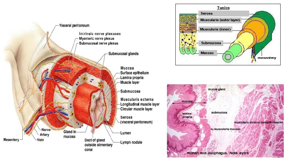
Stomach • Histology of the stomach is similar to the esophagus except that the mucosa is lined with simple columnar epithelium which produces large amounts of mucus Digestion: • Special arrangement of muscular cells in muscularis externa helps to move, churn and mix the food to break it down to smaller fragments (physical) • Chemical breakdown of proteins begins • Stomach lining contains deep gastric pits that secrete gastric juice • Chief cells produce protein digesting enzymes pepsinogens • Parietal cell produce HCL makes stomach acidic and activates enzymes • Low PH of stomach stimulates stomach cells to produce gastrin hormone stimulates stomach glands to produce more protein digesting enzymes, mucus and hydrochloric acid • How is the stomach wall protected from HCL’s corrosive nature? mucous neck cells produce sticky alkaline mucus • After food has been processed in the stomach, it resembles heavy cream and is called chime. The chime then enters the small intestine through the pyloric sphincter
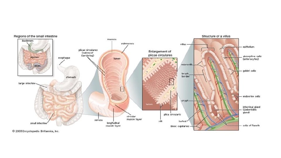
Small Intestine • The body’s major digestive organ • Longest section of the alimentary tube (increase surface area for absorption) • Extends from pyloric sphincter to ileocecal valve (valve that separates small intestine and large intestine) • The large intestine encircles it and frames it in the abdominal cavity • Three subdivisions: • Duodenum • Jejunum (longest part) • Ileum (joins large intestine at ileocecal valve)
Small Intestine cont. • Small intestine can digest only a small amount of food at a time (pyloric sphincter controls food movement from the stomach so that the small intestine is not overwhelmed) • Digestion: • Most of the digestion occurs in small intestine • Enzymes are produced my intestinal cells help in digestion • Enzymes produced by pancreas and ducted into the duodenum through the pancreatic ducts • Bile (formed by liver) enters duodenum through bile duct • Absorption • Most of the food absorption occurs in the small intestine
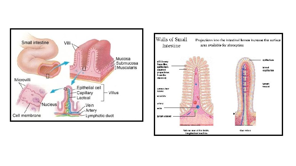
Small Intestine cont. • Structures help increase the absorptive surface: • Microvilli: tiny projections of the plasma membrane of the mucosal cells, which give the cell surface a fuzzy appearance (brush border) • Villi: fingerlike projections of the mucosa. Within each villus is a rich in capillaries and contains a lymphatic capillary called a lacteal • Digested food is absorbed through the mucosa cells into the capillaries and lacteal • Circular folds, called plicae circulares: deep folds of mucosa and sub-mucosa • Peyer’s Patches: • Collection of lymphatic tissue (high in lymphocytes+ other immune cells) found towards the end of the small intestine (remaining undigested food residue is high in bacteria that must be prevented from entering the blood stream)
Large Intestine • Functions • Primary task is absorption of water • Absorption of other substances (e. g : Bacteria in large intestine that produce vitamin K that is absorbed in the LI) • Excretion of wastes • Parts of LI: • Cecum (where small intestine contents enter the large intestine) • Appendix (wormlike structure that hangs from the cecum) • Colon • • Ascending colon (follows the cecum) Transverse colon Descending colon Sigmoid colon (has an S-shaped curve) • Rectum • Anal canal (opens to the exterior) • Has an external voluntary sphincter and internal involuntary sphincter act together to open and close (usually closed except during defecation)
Accessory digestive organs • Pancreas: • Considered both an • Endocrine gland (insulin, glucagon) • Exocrine gland (digestive enzymes): trypsinogen that becomes activated into trypsin in the small intestine to digest proteins into amino acids. • *Endocrine are ductless glands that secret hormones that travel through the blood to regulate a target organ • *Exocrine glands produce secretions that reach their target through a duct • Together insulin and glucagon maintain our blood glucose levels • insulin lowers blood glucose levels, glucagon
Accessory digestive organs • Liver and Gall Bladder: • The liver is the largest gland in the body • Located under the diaphragm • Liver functions include: • Helps in digestion by producing bile (yellow to green watery solution containing bile salts, bile pigments, cholesterol and phospholipids) • Bile salts helps to break down large fat globules into smaller ones to aid the action of fat-digesting enzymes (lipases) • Liver also produces blood clotting factors • The gall bladder stores and secretes bile
Overview of digestion