Anatomy of Duodenum Development of Duodenum Page 79


")
, its cranial ½")

")


- Slides: 32
Anatomy of Duodenum
Development of Duodenum (Page 79)
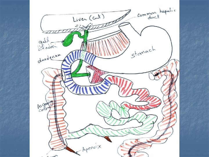
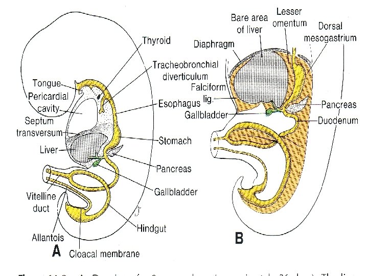
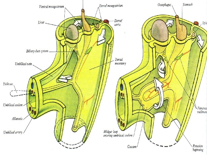
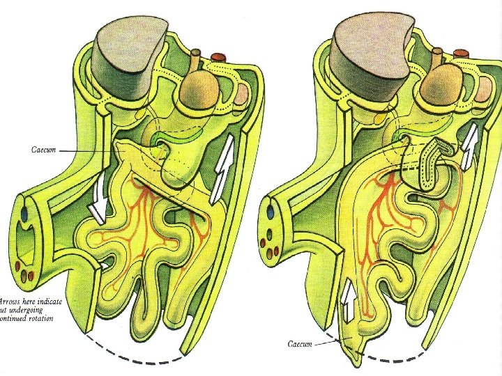
Development of Duodenum n n n Formation of duodenal loop (U-shaped), its cranial ½ belongs to foregut while its caudal ½ belongs to midgut. It gives origin to hepatic and pancraetic buds from its ventral wall in caudal end of foregut. It has dorsal mesoduodenum. It rotates 90 degrees to right with stomach till it rests on posterior abdominal wall with right convexity. Mesoduodenum becomes fused and absorbed to peritoneum of posterior abdominal wall, so it is retro peritoneal. Upper part of duodenum is supplied by coeliac artery, while its lower part by superior mesentric artery.
Congenital Anomalies n n 1 - Duodenal atresia: due to failure of canalization. 2 - Duodenal stenosis: due to defective canalization.
Development of Pancreas (Page 81)
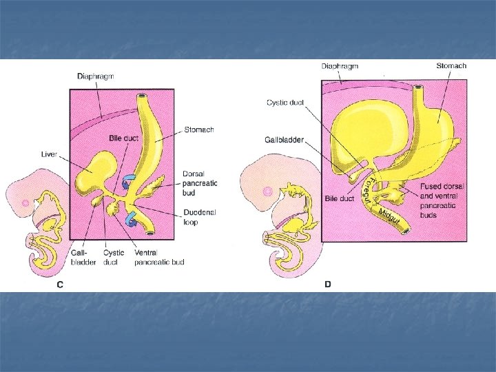
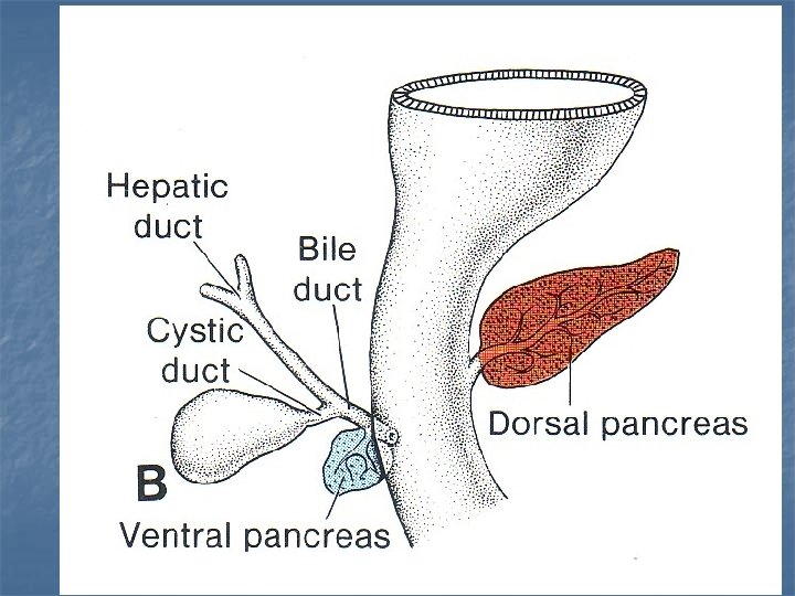
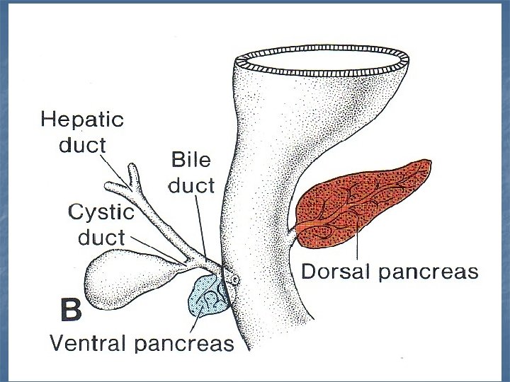
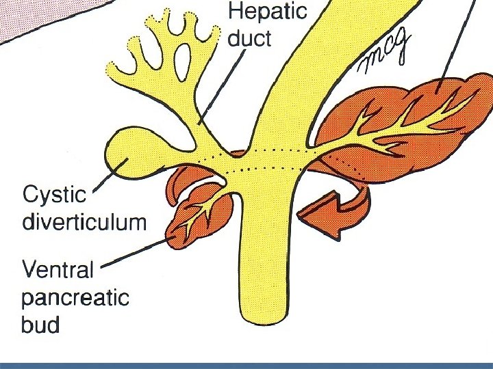
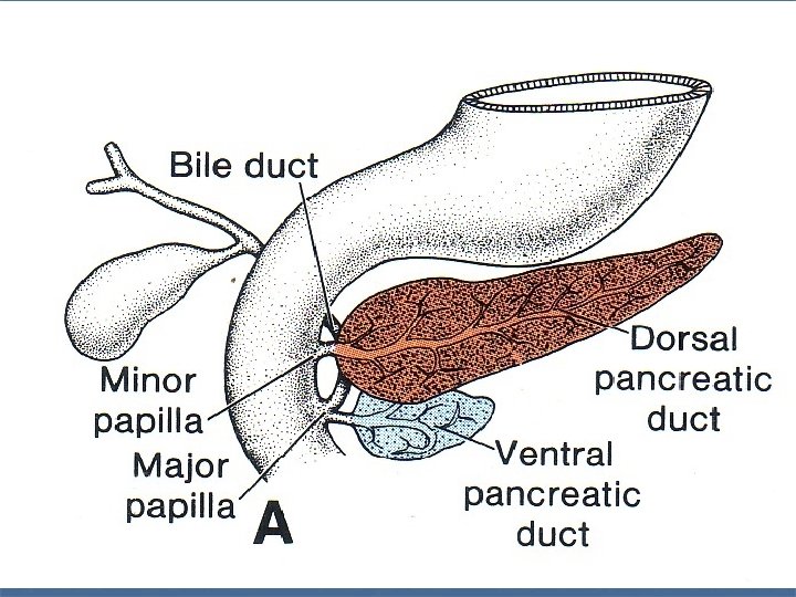
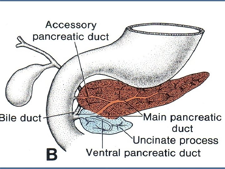
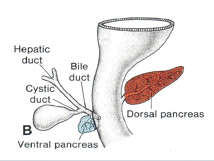
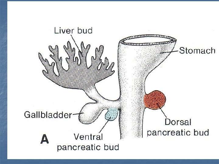
Development of Pancreas n n n It is formed from 2 endodermal buds: ventral & dorsal. Ventral pancreatic bud: It arises from the proximal part of hepatic duct. It migrates together with hepatic duct from ventral wall of foregut to its dorsal wall to lie below dorsal bud. It forms lower part of head of pancreas and uncinate process. Dorsal pancreatic bud: It arises from dorsal aspect of foregut just cranial to hepatic bud. It grows dorsally through mesoduodenum to form upper part of head of pancreas, body and tail.
Development of Pancreatic duct n n Anastomosis between 2 ducts of the pancreatic buds. Definitive pancreatic duct is formed from: 1 - Duct of ventral bud which open in duodenum with common bile duct. n 2 - Anastomosis between 2 ducts. n 3 - Distal part of duct of dorsal pancreatic bud. n 4 - Proximal part of dorsal pancreatic duct may be obliterated or remain as accessory pancreatic duct. n
Anomalies of Pancreas 1 - Annular pancreas: A ring of pancreatic tissue that surrounds the duodenum. n 2 - Persistence of accessory duct: due to persistence of proximal part of dorsal pancreatic bud. n 3 - Accessory pancreas: Pancreatic tissue in abnormal site as in pyloric part of stomach. n
Development of Liver & Gall Bladder
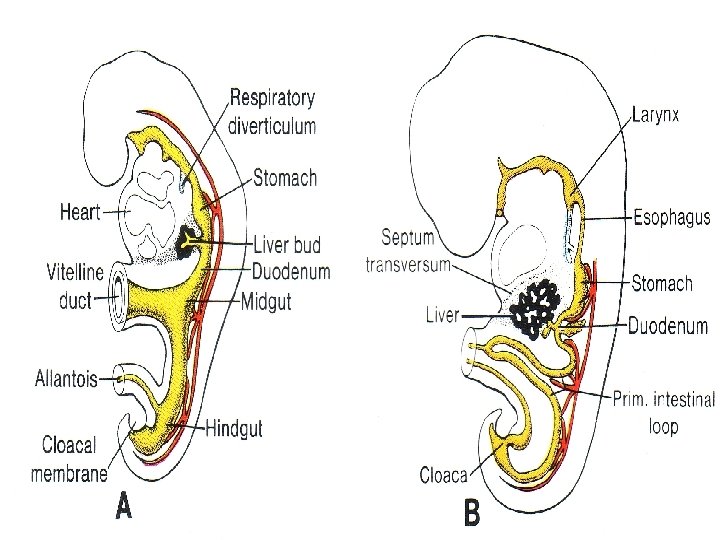
Development of Liver & Gall Bladder n Liver is developed from the followings: 1 - Hepatic bud: form liver cells. n 2 - Vitelline & umbilical veins: form liver sinusoids. n Septum transversum: form fibrous stroma and capsule of the liver. n
Development of Liver - It begins as endodermal diverticulum from convexity of duodenal loop (just above caudal end of foregut) which is called hepatic bud. - Hepatic bud divides into: 1 - Pars cystica: forms gall bladder and cystic duct. 2 - Pars hepatica: forms the liver and its hepatic ducts.
Pars Cystica n It grows to give rise to: n n Gall bladder. Cystic duct.
Pars Hepatica n n It grows cranially and ventrally into ventral mesogastrium between cells of septum transversum. It divides into 2 parts (finally form 2 lobes of liver). Rows of proliferating cells arise from each part. These cells invade vitelline & mbilical veins (inside ventral mesogastrium) to release blood from veins and to form liver sinusoids. Capsule of liver is formed from mesoderm of septum transversum.
-Stem of hepatic bud will migrates from ventral to dorsal wall of duodenum, this is followed by rotation of duodenum to right so finally hepatic dub will open into posteromedial aspect of duodenum. -Firstly, two lobes of liver are equal size but later right lobe grows faster and become larger than left lobe. At birth, liver is relatively big as it acts as haemopoitic organ in fetus. -Fate of ventral mesogastrium: -Part around liver forms its capsule. -Part between liver and stomach forms lesser omentum.
Anomalis n n n 1 - Double gall bladder. 2 - Bile duct atresia. 3 - Absence of gall bladder.