Shigella spp Shigella and Shigellosis Fecaloral transmission persontoperson


: localized infection • Fecal‑oral transmission • Localized infection of intestines")

: Systemic Infection • Fecal-oral transmission • Systemic")


 • 30%")
 – septicemic (blood-borne")


")
 – Basidiomycota (basidiospores) –")


 • Allergy • Hypersensitivity Pneumonitis (HP) and Organic Dust")


 Ringworm of scalp, glabrous skin, and nails caused by a")


 versicolor: A chronic, superficial fungal disease of the skin characterized")


 Primary cutaneous and subcutaneous infections usually the result of a \"barrier")


 – Least harmful, generally considered benefical – Growth")
 – Taste and odor problems; reservoir growth;")
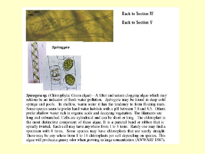
 – Indicators of pollution – Filter cloggers •")
 –")
 • unicellular – many have stiff, patterned plates • many photosynthetic –")

 – Staggering, paralysis, gasping, convulsions, death")
 spp. • ASP-")
 • Capable of 24")


- Slides: 104
Shigella spp.
Shigella and Shigellosis • Fecal-oral transmission – person-to-person, fomites, food, water, ect. • Waterborne and water-washed • Reservoirs: humans and primates • Infectious dose: low; as few as 10 cells to infect • Incubation period: 1 to 7 days; typically, 1 -3 days • Duration of illness: – untreated: severe symptoms for about two weeks
Shigellosis - Illness • Persistent diarrhea with frequent and painful passage of stools consisting mostly of blood, mucus and pus – accompanied by fever and stomach cramps. • Blood and mucus in stools are likely signs of shigellosis • Shigella infect, invade cells lining the large intestine (colon) • Cause breaks (ulcers) in mucous membrane lining of intestine – Inflammation and tissue damage – Causes painful straining to pass stools; can lead to rectal prolapse – Ulcers commonly in the rectum – results in increased production of mucus
Shigellosis - Epidemiology • Four species of Shigella: flexneri, sonnei, dysenteriae, boydii • Major public health problem in many developing countries – causes about 5 to I 0% of childhood diarrhoea – up to 25% of all diarrhea-related deaths can be associated with Shigella Developing countries: • Sh. flexneri is endemic (always present) in most communities • Sh. dysenteriae type 1 often occurs in an epidemic pattern – organism can be absent for a number of years, then reappear and infect a large proportion of the population. • These two species of Shigella generally produce the most severe illness.
Salmonella and Salmonellosis • Belong to Enterobacteriaceae family • Gram-negative bacilli; facultative and flagellated (motile). • 3 major antigens: – "H" or flagellar antigen (phase 1 & 2) – "O" or somatic antigen (part of the LPS moiety) – "Vi" or capsular antigen (called "K" in other Enterobacteriaceae). • Posess LPS endotoxin characteristic of Gramnegative bacteria – composed of an "O” polysaccharide ("O" antigen) – "R" core – endotoxic inner "Lipid A". – Endotoxins evoke fever and can activate
Salmonella spp.
Salmonella gastroenteritis: (S. enteriditis): localized infection • Fecal‑oral transmission • Localized infection of intestines • Damage and inflammation to lamina propria • 0. 5‑ 2 day incubation period • Watery diarrhea, nausea, vomiting, abdominal cramps, low grade fever, lasting several days • Bacteria shed fecally at billions per gram • Infectious dose is relatively high: >103 ID 50 for many strains
Salmonella enteritidis
Typhoid fever: (S. typhi and S. paratyphi): Systemic Infection • Fecal-oral transmission • Systemic infection: – Macrophages, reticuloendothelial system (esp. liver, spleen and bone marrow), gallbladder and intestines as major sites of damage • 1. 5‑ 2 week incubation period • Symptoms: fever, headache, malaise, anorexia, then bloody diarrhea • Mortality rate 10%, if untreated • Carrier state possible – "Typhoid Mary”: infamous food handler; infected hundreds • Fecally shed at billions/gram by ill persons and
Salmonella typhi
Yersinia pestis • Gram Stain: – Small, gram-negative bipolar-stained coccobacilli • Wayson Stain: – Pink-blue cells with a closed safety pin look
Yersinia pestis: Plague • U. S. averages 13 cases/yr (10 in 1998) • 30% of cases are in Native Americans in the Southwest. 15% case fatality rate • Most cases occur in summer
Plague Epidemiology • Three Clinical Types: – bubonic (infected lymph nodes) – septicemic (blood-borne organisms) – pneumonic (transmissible by aerosol; deadliest)
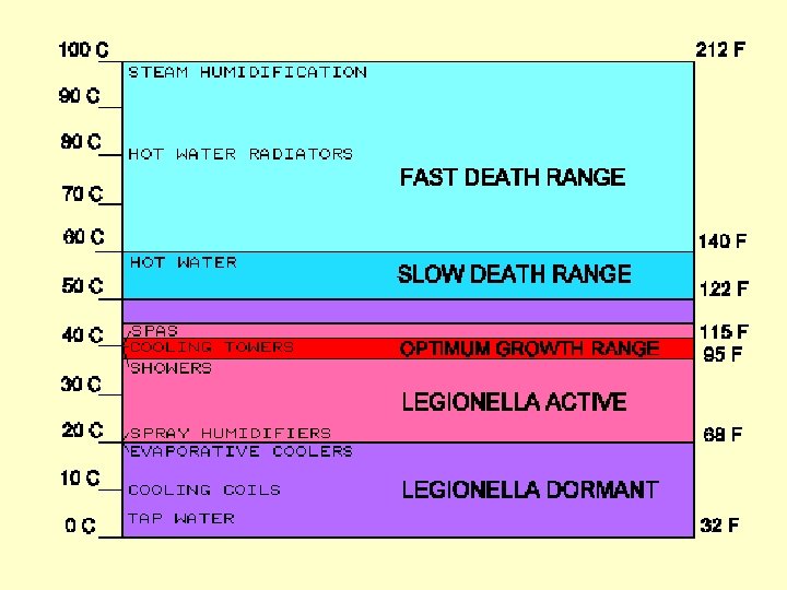
Legionella spp. • • Gram-negative Aerobic Non-sporing Encapsulated ~46 species, 68 serogroups Ubiquitous aquatic organism Thrives in warm environments (32 C-45 C)
Legionella: Legionellosis and Pontiac Fever Reservoirs and amplifiers: • Hot water systems • circulating water ventilation systems (cooling towers) • Plumbing (e. g. , shower heads). • Hot tubs, whirlpools, etc. • Produce fresheners Cleveland Auto plant outbreak, March, 2001: • Plant cooling tower is considered a possible source of the outbreak. • But, more than 100 other internal water sources -favorite breeding grounds for the Legionella bacteria -- were also under investigation….
Legionnaire’s Disease and Pontiac Fever Legionnaire's disease: • Bacterial pneumonia caused by Legionella pneumophila. • A type of pneumonia that affects the lungs and may also affect the stomach and intestines, kidneys, and central nervous system. • Incubation period: 2 -10 days after exposure • Frequently requires hospitalization • Aerosol exposure from contaminated cooling towers, evaporative condensers, whirlpools, shower heads, faucets, & hot water tanks. Pontiac fever: also caused by Legionella. • A "flu-like" illness with fever, chills, headache, myalgia (pain in the muscles), cough, nausea, and breathlessness. • Pneumonia does not occur.
Fungal Pathogens John Scott Meschke Office: Suite 249 N, 4225 Roosevelt Phone: 206 -221 -5470 Email: jmeschke@u. washington. edu
Kingdom Fungi • Major role in recycling of organic matter”Saprophytes” or “Saprobes” • Eukaryotic, unicellular or multicellular structures with rigid chitinous cell wall • No chlorophyll • Asexual and sexual reproduction – Anamorph or Telomorph – Homothallic or heterothallic
FUNGI - Yeasts, Molds, Fleshy fungi Yeasts: spherical to oval cells reproduce by budding aerobic to anaerobic Dimorphic fungi: pathogens yeast in the animal; fungus in the environment
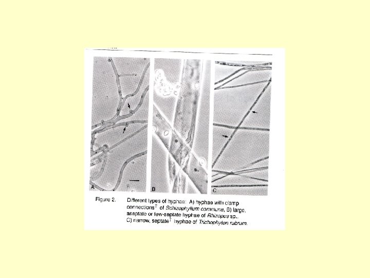
Fungal structure yeasts – single-celled organisms cell wall – chitin typical eukaryotic cell molds – hypha (hyphae) – long thin filaments aggregated hyphae form the mycelium cell wall of chitin
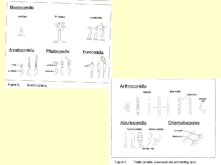
Fungal Structures • Hyphae • Spores • Conidia (asexual spores)
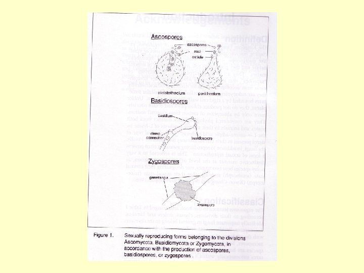
Medically Relevant Fungi • Three major divisions: – Ascomycota (ascospores) – Basidiomycota (basidiospores) – Zygomycota (zygospores) • Fungi Imperfecti – Deuteromycota
Ascomycetes – sac fungi red, brown and blue-green fungi associated with food spoilage plant diseases chestnut blight and Dutch elm disease edible morels and truffles examples: Morchella esculenta common morel Claviceps purpurea – parasite of rye and other grasses disease called ergotism in humans – poisoning in humans St. Anthony’s fire – killed thousands in Middle Ages psychotic delusions, nervous spasms, convulsions ergot alkaloids – LSD
Basidiomycetes – club fungi smuts, rusts, shelf fungi, puffballs, toadstools, mushrooms basidium – club-shaped structure on which spores are produced basidiocarp – mushroom most are saprophytes
Basidiomycetes – club fungi smuts, rusts, shelf fungi, puffballs, toadstools, mushrooms basidium – club-shaped structure on which spores are produced basidiocarp – mushroom most are saprophytes
Basidiomycetes – club fungi examples: smuts & rusts – crop damage in millions Agicarium campestris – multimillion-dollar business Amanita phalloides – toxic two toxins – phalloidin and α-amanitin phalloidin – liver toxin α-amanitin – lining of the intestinal tract Cryptococcus neoformans – cryptococcosis lungs and central nervous system
Zygomycetes most soil organisms a few pathogens of plants, insects, animals form thick-walled zygospores sexual reproduction – fusion of + and - strains examples: bread mold Rhizopus; black spores Mucor – production of sofu from soybeans some pathogens
Deuteromycetes – fungi imperfecti no sexual stage most saprophytes, a few parasites of plants examples: Penicillium – production of antibiotics Aspergillus – production of soy sauce also produce aflatoxins
Yeasts oval, single-celled organisms used in fermentation bread, wine, beer examples: Saccharomyces cerevesiae
Fungal Disease • Infection (mycosis) • Allergy • Hypersensitivity Pneumonitis (HP) and Organic Dust Toxic Syndrome (ODST) • Toxicosis • VOCs? ? ?
Fungal Infections • Mycosis: any fungal Infection • Systemic mycoses: fungal infection deep inside body. Can result from inhalation of spores. Ex: Valley fever (coccidioidomycosis) • Subcutaneous mycoses: infection beneath skin as a result of puncture
Fungal Infections, cont. • Cutaneous mycoses: infections of epidermis, hair, and nails. Direct contact with infected person, animal, hair, or cells is required. • Superficial mycoses: infections of hair shaft and surface (dead) epidermal cells
MEDICALLY IMPORTANT FUNGI Mycosis - fungal infection chronic infection Classification - mode of entry & tissue involvement Systemic - spores inhaled, begins as lung infection Subcutaneous - puncture wound Cutaneous - dermatomycosis epidermis, hair, nails tinea capitis, tinea cruris, tinea pedis Superficial - hair shaft or in epidermal cells Opportunistic - compromised host
MEDICALLY IMPORTANT FUNGI Cutaneous - dermatomycosis epidermis, hair, nails tinea capitis, tinea cruris, tinea pedis Superficial - hair shaft or in epidermal cells
MEDICALLY IMPORTANT FUNGI Opportunistic - compromised host Mucormycosis Aspergillosis enters through respiratory tract
MEDICALLY IMPORTANT FUNGI Opportunistic - compromised host Candidiasis – vaginal infections 10% of blood infections in hospital infections thrush – oral infection
Fungal Toxins • Most fungal toxins produced by: – Aspergillus spp. – Penicillium spp. – Fusarium spp. – Alternaria spp. – Rhizopus – Mucor
Important Mycotoxins • • • Aflatoxins Fumonisins Ochratoxins Trichothicenes Zearalenone and Zearalenoles • Tenuazonic • Patulin • Xanthomegnin and Viomellein • Nephrotoxic glycopeptides • Sterigmatocystin • Fusarochromanone • Ergot alkaloids • Citrinin • 3 -Nitropropionic acid • Roquefortine C • Secalonic acid D • Verrucosidin
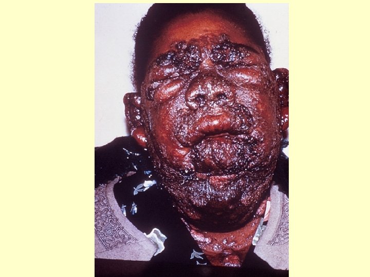
Dermatophytosis (Ringworm and Tinea) Ringworm of scalp, glabrous skin, and nails caused by a closely related group of fungi known as dermatophytes which have the ability to utilize keratin as a nutrient source. The disease process in dermatophytosis is unique for two reasons: (a) No living tissue is invaded; the keratinised stratum corneum is simply colonized. However, the presence of the fungus and its metabolic products usually induces an allergic and inflammatory eczematous response in the host. The type and severity of the host response is often related to the species and strain of dermatophyte causing the infection. (b) The dermatophytes are the only fungi that have evolved a dependency on human or animal infection for the survival and dissemination of their species.
Tinea nigra. Tinea nigra is a superficial fungal infection of skin characterized by brown to black macules, which usually occur on the palmar aspects of hands and occasionally the plantar and other surfaces of the skin. Lesions are noninflammatory and non-scaling. Distribution: World-wide, but more common in tropical regions of Central and South America, Africa, South-East Asia and Australia. Aetiological Agent: Exophiala werneckii a common saprophytic fungus believed to occur in soil, compost, humus and on wood in humid tropical and sub-tropical regions. Familial spread of infection reported.
443
303
Malassezia infections. Pityriasis (tinea) versicolor: A chronic, superficial fungal disease of the skin characterized by well-demarcated white, pink, fawn, or brownish lesions, often coalescing, and covered with thin furfuraceous scales. The colour varies according to the normal pigmentation of the patient, exposure of the area to sunlight, and the severity of the disease. Lesions occur on the trunk, shoulders and arms, rarely on the neck and face, and fluoresce a pale greenish colour under Wood's ultra-violet light. Young adults are affected most often, but the disease may occur in childhood and old age. Distribution: World-wide but more common in tropical than temperate climates. Aetiological Agent: Malassezia furfur a lipophilic yeast forming part of the normal flora of human skin.
350
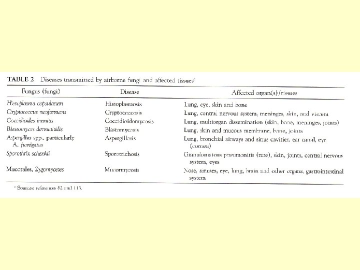
Histoplasmosis An intracellular mycotic infection of the reticuloendothelial system caused by the inhalation of the fungus. Approximately 95% of cases of histoplasmosis are inapparent, subclinical or benign. Five percent of the cases have chronic progressive lung disease, chronic cutaneous or systemic disease or an acute fulminating fatal systemic disease. All stages of this disease may mimic tuberculosis. Distribution: World-wide, especially U. S. A. Sporadic cases do occur in Australia. Aetiological Agent: Histoplasma capsulatum, especially from soil enriched with excreta from chicken, starlings and bats.
322
Coccidioidomycosis Initially, a respiratory infection, resulting from the inhalation of conidia, that typically resolves rapidly leaving the patient with a strong specific immunity to re -infection. However, in some individuals the disease may progress to a chronic pulmonary condition or as a systemic disease involving the meninges, bones, joints and subcutaneous and cutaneous tissues. Distribution: Endemic in south-western U. S. A. , northern Mexico and various centres in South America. Aetiological Agent: Coccidioides immitis, a soil inhabiting fungus.
106
Subcutaneous zygomycosis (Mucormycosis) Primary cutaneous and subcutaneous infections usually the result of a "barrier break" or traumatic implantation of fungal elements. Lesions vary considerably in morphology but include plaques, pustules, ulcerations, deep abscesses and ragged necrotic patches. Most heal with little treatment (debridement and amphotericin B) and they are not usually associated with dissemination. Distribution: World-wide. Aetiological Agents: Cosmopolitan members of the Mucorales including species of Rhizopus, Mucor, Rhizomucor, Absidia, Cunninghamella, Saksenaea and Mortierella.
472
Aspergillosis is a spectrum of diseases of humans and animals caused by members of the genus Aspergillus. These include (1) mycotoxicosis due to ingestion of contaminated foods; (2) allergy and sequelae to the presence of conidia or transient growth of the organism in body orifices; (3) colonization without extension in preformed cavities and debilitated tissues; (4) invasive, inflammatory, granulomatous, necrotizing disease of lungs, and other organs; and rarely (5) systemic and fatal disseminated disease. The type of disease and severity depends upon the physiologic state of the host and the species of Aspergillus involved. Distribution: World-wide. Aetiological Agents: Aspergillus fumigatus, A. flavus, A. niger, A. nidulans and A. terreus.
Cryptococcosis A chronic, subacute to acute pulmonary, systemic or meningitic disease, initiated by the inhalation of the fungus. Primary pulmonary infections have no diagnostic symptoms and are usually subclinical. On dissemination, the fungus usually shows a predilection for the central nervous system, however skin, bones and other visceral organs may also become involved. Distribution: World-wide. Aetiological Agent: Cryptococcus neoformans.
Candidiasis A primary or secondary mycotic infection caused by members of the genus Candida. The clinical manifestations may be acute, subacute or chronic to episodic. Involvement may be localized to the mouth, throat, skin, scalp, vagina, fingers, nails, bronchi, lungs, or the gastrointestinal tract, or become systemic as in septicaemia, endocarditis and meningitis. Distribution: World-wide. Aetiological Agents: Candida albicans, C. glabrata, C. tropicalis, C. krusei. C. parapsilosis, C. guilliermondii and C. pseudotropicalis. All are ubiquitous and occur naturally on humans.
05
065
069
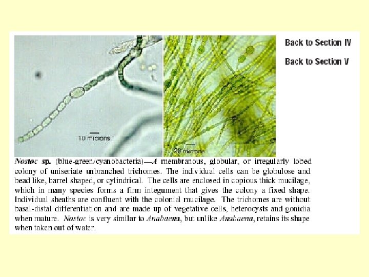
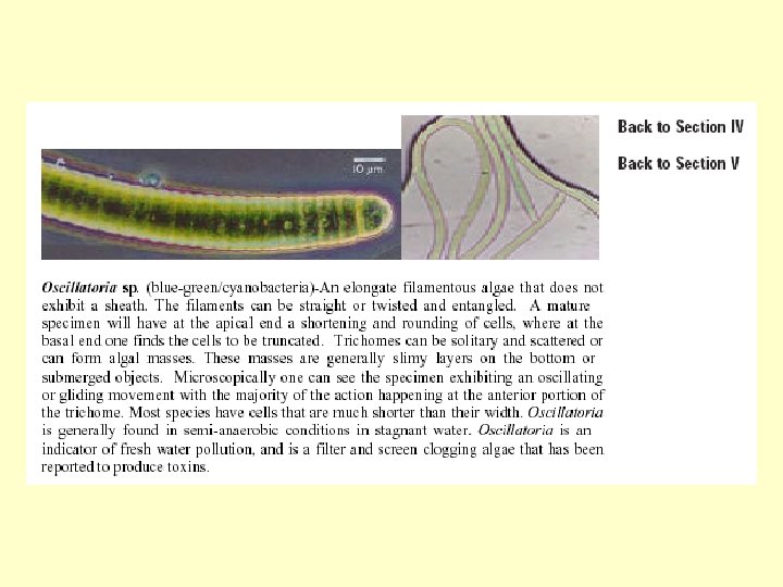
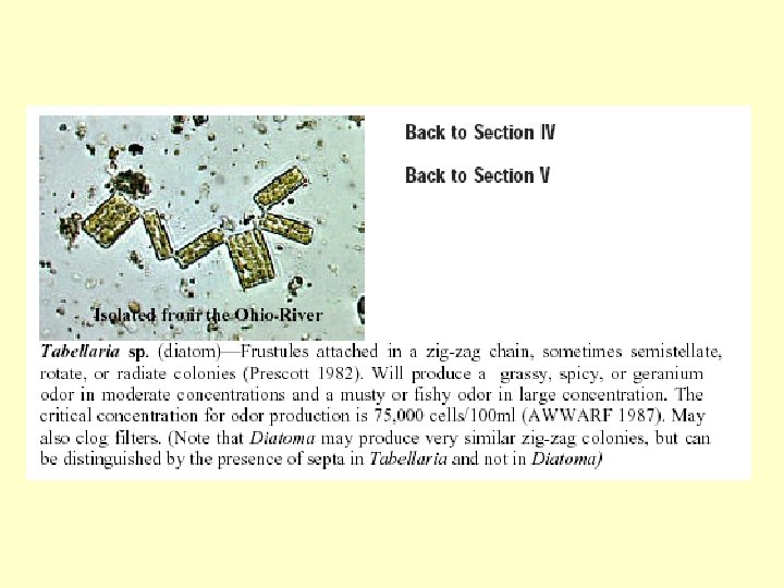
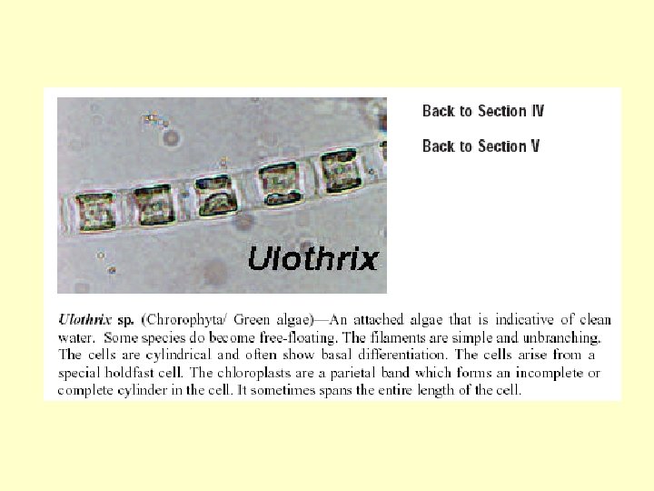
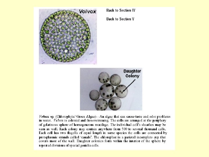
Algae
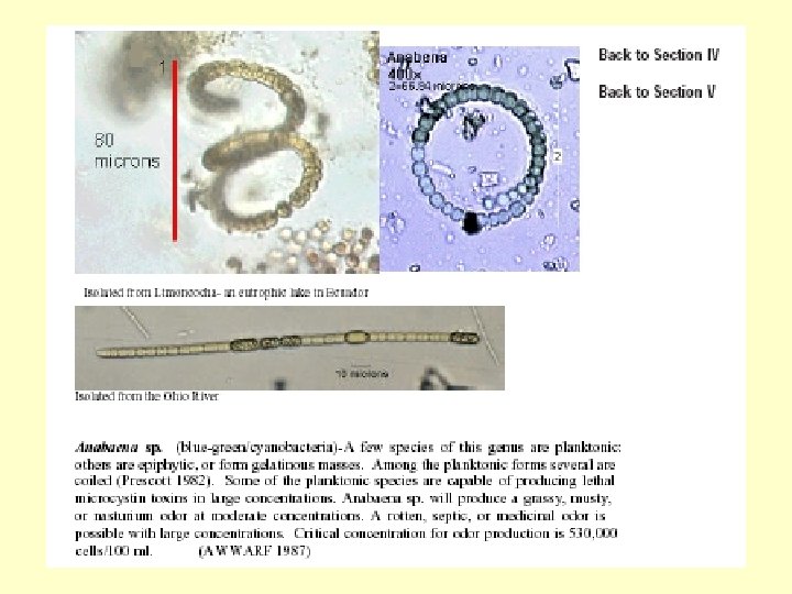
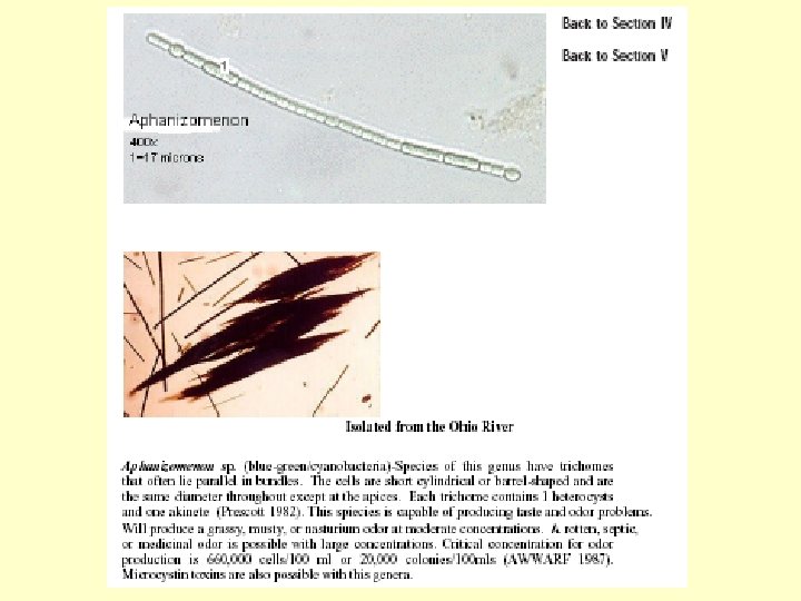
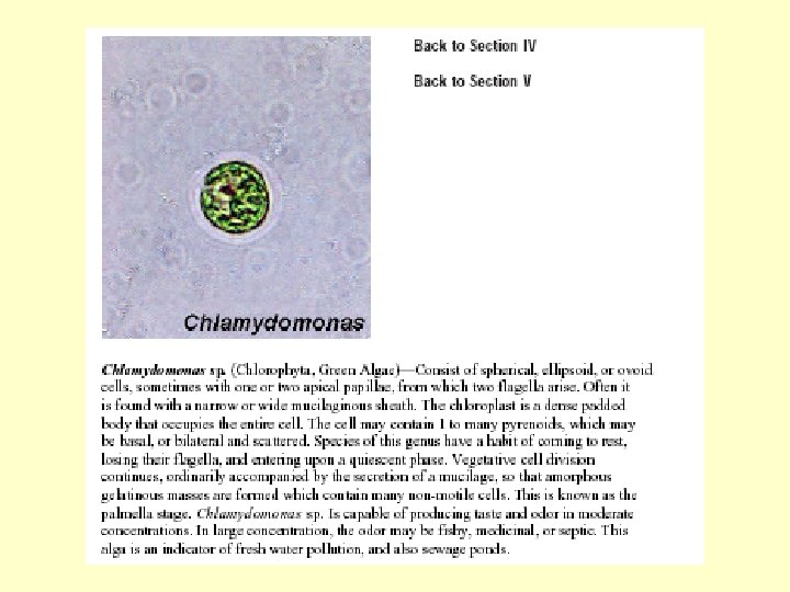
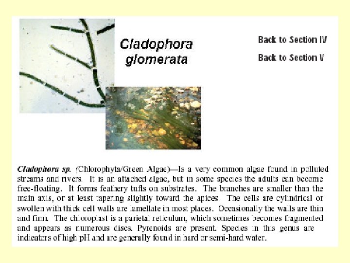
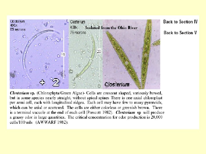
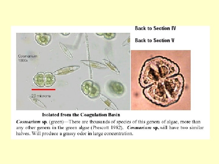
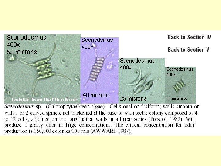
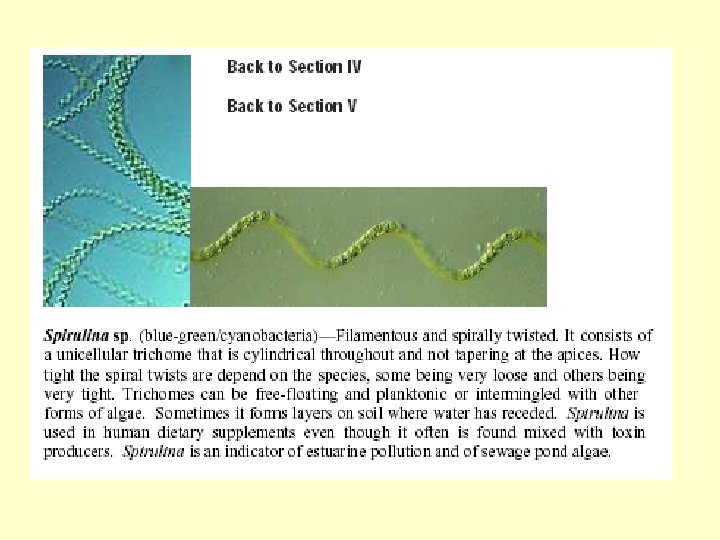
Algae Divisions • Chlorophyta (green algae) – Least harmful, generally considered benefical – Growth in reservoirs; mild taste and odor; some filamentous mat formers • Cyanophyta (blue-green algae) – Prokayotes – Most significant concerns for water quality – Taste and odor problems; filter cloggers; oxygen depletion; toxicity
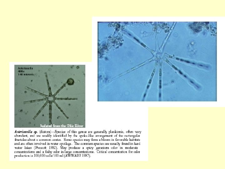
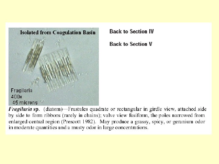
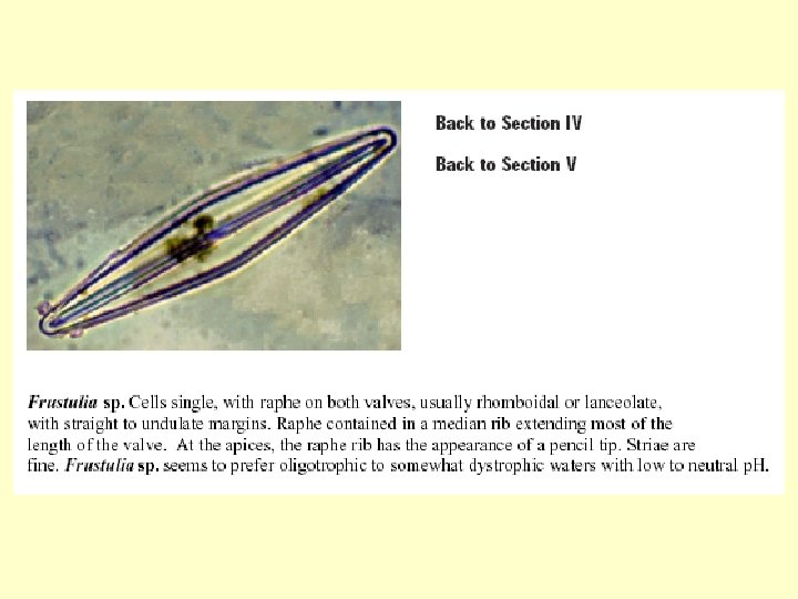
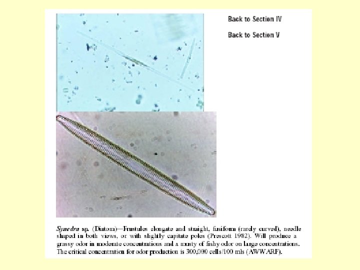
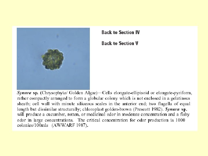
Algae Divisions • Chrysophyta (Yellow-Green/Golden. Brown Algae) – Taste and odor problems; reservoir growth; filter cloggers – Frustules used for filtration • Pyrrhophyta (dinoflagellates) – Taste and odor problems – Red tide problems
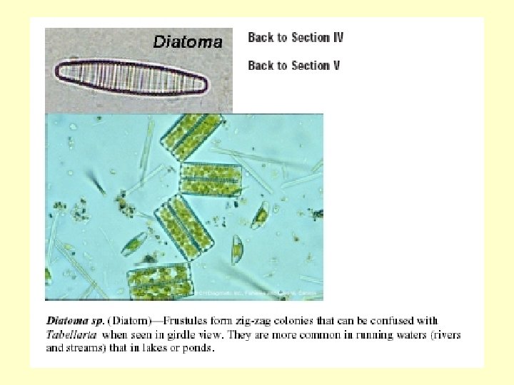
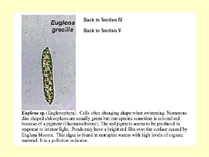
Algae Divisions • Euglenophyta (protozoan-like algae) – Indicators of pollution – Filter cloggers • Crytptophyta (crytomonads) – Taste and odor problems • Rhodophyta (red algae) – Growth on reservoir walls and irrigation ditches
Taste and Odor • Dirty or Musty – Geosmin and MIB (2 -methylisoborneol) – blue-green algae, actinomycetes • Fishy, Cod liver Oil – Chrysophyta, Pyrrhophyta • Septic Odor – Pryyhophyta • Cucumber Odor – Chrysophyta
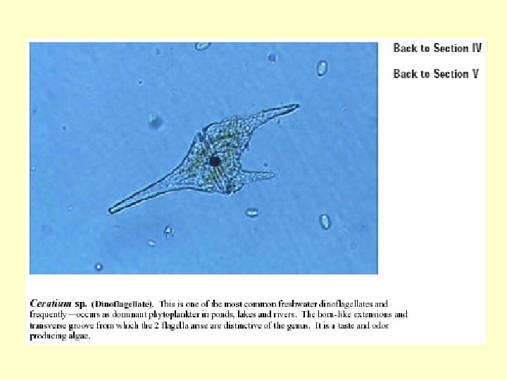
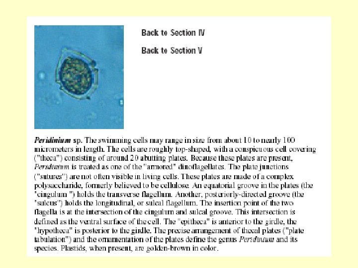
Pyrrhophyta (Dinoflagellates) • unicellular – many have stiff, patterned plates • many photosynthetic – some ingest other cells • most are marine – some cause phosphorescence in ocean waters – some endosymbionts • zooxanthellae – unarmored form seen in host
thecae propels cell forward while causing it to spin Figure 26. 9 acts as rudder
Freshwater Algal Toxins • Anatoxin (e. g. Anabaena) – Staggering, paralysis, gasping, convulsions, death – 200 μg/kg LD 50 • Microcystin (e. g. Anabaena, Microcystis, Oscillatoria) – Jaundice, shock, abdominal pain/distention. Weakness, nausea, vomiting, severe thirst, rapid/weak pulse, death – 300 -600 μg/kg LD 50 • Saxitoxin/Neosaxitoxin (e. g. Anaphnizomenon) – Weakness, staggering, loss of muscle coordination, difficulty in swallowing, labored respiration, muscle paralysis, death, tingling around mouth or fingertips, slurred speech – 9 μg/kg LD 50 • Hepatotoxin (e. g. Gleotrichia) – Jaundice, abdominal pain/distention, weakness, nausea/vomiting • Cytotoxin (e. g. Gleotrichia) – Skin irritation, gastrointestinal upset
Marine Algal Toxins • PSP- Saxitoxins, Gonyautoxins, C-Toxins- Alexandrium (formerly Gonyaulax) spp. • ASP- Domoic Acid – Pseudonitchia spp. • DSP- Okadaic acid, dinophysiotoxins, pectenotoxins, and yessotoxin- Dinophysis spp. and Prorocentrum spp. • Ciguatera toxin, Maitotoxin, & Scaritoxin- Gambierdiscus toxicus • NSP/Brevetoxin- Karenia brevis (formerly Ptychodiscus or Gymnodinium breve) • Pfiesteria-like Toxins- Three putative toxins- lipid soluble dermonectrotic toxin, lipid-soluble lethality factor, watersoluble neurotoxin
Pfiesteria piscicida • Pfiesteria piscicida - colorless, singlecelled organism (dinoflagellate) • Capable of 24 distinct forms depending on water characteristics, major ones: – Amoeba forms – Zoospores – Gametes – Planozygote – Cyst forms
Pfiesteria and Fish • Toxic zoospores excrete toxins that drug fish and destroy skin • Gametes, zoospores and planozygotes feed on substances from sores on fish • Amoeba forms feed on dead fish • Toxins affect liver, brain, kidney and pancreas of fish • Toxins affect other aquatic life as well
Pfiesteria Toxins and Humans • Can affect humans by touching or ingesting water or breathing aerosolized water containing toxins • Respiratory problems • Headaches • Moodiness • Aching joints and muscles • Disorientation • Memory loss