Sex linkage and Pedigrees Sex determination in mammals


 and")


- Slides: 23
Sex linkage and Pedigrees
Sex determination in mammals l In humans and some other organisms, X and Y chromosomes determine the sex of an individual. l This is because they carry certain genes that are critical in sex determination, such as the SRY gene on the mammalian Y chromosome, which controls testis formation. ¡ Individuals with two similar sex chromosomes are the homogametic sex. (i. e. women XX) ¡ Individuals with different sex chromosomes are the heterogametic sex. (i. e. men XY) l During the growth and development of females’ cells, one X chromosome can be inactivated in body cells. The inactivated X chromosome is visible in a female’s cells as a Barr body.
Sex determination in other organisms WZ Chromosome System l Males are homogametic (ZZ) and females are heterogametic (ZW). l Birds and strawberries are examples of the W/Z determination. XO Chromosome System l Only one sex chromosome. l Females are XX but males are XO, where the O refers to the absence of a matching sex chromosome. l In the XO chromosome system diploid number is therefore even in females and odd in males. Sex Determination by haplodiploidy l Males develop from unfertilised eggs and are therefore haploid. l Females develop from fertilised eggs and are diploid. Examples include wasps and bees. Sex Determination by environmental factors l Environmental sex determination may depend on: ¡ temperature (e. g. turtles, crocodiles) ¡ day length (shrimp) ¡ richness and availability of food resources (e. g. nematodes).
X linked inheritance l Males receive their X chromosome from their mother – so they inherit all X-linked traits from their mothers. The alleles on this chromosome will determine the phenotype of X-linked traits regardless of whether the trait is dominant or recessive in heterozygous females. l All females inherit an X chromosome from each parent. The random nature of X chromosome inactivation means that heterozygous females express different alleles in different cells.
X linked inheritance l X-linked Recessive disorders ¡ Show a pattern of transmission of the mutant phenotype from the female parent to male offspring. ¡ Only females may be carriers of an X-linked recessive trait. ¡ Examples of X-linked recessive disorders include haemophilia A, haemophilia C and red-green colour blindness.
X linked inheritance l X-linked Dominant disorders ¡ Show a pattern of transmission of the mutant phenotype from an affected male parent to all female offspring, and from an affected heterozygous female parent to 50% of all offspring. ¡ Examples of X-linked dominant disorders include Vitamin D resistant rickets and fragile X syndromes
Y linked inheritance l There are far fewer Y-linked than X-linked genetic disorders l This is not surprising given that the Y chromosome is smaller and has many less genes than the X chromosome. l Y-linked inheritance shows a pattern of transmission of the mutant phenotype from father to son, and it is never observed in females. l An example of a Y linked phenotypic trait is hairy ears.
Sex limited inheritance l Y-linked inheritance is often confused with sexlimited inheritance. l Sex-limited traits can only occur in one sex because the feature affected is unique to that sex. l For example, premature baldness is an autosomal dominant trait, but presumably as a result of female sex hormones, the condition is rarely expressed in the female, and then usually only after menopause.
X-inactivation l During the growth and development of females’ cells, one X chromosome can become inactivated in each body cell. l The inactivated X chromosome is visible in a female’s cells as a Barr body. l Which of the two X chromosomes becomes inactive in a cell is a matter of chance, therefore heterozygous females express different alleles in different cells. l This is generally noticeable in the phenotype – for example a woman heterozygous for the recessive condition haemophilia A will produce sufficient clotting factor VIII. l Tortoise shell cats are an example where X inactivation is visible in the phenotype as one of the genes which controls coat colour is sex-linked.
X-inactivation l l One of the genes that controls coat colour in cats is sex-linked. It has alternative alleles Xo (orange) and Xb (black) Patches of cells in which the Xo are inactivated will produce dark fur. Patches of cells in which the Xb is inactivated will produce orange fur. Barr body
Pedigree Analysis l Is the technique of looking through a family tree (of humans or other organisms) for the occurrence of a particular characteristic in one family over a number of generations. l Can be used to determine the likely mode of inheritance: ¡ ¡ Autosomal dominant Autosomal recessive X-linked dominant X-linked recessive l When looking at pedigrees, incomplete penetrance is occasionally observed. ¡ Incomplete penetrance describes the situation where a proportion of a population with a particular genotype does not show the expected phenotype. ¡ Complete penetrance of a phenotype means that all individuals with a particular genotype will show the affected phenotype.
Symbols used in drawing pedigrees
Autosomal Dominant Pattern l An idealised pattern of inheritance of an autosomal dominant trait includes the following features: ¡ both males and females can be affected ¡ all affected individuals have at least one affected parent ¡ transmission can be from fathers to daughters and sons, or from mothers to daughters and sons ¡ once the trait disappears from a branch of the pedigree, it does not reappear ¡ in a large sample, approximately equal numbers of each sex will be affected. Examples include: l Huntington disease l Achondroplasia (a form of dwarfism) l Familial form of Alzheimer disease l Defective enamel of the teeth l Neurofibromatosis (the ‘Elephant man’ disease)
Autosomal Recessive Pattern l An idealised pattern of inheritance of an autosomal recessive trait includes the following features: ¡ ¡ both males and females can be affected two unaffected parents can have an affected child all the children of two persons with the condition must also show the condition the trait may disappear from a branch of the pedigree, but reappear in later generations ¡ over a large number of pedigrees, there approximately equal numbers of affected females and males. Examples include: l l l Albinism Cystic fibrosis Thalassaemia Tay-Sachs disease Phenylketonuria Red hair colour
X linked Dominant Pattern l An idealised pattern of inheritance of an X-linked dominant trait includes the following features: ¡ ¡ ¡ a male with the trait passes it on to all his daughters and none of his sons a female with the trait may pass it on to both her daughters and her sons every affected person has at least one parent with the trait if the trait disappears from a branch of the pedigree, it does not reappear over a large number of pedigrees, there are more affected females than males Examples include: l Vitamin D resistant rickets l Incontinentia pigmenti, a rare disorder that results in the death of affected males before birth
X linked Recessive Pattern l An idealised pattern of inheritance of an X-linked recessive trait includes the following features: ¡ all the sons of a female with the trait are affected ¡ all the daughters of a male with the trait will be carriers of the trait and will not show the trait; the trait can appear in their sons ¡ none of the sons of a male with the trait and an unaffected female will show the trait, unless the mother is a carrier ¡ all children of two individuals with the trait will also show the trait ¡ in a large sample, more males than females show the trait. Examples include: l Ichthyosis, an inherited skin disorder l One form of red–green colour-blindness l One form of severe combined immunodeficiency disease l Haemophilia l Fragile X syndrome l Duchenne muscular dystrophy
Is the condition observed in each generation of a family in which it occurs? NO YES If daughters have the condition does their father also have it? NO Autosomal recessive Do males with the condition who mate with a normal female have all daughters, but no sons with the condition? ON Is the condition mainly in males? Do only males have condition, passing it from father to son? NO YES Sex-linked recessive Autosomal dominant YES Sex-linked dominant Y linkage
Linked Genes l Linkage is the tendency for two or more genes located on the same chromosome to be inherited together. l The closer they are, the more likely they are to be inherited together. l Linkage is never complete because of crossing over during meiosis. l Linkage and the phenomenon of crossing over can be used to determine the distance between genes on a chromosome. l Linked genes can be denoted in the following ways when the allele arrangements are known. These include : A b Ab/a. B A b a. B
Inheritance of linked genes l Assume two genes P and V are linked: gametes ½ PV ½ pv ½ PV ¼ ¼ PV/PV PV/pv ½ pv ¼ ¼ PV/PV pv/pv ¡ Parental cross l PV/PV x pv/pv ¡ F 1 l All PV/pv l Will have dominant phenotype ¡ F 2 l See punnet square to right l When crossing two F 1 heterozygotes for two linked gens, and disregarding crossing over, the ratio of the phenotypes is close to 3: 1
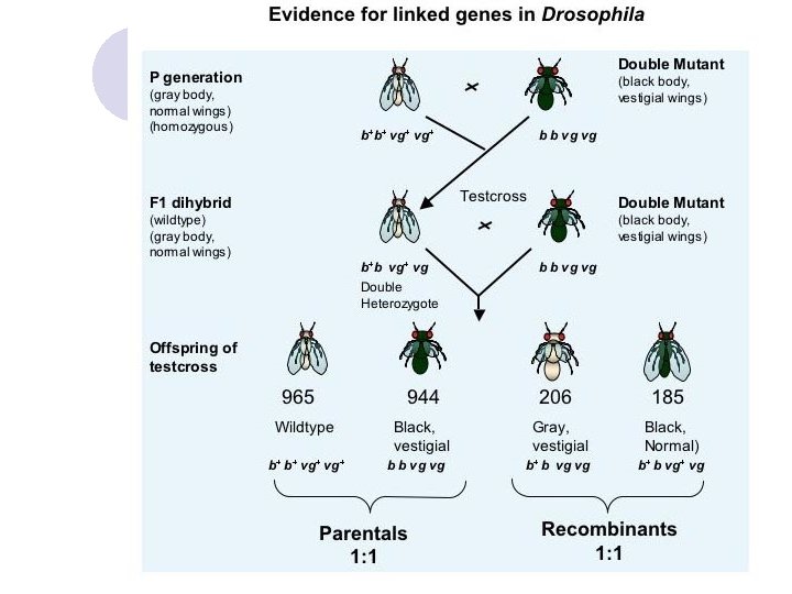
Detecting Linkage l The results of test crosses between known double heterozygote with a double homozygous recessive allow us to determine if two gene loci are linked. l If the two gene loci are not linked, the genes will assort independently and the outcome of the test cross will be four classes of offspring in equal proportions l If the two gene loci are linked, there will be four classes of offspring but the proportions of these will not be equal. Instead, there will be an excess of offspring from parental gametes and a deficiency of offspring from recombinant gametes. l Genes are ‘linked’ when the percentage of recombinant gametes falls below 50%.
Estimating the distance between linked genes l From the results of a test cross with linked genes, it is possible to estimate the distance between the gene loci. l This estimate is based on the percentage of recombinant offspring. Distance between loci = 100 × number of recombinant offspring total number of offspring l The percentage of recombinant offspring corresponds to the number of map units separating the two genes.
Estimating the distance between linked genes l There are 12 per cent total recombinant offspring, therefore the loci of the two genes are separated by about 12 map units. l The chromosome make-up of the cross can be represented as: