Neurons and Synapses Essential idea Neurons transmit the


 • Axon terminals • Pre-synaptic membrane •")


")


. There is localized hypo-polarization due to Na+")


- Slides: 29
Neurons and Synapses Essential idea: Neurons transmit the message, synapses modulate the message.
Synapses are junctions between neurons and receptor or effector cells. The Synapse • This is the name of the space between the axon of the pre-synaptic neuron and the dendrite (or cell body) of the post -synaptic neuron that is receiving the signal. synapse
Definition: It is the site of functional contact b/w two neurons at which an electric impulse is transmitted from one neuron to another.
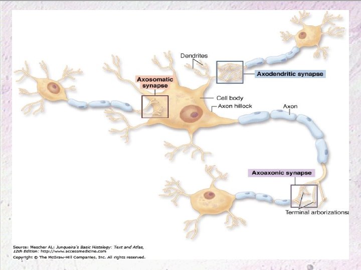
Types of synapses- on the basis of site of contact • 1. Axodendritic synapses (most common type) • 2. Axosomatic synapses • 3. Dendrodenritic synapses • 4. axosaxonic synapses
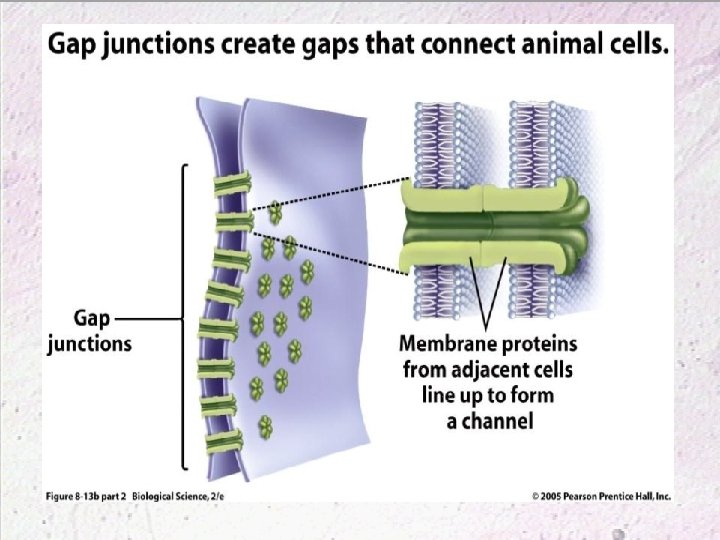
Types of synapses- on the basis of method of signal transmission • Chemical synapses: • • Most common type • • Signal transmission is delayed for about 0. 5 msec in these synapses. • Electrical synapses (nexus): • • Less common • • Flow of ions from one neuron to another via gap junctions.
Anatomy of a typical synapse (synaptic morphology) • Axon terminals • Pre-synaptic membrane • Post-synaptic membrane • Synaptic cleft (20 -30 nm wide) • Synaptic vesicles
When presynaptic neurons are depolarized they release a neurotransmitter into the synapse. What happens at a synapse? • An action potential cannot cross the synaptic cleft, and instead the nerve impulse is carried by chemicals called neurotransmitters. • Neurotransmitters are made by the neuron that is sending the impulse (the pre-synaptic neuron) and stored in synaptic vesicles at the end of the axon. • The cell that is receiving the nerve impulse (the postsynaptic neuron) has chemical-gated ion channels in its membrane, called neuroreceptors. • These have specific binding sites for the neurotransmitters
When presynaptic neurons are depolarized they release a neurotransmitter into the synapse. Synapse
Neurons pump sodium and potassium ions across their membranes to generate a resting potential Overview of Process
When presynaptic neurons are depolarized they release a neurotransmitter into the synapse. How it works • 1. At the end of the pre-synaptic neuron there are voltage-gated calcium channels. When an action potential reaches the synapse these channels open, causing calcium ions to flow into the cell. • 2. The calcium ions cause the synaptic vesicles to fuse with the cell membrane, releasing their contents (the neurotransmitter chemicals) by exocytosis. • 3. The neurotransmitters diffuse across the synaptic cleft.
When presynaptic neurons are depolarized they release a neurotransmitter into the synapse. How it works • 4. The neurotransmitter binds to the neuroreceptors in the post-synaptic membrane, causing the channels to open. In the example shown these are sodium channels, so sodium ions flow in. • 5. This causes a depolarisation of the post-synaptic cell membrane, which may initiate an action potential.
When presynaptic neurons are depolarized they release a neurotransmitter into the synapse. How it works 6. The neurotransmitter is broken down by a specific enzyme in the synaptic cleft. The breakdown products are absorbed by the pre-synaptic neuron by endocytosis and used to re-synthesize more neurotransmitter, using energy from the mitochondria. This stops the synapse being permanently on.
Events occurring at a chemical synapse during signal transmission (Synaptic Transmission Mechanism)
Secretion and reabsorption of acetylcholine by neurons at synapses. • acetylcholine is a neurotransmitter found between neurons and muscle cells (and other places) • 2 parts: – acetyl (from respiration) – choline (from diet) • it travels across the synapse to bind its receptor • however, the enzyme acetylcholinesterase rapidly breaks down acetylcholine in the synapse (into choline and acetate) • Choline is absorbed by pre-synaptic neuron and re-used to make more acetylcholine.
Blocking of synaptic transmission at cholinergic synapses in insects by binding of neonicotinoid pesticides to acetylcholine receptors. • neonicotinoids = an insecticide – synthetic chemical similar to nicotine • binds the acetylcholine receptor irreversibly • leads to paralysis and death in insects • neonicotinoids very widely used (because safe for mammals) • but it’s non-specific, so it also kills helpful insects (like bees).
EPSP and IPSP • Depending on type of neurotransmitter & type of change in permeability of post-synaptic membrane, post-synaptic neuron is either excited or inhibited. • Neuro-transmitter binds with receptor on post -synaptic membrane opening of ion channels localized change in membrane potential post-synaptic membrane potential (PSP) • 2 types : Excitatory (EPSP), Inhibitory (IPSP).
EPSP • Resembles EPP (end plate potential). There is localized hypo-polarization due to Na+ influx. • Resting potential of cell body of neuron is • -65 m. V. • When EPSP is produced hypopolarization potential becomes less negative reach threshold of excitation (-45 m. V) ACTION POTENTIAL in cell body.
Purpose of EPSP • To bring potential of membrane to threshold • (-45 m. V) • It is graded like EPP (directly proportional to amount of neurotransmitter released).
IPSP: • Produced when post-synaptic neuron is inhibited. • Neuro-transmitter is of inhibitory type (GABA. Glycine) • It binds with receptors on postsynaptic membrane change in permeability of membrane for K+ or Cl- (there is opening of K+ or Clchannels efflux of K+ cell becomes more negative hyperpolarization / IPSP. • Opening of Cl- channels extracellular Cl- moves into the cell more negative hyper-polarization / IPSP.
Effect of IPSP: • Because of IPSP, resting potential which is • -65 m. V, becomes -70 to -75 m. V Post-synaptic neuron is inhibited POST-SYNAPTIC INHIBITION. PRE-SYNAPTIC INHIBITION: • Synaptic knob has additional synapse with other nerve terminals release of inhibitory neurotransmitter from additional synapse synaptic knob is inhibited no further transmission from synapse now to post-synaptic neuron
Properties of Synaptic Transmission DALE’S LAW: • At a given synapse, only 1 type of neurotransmitter is released, it may be excitatory or inhibitory. • Later on it was found that in certain cases, release of additional substances at a given synapse • e. g. , in noradrenergic synapses: along with norepinephrine, some dopamine, neuropeptide Y & prostaglandins are also released.
LAW OF FORWARD CONDUCTION: • Through synapses, impulses are conducted always from pre-synaptic to post synaptic neuron, never in backward direction. • (NO REVERSE GEAR!!) SYNAPTIC DELAY: • At a synapse, there is delay due to time taken in events during synaptic transmission. Through each synapse, there is delay of 0. 5 milli seconds.
FATIGUE OF SYNAPTIC TRANSMISSION • If impulses are conducted through a synapse repeatedly fatigue due to exhaustion of stores or progressive inactivation of receptors on post-synaptic membrane. • Significance of fatigue? ? • Fatigue of synaptic transmission is protective in nature termination of epileptic fit
IN UNITY RESTS STRENGTH! SUMMATION: • Adding up of effects of stimuli particularly if stimuli are sub threshold. • On a single motor neuron, thousands of synaptic knobs terminate to form synapses. • About 80% of these synapses are on dendrites, remaining on cell body & few on axons. • So, single impulse coming to motor neuron through a synapse, can’t excite a motor neuron & there must be summation of effects of stimuli.
TEMPORAL • Impulses transmit through 1 or few synaptic knobs repeatedly effects on post-synaptic neurons are added stimulation. • Second stimulus must fall when effect of 1 st one is still there
SPATIAL • Impulses are conducted along a number of synapses simultaneously effects on postsynaptic neuron are added excitation.
ALKALOSIS INCREASE EXCITABILITY OF SYNAPSES, ACIDOSIS DEPRESSES SYNAPTIC TRANSMISSION Increase excitability • Caffeine (cerebral stimulant) • Theophylline • Strychnine • Decreased calcium (tetany) Decrease excitability • Anesthetics • Hypoxia • Increased calcium (stabilize the membrane)