Neurons Synapses Signaling Neurons are nerve cells that


- Slides: 27

Neurons, Synapses, & Signaling § Neurons are nerve cells that transfer information within the body § Neurons use two types of signals to communicate: electrical signals (long-distance) and chemical signals (short-distance) § A neuron’s structure is closely tied to its function © 2014 Pearson Education, Inc.

Neuron Structure and Function § Most of a neuron’s organelles are in the cell body § Most neurons have dendrites, highly branched extensions that receive signals from other neurons § The axon is typically a much longer extension that transmits signals to other cells at synapses § The terminal branches of an axon pass information across the synapse in the form of chemical messengers called neurotransmitters § A synapse is a junction or space between two neurons © 2014 Pearson Education, Inc.

A Typical Neuron Cell Dendrites – receive signals Nucleus Cell body Presynaptic Cell = 1 st neuron Synapse Neurotransmitter © 2014 Pearson Education, Inc. Axon – sends the signal down the neuron to the Terminal branches at the end Terminal Branches Postsynaptic cell = 2 nd neuron

§ Information is transmitted along a pathway from a presynaptic neuron cell to a postsynaptic neuron cell (or effector cell) § Chemical signals called neurotransmitters cross the space = synapse between neurons. § Nervous systems process information in three stages involving the 3 types of neurons § sensory input (sensory neurons in PNS) § integration (associative neurons in CNS) § motor output (motor neurons in PNS) © 2014 Pearson Education, Inc.

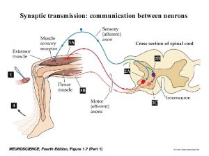
Figure 49. 7 Cell body of sensory neuron in dorsal root ganglion CNS Quadriceps muscle Hamstring muscle Key Motor neurons Sensory neuron Motor neuron Interneuron © 2014 Pearson Education, Inc.

Nerve signals travel along a Specific Pathway: STIMULS: Receptor Sensory Neurons CNS motor neurons effectors: RESPONSE Central nervous system (CNS) Brain Cranial nerves Spinal cord Spinal nerves Peripheral nervous system (PNS) © 2014 Pearson Education, Inc.

Nerve signals = Action potential signals are sent to different regions of the Brain Eye Reticular formation Input from touch, pain, and temperature receptors © 2014 Pearson Education, Inc. Input from nerves of ears

§ Sensors detect external stimuli and internal conditions and transmit information along sensory neurons to the CNS § In the CNS (brain & spinal cord) interneurons = associative neurons integrate the information § Motor output leaves the CNS via motor neurons, which trigger muscle or gland activity = response © 2014 Pearson Education, Inc.

§ We have a complex nervous system that consists of § A central nervous system (CNS) where integration takes place; this includes the brain and a nerve cord § A peripheral nervous system (PNS), which carries information into and out of the CNS © 2014 Pearson Education, Inc.

Neurons send signals using ion pumps and ion channels, altering the resting potential § Every cell has a voltage (difference in electrical charge) across its plasma membrane called a membrane potential § The resting potential is the membrane potential of a neuron that’s not sending signals § Changes in membrane potential act as signals © 2014 Pearson Education, Inc.

At Resting Potential, Neurons are NOT transmitting Signals § In a human neuron at resting potential, the concentration of K+ is highest inside the cell, while the concentration of Na+ is highest outside the cell § The inside of the membrane has a negative charge; the outside has a positive charge § Sodium-potassium pumps use the energy of ATP to maintain these K+ and Na+ gradients across the plasma membrane © 2014 Pearson Education, Inc.

Neuron Membrane at Resting Potential © 2014 Pearson Education, Inc.

A Nerve Signal = Action Potential. Sequence of Events: 1. A stimulus causes the opening of Na+ ion channels in the plasma membrane. 2. Na+ rushes in the cell = Depolarization occurs. This is the start of the action potential 3. Depolarization causes K+ ion channels to open and K+ rushes out = Repolarization occurs 4. Now, the Na+ K+ pump restores normal resting potential © 2014 Pearson Education, Inc.

Depolarization Action Potential Ions A stimulus causes Na+ ion channel gate to open Ion channel Gate closed: No ions flow across membrane. © 2014 Pearson Education, Inc. Gate open: Na+ ions flow in through channel.

§ Opening of Na+ ion channels triggers a depolarization because Na+ diffuses into the cell § An action potential has begun. This depolarization is a change in membrane’s polarity: + inside and outside § Action potentials have a constant magnitude or size because they are triggered by an all-or-none; either the ion gated channel opened or it didn’t § If the stimulus is strong enough to open the ion gated channels, threshold has been reached an action potential will begin © 2014 Pearson Education, Inc. -

Figure 48. 10 c Strong depolarizing stimulus The polarity of the Membrane switches: Becomes Positive on the inside and Negative on the outside +50 Membrane potential (m. V) Action potential is triggered by a depolarization that reaches the threshold. 0 − 50 − 100 © 2014 Pearson Education, Inc. Action potential Threshold Resting potential 0 1 2 3 4 5 6 Time (msec)

An action potential moves in a series of stages: 1. At resting potential most sodium (Na+) and potassium (K+) channels are closed 2. Gated Na+ channels open first and Na+ flows into the cell 3. The threshold is crossed, and depolarization has begun producing an action potential 4. During the falling phase, Na+ channels close and K+ channels open, and K+ flows out of the cell = repolarization occurs © 2014 Pearson Education, Inc.

Figure 48. 11 f Membrane potential (m. V) +50 3 0 − 50 − 100 © 2014 Pearson Education, Inc. Action potential 2 4 Threshold 1 Resting potential Time 5 1

§ During the refractory period after an action potential, a second action potential cannot be initiated until the Na+ K+ pump restores normal § Na+ is outside and K+ is inside = resting potential § Action potentials travel in only one direction: down the axon toward the synaptic terminals § At the terminal the action potential triggers the release of neurotransmitters into the synapse © 2014 Pearson Education, Inc.

Figure 48. 12 -1 Axon Action potential Na+ © 2014 Pearson Education, Inc. Plasma membrane Cytoplasm

Figure 48. 12 -2 Axon Plasma membrane Action potential Cytosol Na+ K+ Action potential Na+ K+ © 2014 Pearson Education, Inc.

Figure 48. 12 -3 Axon Plasma membrane Action potential Cytosol Na+ K+ Action potential Na+ K+ © 2014 Pearson Education, Inc.

§ Some axons are wrapped in myelin sheaths that have spaces called nodes of Ranvier § Action potentials in myelinated axons jump between the nodes of Ranvier in a process called saltatory conduction § This allows action potential signals to be transmitted faster © 2014 Pearson Education, Inc.

Figure 48. 14 Schwann cell makes the myelin sheath Depolarized region (node of Ranvier) Myelin sheath Cell body Axon © 2014 Pearson Education, Inc.

Neurons communicate with other neuron cells at synapses § Action potentials are electrical signals along neuron membranes. § The action potential causes the release of the neurotransmitter by the axon terminal branches. § The chemical signal = neurotransmitter diffuses across the synapse and stimulates dendrites of the next neuron (postsynaptic = after the synapse) § A new action potential begins on the membrane of this postsynaptic neuron © 2014 Pearson Education, Inc.

Figure 48. 16 Presynaptic cell 1 Axon Postsynaptic cell Synaptic vesicle containing neurotransmitter Synaptic cleft Postsynaptic membrane Presynaptic membrane 3 K+ 4 Ca 2+ 2 Voltage-gated Ca 2+ channel © 2014 Pearson Education, Inc. Ligand-gated ion channels Na+

§ A number of toxins can disrupt a neurotransmission § These include the nerve gas, sarin, and the botulism toxin produced by certain bacteria § Death can result if signal transmission stops © 2014 Pearson Education, Inc.
 Chapter 48 neurons synapses and signaling
 Chapter 48 neurons synapses and signaling
 Insidan region jh
 Walmart thất bại ở nhật
 Gây tê cơ vuông thắt lưng
 Block nhĩ thất cấp 1
 Tìm độ lớn thật của tam giác abc
 Sau thất bại ở hồ điển triệt
 Thể thơ truyền thống
 Con hãy đưa tay khi thấy người vấp ngã
 Thơ thất ngôn tứ tuyệt đường luật
 Tôn thất thuyết là ai
 Phân độ lown ngoại tâm thu
 Nervous cell
 Summation of postsynaptic potentials
 Synapses
 Synapses telecom
 Bioflix activity: how synapses work -- synapse structure
 Synapse functions
 Chemical synapses
 2 types of synapses
 Synapses
 Electrical synapse vs chemical synapse
 Synapse functions
 Trigeminal nerve which cranial nerve
 Pseudostratified vs simple columnar
 Loop of henle
 Cell substance
