Hookworms Ancylostoma spp and Necator spp Human hookworm


 Faecal - soil")


 of the dog and cat hookworms can infect humans, but the juvenile")


 can be")
- Slides: 32
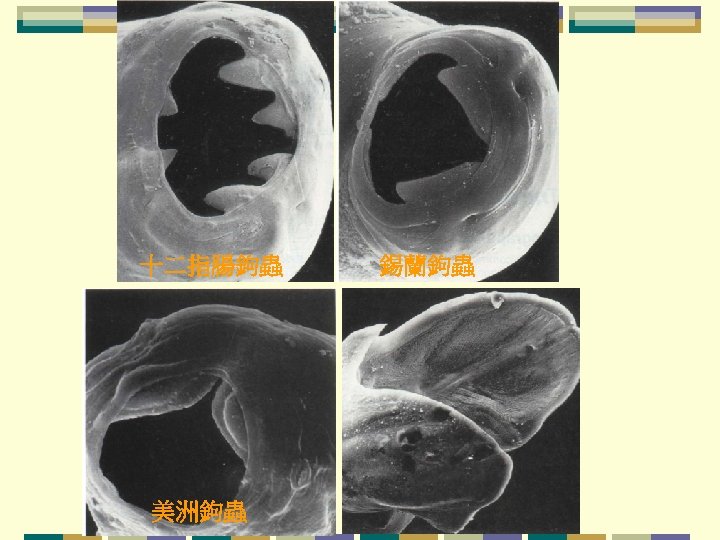
Hookworms鈎蟲 Ancylostoma spp. and Necator spp. Human hookworm Necator americanus and Ancylostoma duodenale. 美洲鈎蟲 十二指腸鈎蟲 Cat hookworm Ancylostoma ceylanicum, also reported from man 錫蘭鈎蟲
Geographical Distribution N. americans Mainly in moist tropical regions : S. E. Asia, sub-Sahara Africa and tropical America. A. duodenale Mainly in dryer and colder areas : North India, Pakistan, North China, Middle East and North Africa. Ancylostoma duodenale and Necator americanus, which infect an estimated 800, 000 persons, the dog and cat hookworms, A. caninum and A. braziliense, respectively.
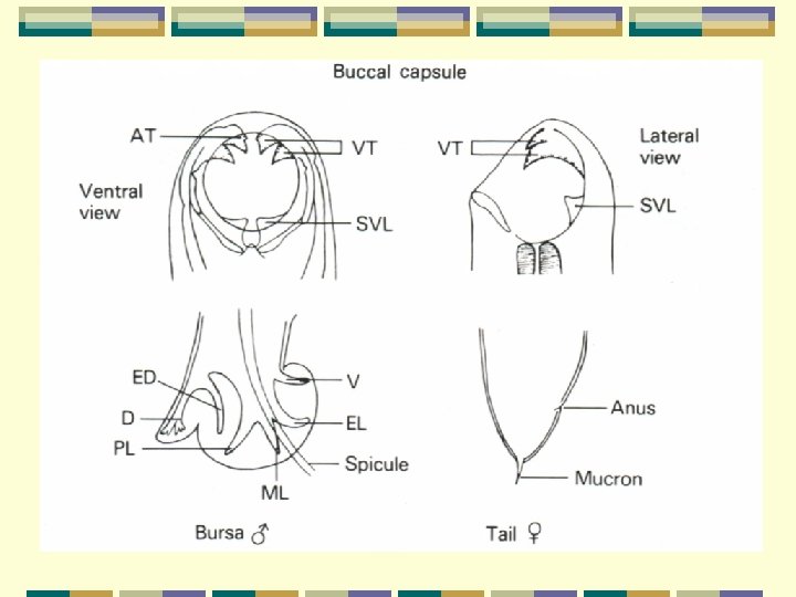
Morphology The adults: cylindrical with the head bent sharply backwards giving them a hooked appearance about 10 mm in length The males: bursa at posterior end; smaller than the females The hookworm species are mainly differentiated by their buccal capsule and the arrangement of rays 肋 in the bursa 美洲鉤蟲
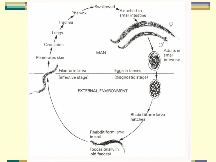
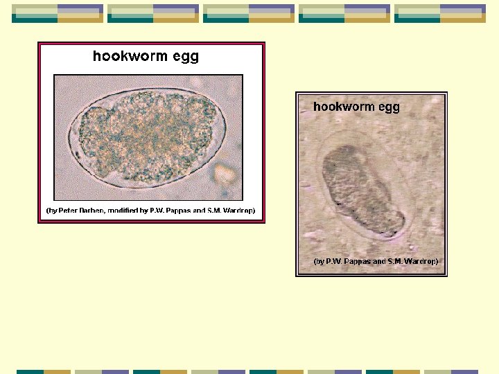
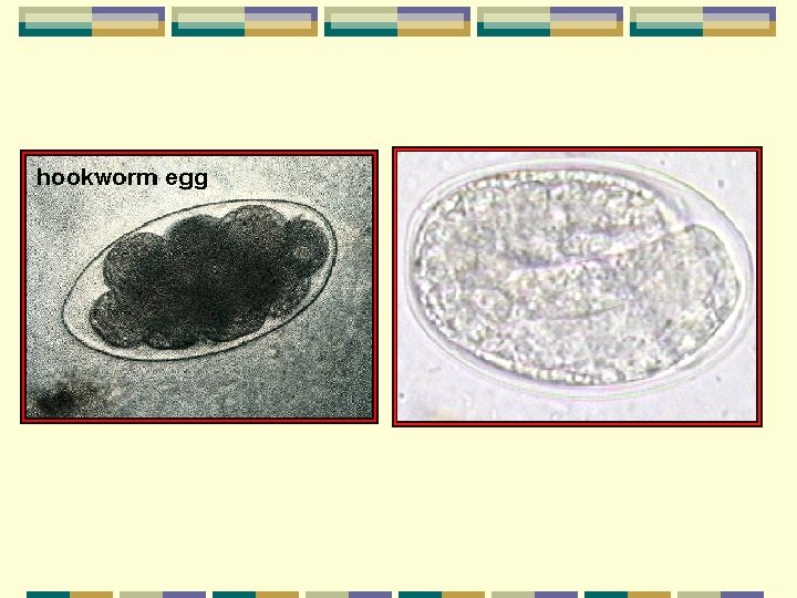
The eggs: ovoid with a thin transparent shell; easy exit for larva measure 74 to 76 x 36 to 40μm various species are indistinguishable Female can produce 10, 000 -25, 000 eggs per day Depending on the species When passed in the host intestine they are single celled They are discharged in the faeces: often at the 4 - or 8 -cell stage this is the diagnostic stage of the parasite Within several hours of being passed, a multicell stage develops and then a rhabditiform larva (first stage) forms桿狀線蟲.
The rhabditiform larva: Differentiated from species Various morphological difference It is possible to differentiate third stage larva of N. americanus from A. duodenale by certain morphological features
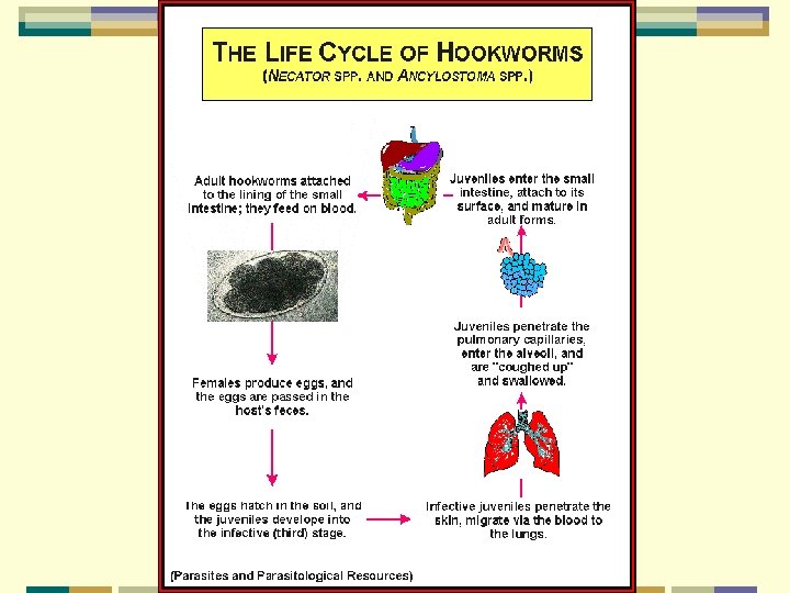
Life Cycle First stage larva: hatches out from the egg in 24 to 48 hours Actively feeds on organic debris and bacteria in the soil 2 nd stage larva: on the third day 3 rd stage larva: on the fifth day filariform絲狀蟲; nclosed in a sheath does not feed; actively motile. The third stage larva initiates the infection by penetrating the skin and passing into blood circulation. Through the blood it is carried to the right heart and then to pulmonary blood vessels. It soon breaks out of the pulmonary blood vessels into the alveoli.
It then crawls up the trachea and is swallowed with saliva to reenter the intestinal tract. Finally, it attaches itself to the mucous membrane of the small intestine to mature into adults. During copulation the male firmly encircles the female vulva with its bursa and the gravid female starts egg laying 5 -7 weeks after infection. The adult worm survives for 1 -14 years. In the case of Ancylostoma duodenale, infection can also be established by swallowing filariform larvae on leafy vegetables and other food contaminated with soil. Another important recent finding is that A. duodenale larvae can persist in somatic tissues of man. This kind of arrested development apparently helps the survival of the worm in case of a seasonably unfavourable external environment.
Clinical Aspects Ground itch: The stage of skin penetration Intense itching and dermatitis at the site of entry; between the toes Pneumonitis and bronchitis The larval migration through the lungs This is less severe than the Loeffler's syndrome seen during the migration of Ascaris larvae. The final stage of development and attachment to the intestine is often asymptomatic. Heavy infections: induce microcytic hypochromic anaemia The blood loss is from sucking by the worm and also from continuing haemorrhage at the site of attachment.
Clinical Aspects One Necator worm results in the loss of 0. 03 to 0. 05 ml of blood/day An Ancylostoma worm results in the loss of 0. 15 ml of blood/day It is important that there is a difference between hookworm disease and hookworm infection. Hookworm infection does not necessarily mean that the disease is present, as blood loss produced by light infections can be compensated by adequate food intake. Disease occurs when the intake of food is unable to compensate for the blood loss Infected children are often oedematous indicating that these is a protein-losing enteropathy as well.
Diagnosis Stool examination This is based on finding the eggs in the faeces. An estimation of worm load is often necessary for any correlation of the anaemia with the hookworm infection.
Treatment Anthelmintic treatment: Bephenium hydroxynaphthoate: for Ancylostoma duodenale The dose for adults is 5 g (2. 5 g base) as a single dose and Children: under 2 years 2. 5 g (1. 25 g base) / a single dose. The drug is available in granule form and is given before breakfast with a drink. Bephenium is poor in cases of N. americanus infection Side effect : Bephenium salts often produce nausea, vomiting and gastrointestinal disturbances.
Treatment Pyrantel pamoate: N. americanus: is more efficacious A single dose of pyrantel pamoate (10 mg/kg body weight) is generally sufficient in cases of A. duodenale infection, and light infections of N. americanus. In heavy infections of N. americanus, treatment may have to be repeated after 3 days. Mebendazole : also a good antihookworm drug It is given as 100 mg twice daily for 3 days, irrespective of age. However, it should not be given to pregnant women. Iron and folic acid: Anaemia patient should be given
Prevention and Control 3 interaction factors determine the hookworm epidemiology (a) Faecal - soil contact. (b) Infected soil - human contact. (c) Suitability of soil and climate for the development of eggs and larvae. Faecal-soil contact. Deposition of faeces containing eggs contaminates the soil The key to proper control is provision of latrines and sanitary disposal of faeces. If fresh human faeces is used as a fertilizer, it should be treated by composting堆肥 or chemicals to kill the larvae
Infected soil - human contact. Wearing of protective footwear Cooking or washing of vegetables which may be contaminated with larvae. Suitability of soil and climate for the development of eggs and larvae – A. duodenale eggs are able to withstand desiccation better than N. americanus, and in suitable soil the larvae can survive for several months. No simple method available for destroying the larvae in soil. Health education and mass chemotherapy An important part of control Have been tried with considerable success in some parts of the world.
Hookworms average about and live in the small intestine of the host.
About two days after passage the hookworm egg hatches, and the juvenile worm (or larva) develops into an infective stage in about five days. The next host is infected when an infective larva penetrates the host's skin. The juvenile worm migrates through the host's body and finally ends up in the host's small intestine where it grows to sexual maturity. The presence of hookworms can be demonstrated by finding the characteristic eggs in the feces; the eggs can not, however, be differentiated to species
The mouthparts of hookworms are modified into cutting plates. Attachment of hookworms to the host's small intestine causes hemorrhages, and the hookworms feed on the host's blood. Hookworm disease can have devastating effects on humans, particularly children, due to the loss of excessive amounts of blood.
Juveniles (larvae) of the dog and cat hookworms can infect humans, but the juvenile worms will not mature into adult worms. Rather, the juveniles remain in the skin where they continue to migrate for weeks (or even months in some instances). This results in a condition known as "cutaneous" or "dermal larval migrans" or "creeping eruption. " Hence the importance of not allowing dogs and cats to defecate indiscriminately.
The following image provides an excellent example of how hookworms are attached to and embedded in the epithelium of the host's gastrointestinal tract A histological section of a hookworm in the host's small intestine.
Ancylostoma braziliense, a common hookworm of cats. The oral opening contains one large (and one smaller) cutting "tooth" one each side. One of the large "teeth" is marked (*). Juveniles (larvae) of this species are an important source of cutaneous larval migrans.
The anterior end of Ancylostoma caninum, a common parasite of dogs. This species is characterized by the presence of three cutting "teeth" on each side of the oral opening (not all of them can be seen in this image).
Another image of the anterior end of Ancylostoma caninum. The cutting "teeth" (6 total, 3 on each side) can be seen clearly.
The anterior end of Necator americanus, one of two species of hookworms infecting humans. The oral opening of this species contains cutting "plates" as opposed to "teeth. " The muscular esophagus is labeled in this image (*).
The anterior end of Uncinaria stenocephala, a caninum hookworm. This species, like Necator americanus, has cutting "plates. "
Scanning electron micrograph of the oral opening of Ancylostoma duodenale, another species of human hookworm. Note the presence of four cutting "teeth, " two on each side.
In this image, the bursa of the male canine hookworm (A. caninum) can be seen wrapped around the female hookworm during the act of copulation.