
SHIGELLOSIS Dr Majid Asgharzadeh ETIOLOGY family Enterobacteriaceae gramnegative

SHIGELLOSIS �Dr Majid Asgharzadeh
ETIOLOGY �family Enterobacteriaceae �gram-negative bacilli �facultative aerobic �Somatic antigen (or O antigen) side chains that determine serotype and serogroup �nonmotile they lack the H antigen[flagellar] �four species or subgroups �(A, B, C, and D) � 43 serotypes A as S. dysenteriae B as S. flexneri C as S. boydii D as S. sonnei
�In developed countries �Shigella sonnei 85% �Shigella flexneri 14% �Shigella boydii, 1% �other species less than 1% �resource-limited countries pattern is reversed �S flexneri 66% �S dysenteriae �S. sonnei 24%
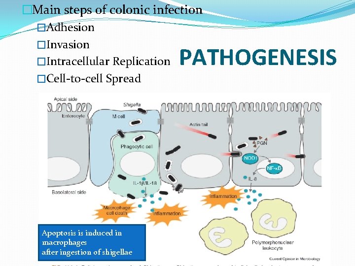
�Main steps of colonic infection �Adhesion �Invasion �Intracellular Replication �Cell-to-cell Spread PATHOGENESIS Apoptosis is induced in macrophages after ingestion of shigellae

Virulence Factors �genetic basis of virulence �molecular apparatus called a type III secretion system (T 3 SS) �capable of injecting bacterial proteins through bacterial and host membranes into host cells (translocation) �the extracellular milieu (secretion) to influence host biochemistry and cell physiology directly
�A chromosomal locus in S. dysenteriae serotype 1 encodes a Protein Synthesis-inhibiting Exotoxin (Shiga toxin, Stx) that is a major virulence factor �Plesiomonas shigelloides �hypothesized to protect populations with poor water supplies against S. sonnei
EPIDEMIOLOGY �Humans are the natural host �fecal-oral route �via contact with a contaminated inanimate object � ingestion of contaminated food or water �sexual contact �Houseflies also may be vectors � 10 organisms �Prolonged organism survival �water (up to 6 months) � food (up to 30 days)
�incubation period varies �from 1 to 7 days �ranges from 12 hours to a few days �but typically is 1 to 3 days
Risk Factors for Severe Disease �Infants and adults >50 y �Children who are not breastfed �Children recovering from measles �Patients with HIV �Malnourished children and adults �Any patient who develops �dehydration, �unconsciousness �hypothermia or hyperthermia �history of convulsion with first seen
Increased Risk Of Infection �Children 5 years or younger �child care settings �crowded conditions �Men who have sex �Travel to resource-limited countries �inadequate sanitation
CLINICAL MANIFESTATIONS �primarily infect the large intestine �Onset of high fever, toxicity, and crampy abdominal pain is sudden and typically precedes the onset of diarrhea. � During the first 48 hours, high-volume watery diarrhea may occur (small bowel phase of disease); subsequently, low-volume, bloody, and mucous diarrhea develops in association with urgency and tenesmus (large bowel or “dysenteric” disease)
�clinical manifestations range �from �watery or loose stools �with minimal or no constitutional symptoms �to �more severe symptoms, including � high fever, � abdominal cramps or tenderness, � tenesmus, � mucoid stools � with or without blood
�approximately half of children do not develop bloody diarrhea during the course of their disease �The presence of only watery diarrhea does not exclude the possibility of shigellosis, especially in an ill patient with high fever
� Septicemia � by Shigella organisms or � by other gut flora � � � Neonates malnourished children people with S dysenteriae serotype 1 infection but may occur in healthy children with nondysenteriae � shigellosis �Generalized seizures � self-limited � associated with high fever or electrolyte abnormalities �Reactive arthritis � HLA-B 27 � rare � weeks or months after
Shigella dysenteriae serotype 1 �often causes a more severe illness than other shigellae with a higher risk of complications �septicemia �pseudomembranous colitis �toxic megacolon �Intestinal perforation �HUS �rare in industrialized countries
�Physical examination shows � fever �signs of toxicity � tenderness over the lower abdominal quadrants �hyperactive bowel sounds �Rectal prolapse may be present, particularly when diarrhea is associated with malnutrition
�Children with Shigella encephalopathy have significantly higher case-fatality rates than children with shigellosis without encephalopathy. �The pathogenesis of neurologic signs and symptoms during episodes of shigellosis is unclear. �Hypoglycemia an electrolyte abnormalities are found in a few patients. � Direct invasion of the central nervous system during Shigella bacteremia is very rare.
Severe toxic encephalopathy EKIRI SYNDROME �described in Japan �Dysentery with hyperpyrexia �convulsions �sensory disturbances �rapid progression to death. � Mild hyponatremia � The children died of cerebral edema early in the course of disease (6 to 48 hours after onset). �NOT have �sepsis �DIC �HUS �Severe dehydration �This toxic encephalopathy is rare. �pathogenesis is unclear.
HUS �Shiga toxin �a potent cytotoxin �produced by S dysenteriae serotype 1 �small number of strains �S flexneri type 2 a �S dysenteriae type 4, �S sonnei �HUS has not been associated with infections attributable to these serotypes
�The course without therapy typically lasts 5 to 7 days �many enteric infections exacerbate undernutrition, initiating a “viscous cycle” of diarrhea and undernutrition �Even without antimicrobial therapy, the �carrier state usually ceases within 1 to 4 weeks after onset of illness; �long-term carriage is uncommon and does not correlate with underlying intestinal dysfunction.
Complications of Shigellosis � � � Abdominal Persistent diarrhea Postdysenteric irritable bowel syndrome Ileus, toxic megacolon, intestinal perforation Protein-losing enteropathy, malnutrition Surgical complications: intestinal perforation and obstruction, appendicitis, intraabdominal abscesses Neurologic Seizures Headache, lethargy, disorientation, hallucinations Coma Severe toxin encephalopathy or ekiri syndrome � Bacteremia � In malnourished children, young infants, and HIV-AIDS patients � Hemolytic-Uremic Syndrome � Only with Shigella dysenteriae serotype 1 � Urogenital � Vulvovaginitis, urinary tract infections � Other � Conjunctivitis, keratitis, corneal ulcers � Reactive arthritis � Reiter syndrome � Hepatitis � Myocarditis
DIAGNOSTIC TESTS �presence of fecal lactoferrin �Fecal leukocytes sensitive for colitis not specific for shigellosis �blood leukocyte count in patients with shigellosis often is normal �although leukopenia or leukocytosis may occur �A leukemoid reaction has been reported in 10% of patients infected with the Shiga bacillus

Plated lightly � Isolation of Shigella organisms � feces specimens � rectal swab specimens �At least two different culture media Mac. Conkey xylose-lysine-deoxycholate, Hektoen enteric agar Heavier plating more inhibitory Shigella. Salmonella medium � stool as soon as possible main determinant of success in isolating � not survive in fecal specimens during transit, � transport media Cary-Blair � transported at 4°C. (can’t be transported to lab in 2 hrs) � Enrichment Broth Mediadoes not significantly impact isolation rates � selective agar plate � Recovery of � shigellae is easier early in the course of the disease
PCR �detection of multiple bacterial, viral, and parasitic pathogens including Shigella have high sensitivity �but may yield false-positive results �(eg, detecting nonviable organisms)
�Serologic studies are not helpful in establishing the diagnosis �humoral antibodies develop after clinical recovery. �Serologic studies helpful in epidemiologic studies �differentiate Shigella from Escherichia species �Biochemical profiling �Serogrouping �mass spectrometry of cellular components
�B/C Should done in � severely ill �immunocompromised �malnourished children

�Thanks for your attention
- Slides: 28